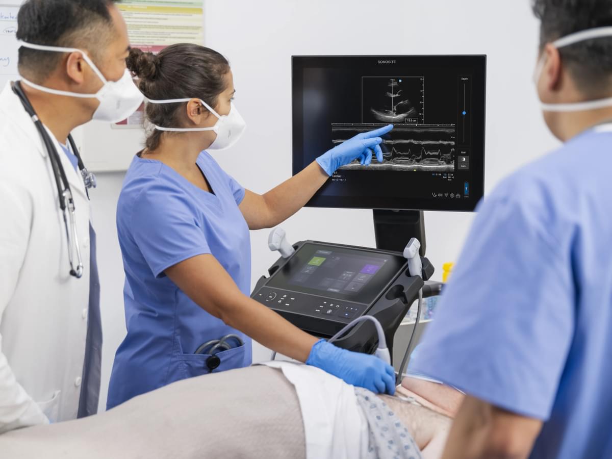
Heart disease prevalence continues to climb1, claiming millions of lives every year and remaining the leading cause of death worldwide2. Diagnosis is still challenging, creating avoidable delays between presentation and treatment3, calling for improvements to the speed and accuracy of current methods. Ultrasound – one of the most inexpensive and widely available imageing modalities – allows for direct visualisation of cardiac structure and function, representing a reasonable first step in cardiac health assessment4. Cardiac ultrasound is considered a first-line tool in heart disease diagnosis and management precisely because of its simplicity. Its inherent advantages – the use of harmless, painless sound waves, overall affordability, and ease of use – combine convenience with high-resolution imageing without ionising radiation, bringing returns on diagnostic information in real-time5. Today’s cardiac ultrasound technology offers accurate diagnosis and enables development of treatment modalities, making it an ideal approach for frontline diagnostics and image-guided procedures.
Cardiac Imageing at the Point of Care
Although clinical application of echocardiography can be traced back to the early 1950s6, the emergence of portable ultrasound systems truly revolutionised its use and propelled its expansion beyond traditional settings and within medical training programmes7. Point-of-care ultrasound (POCUS) has become a practise-changing technology for emergent and critically ill patient care. In primary care, access to echocardiography can accelerate specialist input and implementation of disease-modifying therapy8, enhancing operational efficiency and improving patient outcomes. In the current health care delivery landscape, POCUS can expedite triage and diagnosis, direct consultations, and guide clinical therapy and patient disposition, significantly decreasing morbidity and mortality in critically ill patients9.
Indications
Noninvasive, transthoracic echocardiography (TTE) can provide detailed structural and hemodynamic information for identification of life-threatening conditions such as major valve failure, severe reduction in biventricular function, cardiac tamponade, pulmonary embolism, and aortic dissection10. Cardiac POCUS can also aid in differentiating shock states and guiding resuscitative measures11.
Chest Pain - Focused cardiac ultrasound improves diagnostic specificity for evaluation of chest pain when differentials such as global cardiac dysfunction, aortic dissection, pericardial effusion, cardiac tamponade, pulmonary embolism (PE), or several primary pulmonary processes must be excluded.12 13 14 In the appropriate clinical setting, left ventricle (LV) wall motion abnormalities detected by ultrasound can indicate acute coronary syndrome;11 RV dilation may be suggestive of PE.15
Dyspnea – Shortness of breath can have cardiac, pulmonary, or a combination of both etiologies; POCUS can help make this important distinction. Cardiac ultrasound can assess the presence of pericardial effusion16, myocarditis, congestive heart failure, endocarditis17, acute coronary syndrome, or valvular heart disease18. In addition, POCUS lung ultrasound can evaluate pleural line integrity, consolidation, and pneumothorax.19 Bedside TTE has a specificity of 83% and a sensitivity of 53% for pulmonary embolism assessment20 and is an adequate rule-in test for unstable patients that are unable to get other confirmatory studies.
Hypotension – Shock represents up to one-third of all intensive care unit admissions and is associated with high morbidity and in-hospital mortality.21 Early recognition can improve patient outcomes,22 but traditional physical examination and hemodynamic monitoring alone cannot accurately establish circulatory failure etiology.23 Ultrasound protocols for standardised assessment of the inferior vena cava (IVC), gross LV function, lung, and abdomen can drastically increase overall diagnostic accuracy in various patient environments,24 allowing for rapid differentiation between septic, cardiogenic, hypovolemic, and obstructive shock.25 In addition, POCUS enables real-time evaluation of patients' response to interventions, helping guide diagnostic procedures.12
Cardiac Arrest – Approximately 90% of sudden cardiac arrest events are fatal, with 50% of victims presenting no prior symptoms or previous history of heart disease, and accounting for an estimated 325,000 deaths each year.26 POCUS can assist in timely detection of reversible anatomic causes such as pericardial effusion, cardiac tamponade, tension pneumothorax, or pulmonary embolism, and identify conditions that present similarly but have opposing treatments, like asystole and fine ventricular fibrillation.27 Ultrasound can also guide the application and location of force during resuscitation to elicit higher quality compression for improved survival. Combined compression effectiveness and cardiac activity measures have added prognostic significance: organised cardiac activity seen on ultrasound following pulseless electrical activity (PEA) arrest or asystole is associated with increased survival compared to disorganised activity,28 intra-arrest knowledge that can help inform the resuscitation timeline.
Penetrating or Significant Blunt Trauma – Rapid hemopericardium evaluation in thoracic trauma cases represents the earliest use of limited cardiac ultrasound by non-cardiologists,29 a practise that sparked debate and has since undergone considerable change. Today, ultrasound in trauma is represented by "extended focused assessment with sonography for trauma" (eFAST), a constellation of images used for detection of intraperitoneal haemorrhage, pericardial tamponade, and haemothorax or pneumothorax.30
Invasive ultrasound may be required in specific image-guided procedures. Transesophageal ultrasound (TUE) provides superior imageing of the posterior heart chambers and valvular structures when compared to TTE, and can be used for better visualisation during compression,31 coronary artery bypass graft surgery, transcatheter device placement, or to identify valvular prolapse, rupture, and vegetation in infective endocarditis.32 Intravascular ultrasound (IVUS) may be performed during cardiac catheterization to directly visualise atherosclerosis within the vessel walls when the extent and severity of stenosis are indeterminant during percutaneous coronary intervention (PCI).33
Techniques
- The subxiphoid view is the preferred scan to evaluate the entire heart and assess for pericardial fluid as part of the FAST/eFAST exam. Pericardial effusions are anechoic and typically encircle the heart when clinically significant.
- The parasternal long-axis view enables examination of the morphology and motion of the interventricular septum, left atrium, LV, MV, AV, aortic root, and ascending aorta. LV function can be directly assessed via fractional shortening, or indirectly estimated via E-point septal separation (EPSS). A dilated ascending aorta may indicate type A aortic dissection in the appropriate clinical context.
- The parasternal short-axis view at the level of the LV is a circular look at the walls of the LV and is ideal for evaluation of wall motion and overall contractility of the heart muscle.
- The apical 4 chamber view is ideal for comparison of the left and right sides of the heart. In this view it is possible to identify pericardial effusion and cardiac tamponade.
Ultrasound-guided pericardiocentesis is typically performed using subxiphoid view, but apical 4-chamber views may also be used, depending on patient habitus, positioning, and which axis of the heart is optimally viewed.
Fujifilm Sonosite: Innovators in the Cardiac Ultrasound Space
At FUJIFILM Sonosite, we understand the need to balance the demands of increasing cardiovascular disease with the constant pressure to improve department efficiency. Our POCUS systems are designed to deliver instant and reproducible results at the patient’s bedside with profound diagnostic, prognostic, and treatment implications, helping triage, determine clinical pathways, and guide procedures and resuscitation. They are routinely used in TTE goal-directed echocardiography, IVC collapsibility/stroke volume assessments to determine fluid responsiveness, RUSH exam for shock and hypotension, and TEE echocardiography for shock and cardiac arrest, and feature:
- Improved 2D and colour doppler imageing for enhanced speed and confidence in clinical interpretation;
- Intuitive user interface that reduces administration tasks, coordinates workflows, and automates data across the POCUS service line;
- Quick boot-up (under 25 seconds) from cold start to live scanning;
- Improved ergonomics and fluid-resistant surfaces for simplified cleaning and disinfection.
Learn how FUJIFILM Sonosite products can enhance your practise:
- Sonosite LX: Advanced image clarity on an expansive touchscreen display that allows rotation and extension for easy viewing with clinical team members and consultation with patients.
- Sonosite PX: combines Sonosite’s most advanced image clarity with improved colour sensitivity for accurate blood flow or inflammation assessment, and adaptable horizontal-to-vertical work surface for optimal bedside ergonomics.
- Sonosite SII: designed to maximize productivity, this Continuous Wave and Pulsed Wave Doppler imageing system can be used across multiple hospital environments, including a zero-footprint option for space-constrained rooms.
- Sonosite Edge II: Truly portable (can be used on or off the optional Edge Stand) with a simplified interface for intuitive access to frequently used functions.
- Sonosite M Turbo: Engineered for portability and unmatched durability, can be used for a wide range of diagnostic and procedural applications.
1. Roth GA, Mensah GA, Johnson CO, Addolorato G, Ammirati E, et al; GBD-NHLBI-JACC Global Burden of Cardiovascular Diseases Writing Group. 2020. Global burden of cardiovascular diseases and risk factors, 1990-2019: Update from the GBD 2019 study. J Am Coll Cardiol 76(25):2982-3021, PMID: 33309175, https://doi.org/10.1016/j.jacc.2020.11.010.
2. Vaduganathan M, Mensah GA, Turco JV, Fuster V, Roth GA. 2022. The global burden of cardiovascular diseases and risk: A compass for future health. J Am Coll Cardiol 80(25):2361-2371, PMID: 36368511, https://doi.org/10.1016/j.jacc.2022.11.005.
3. Hayhoe B, Kim D, Aylin PP, Majeed FA, Cowie MR, Bottle A. 2019. Adherence to guidelines in management of symptoms suggestive of heart failure in primary care. Heart 105(9):678-685, PMID: 30514731, https://doi.org/10.1136/heartjnl-2018-313971.
4. Kimura BJ. 2017. Point-of-care cardiac ultrasound techniques in the physical examination: better at the bedside. Heart 103(13):987-994, PMID: 28259843, https://doi.org/10.1136/heartjnl-2016-309915.
5. Aly I, Rizvi A, Roberts W, Khalid S, Kassem MW, et al. 2021. Cardiac ultrasound: An anatomical and clinical review. Transl Res Anat 22:100083, https://doi.org/10.1016/j.tria.2020.100083.
6. Edler I, Hertz CH. 1954. The use of ultrasonic reflectoscope for the continuous recording of the movements of heart walls. Clin Physiol Funct Imageing (2004) 24(3):118-36, PMID: 15165281, https://doi.org/10.1111/j.1475-097X.2004.00539.x.
7. Yamada H, Ito H, Fujiwara M. 2022. Cardiac and vascular point-of-care ultrasound: current situation, problems, and future prospects. J Med Ultrason 49(4):601-608, PMID: 34997377, https://doi.org/10.1007/s10396-021-01166-3.
8. Potter A, Pearce K, Hilmy N. 2019. The benefits of echocardiography in primary care. Br J Gen Pract 69(684):358-359, PMID: 31249096, https://doi.org/10.3399/bjgp19X704513.
9. Rooney KD, Schilling UM. 2014. Point-of-care testing in the overcrowded emergency department- can it make a difference? Crit Care 18(6):692, PMID: 25672600, https://doi.org/10.1186/s13054-014-0692-9.
10. Herbst MK, Velasquez J, Adnan G, O'Rourke MC. 2022. Cardiac Ultrasound. In StatPearls [Internet]. Treasure Island (FL): StatPearls Publishing, PMID: 29262211.
11. Arntfield RT, Millington SJ. 2012. Point of care cardiac ultrasound applications in the emergency department and intensive care unit- A review. Curr Cardiol Rev 8(2):98-108, PMID: 22894759, https://www.eurekaselect.com/article/44380
12. Amsterdam EA, Kirk JD, Bluemke DA, et al. 2010. Testing of low-risk patients presenting to the emergency department with chest pain: a scientific statement from the American Heart Association. Circulation 122(17):1756-76, PMID: 20660809, https://doi.org/10.1161/CIR.0b013e3181ec61df. Erratum in: Circulation 122(17):e500-1.
13. Labovitz AJ, Noble VE, Bierig M, et al. 2010. Focused cardiac ultrasound in the emergent setting: a consensus statement of the American Society of Echocardiography and American College of Emergency Physicians. J Am Soc Echocardiogr 23:1225-30, PMID: 21111923, https://doi.org/10.1016/j.echo.2010.10.005.
14. Melgarejo S, Schaub A, Noble VE. Expert Analysis- Point of care ultrasound: An overview. [Website]. Washington, DC: American College of Cardiology. https://www.acc.org/latest-in-cardiology/articles/2017/10/31/09/57/point-of-care-ultrasound [accessed 24 Feb 2023].
15. Chung-Esaki H, Knight R, Noble J, Wang R, Coralic Z. 2012. Detection of acute pulmonary embolism by bedside ultrasound in a patient presenting in PEA arrest: A case report. Case Rep Emerg Med 2012:794019, PMID: 23326723, https://doi.org/10.1155/2012/794019.
16. Blaivas M. 2001. Incidence of pericardial effusion in patients presenting to the emergency department with unexplained dyspnea. Acad Emerg Med 8(12):1143-6, PMID: 11733291, https://doi.org/10.1111/j.1553-2712.2001.tb01130.x.
17. Iung B, Rouzet F, Brochet E, Duval X. 2017. Cardiac imageing of infective endocarditis, echo and beyond. Curr Infect Dis Rep 19(2):8, PMID: 28233189, https://doi.org/10.1007/s11908-017-0560-2.
18. Buck T, Bösche L, Plicht B. 2017. Real-time 3D echocardiography for estimation of severity in valvular heart disease: Impact on current guidelines. Herz 42(3):241-254 [German], PMID: 28229203, https://link.springer.com/article/10.1007/s00059-017-4540-y
19. Milos R, Bartha C, Röhrich S, Heidinger BH, Prayer F, et al. 2023. Imageing in patients with acute dyspnea when cardiac or pulmonary origin is suspected. BJR Open 5: 20220026, https://academic.oup.com/bjro/article/5/1/20220026/7468308
20. Fields JM, Davis J, Girson L, Au A, Potts J, et al. 2017. Transthoracic echocardiography for diagnosing pulmonary embolism: A systematic review and meta-analysis. J Am Soc Echocardiogr 30(7):714-723.e4, PMID: 28495379, https://doi.org/10.1016/j.echo.2017.03.004.
21. Berg I, Walpot K, Lamprecht H, Valois M, Lanctôt JF, et al. 2022. A systemic review on the diagnostic accuracy of point-of-car.e ultrasound in patients with undifferentiated shock in the emergency department. Cureus 14(3):e23188, PMID: 35444920, https://doi.org/10.7759/cureus.23188.
22. Sebat F, Musthafa AA, Johnson D, Kramer AA, Shoffner D, et al. 2007. Effect of a rapid response system for patients in shock on time to treatment and mortality during 5 years. Crit Care Med 35(11):2568-75, PMID: 17901831, https://doi.org/10.1097/01.CCM.0000287593.54658.89.
23. Wo CC, Shoemaker WC, Appel PL, Bishop MH, Kram HB, Hardin E. 1993. Unreliability of blood pressure and heart rate to evaluate cardiac output in emergency resuscitation and critical illness. Crit Care Med 21(2):218-23, PMID: 8428472, https://doi.org/10.1097/00003246-199302000-00012.
24. Jones AE, Tayal VS, Sullivan DM, Kline JA. 2004. Randomised, controlled trial of immediate versus delayed goal-directed ultrasound to identify the cause of nontraumatic hypotension in emergency department patients. Crit Care Med 32(8):1703-8, PMID: 15286547, https://doi.org/10.1097/01.ccm.0000133017.34137.82.
25. Volpicelli G, Lamorte A, Tullio M, Cardinale L, Giraudo M, et al. 2013. Point-of-care multiorgan ultrasonography for the evaluation of undifferentiated hypotension in the emergency department. Intensive Care Med 39(7):1290-8, PMID: 23584471, https://doi.org/10.1007/s00134-013-2919-7.
26. Tsao CW, Aday AW, Almarzooq ZI, Alonso A, Beaton AZ, et al. 2022. Heart disease and stroke statistics- 2022 update: A report from the American Heart Association. Circulation 145(8):e153-e639, PMID: 35078371, https://doi.org/10.1161/CIR.0000000000001052. Erratum in: Circulation 146(10):e141.
27. Ávila-Reyes D, Acevedo-Cardona AO, Gómez-González JF, Echeverry-Piedrahita DR, Aguirre-Flórez M, et al. 2022. Point-of-care ultrasound in cardiorespiratory arrest (POCUS-CA): narrative review article. Ultrasound J 13(1):46, PMID: 34855015, https://doi.org/10.1186/s13089-021-00248-0.
28. Lee L, DeCara JM. 2020. Point-of-care ultrasound. Curr Cardiol Rep 22(11):149, PMID: 32944835, https://doi.org/10.1007/s11886-020-01394-y.
29. Nordenholz KE, Rubin MA, Gularte GG, Liang HK. 1997. Ultrasound in the evaluation and management of blunt abdominal trauma. Ann Emerg Med 29(3):357-66, PMID: 9055775, https://doi.org/10.1016/s0196-0644(97)70348-2.
30. Bloom BA, Gibbons RC. 2023. Focused assessment with sonography for trauma. In: StatPearls [Internet]. Treasure Island (FL): StatPearls Publishing, PMID: 29261902.
31. Gaspari R, Weekes A, Adhikari S, Noble VE, Nomura JT, et al. 2016. Emergency department point-of-care ultrasound in out-of-hospital and in-ED cardiac arrest. Resuscitation 109:33-39, PMID: 27693280, https://doi.org/10.1016/j.resuscitation.2016.09.018.
32. Hahn RT, Abraham T, Adams MS, Bruce CJ, Glas KE, et al. 2014. Guidelines for performing a comprehensive transesophageal echocardiographic examination: recommendations from the American Society of Echocardiography and the Society of Cardiovascular Anaesthesiologists. Anesth Analg 118(1):21-68, PMID: 24356157, https://doi.org/10.1213/ANE.0000000000000016.
33. Otake H, Kubo T, Shinke T, Hibi K, Tanaka S, et al. 2020. OPtical frequency domain imageing vs. INtravascular ultrasound in percutaneous coronary InterventiON in patients with Acute Coronary Syndrome: Study protocol for a randomised controlled trial. J Cardiol 76(3):317-321, PMID: 32340781, https://doi.org/10.1016/j.jjcc.2020.03.010.

