

Central line complications: they’re risky, costly, and all-around burden to healthcare. Whether it’s CLABSIs or iatrogenic pneumothoraces, CVC complications are not just terrifying for patients and unnerving for doctors; they are also costly for medical facilities.
Fortunately, there are tested methods for Iatrogenic Pneumothorax Prevention with POCUS that can drastically reduce complication rates (and associated costs) for critically ill patients who require venous catheterization.
1. If possible, aim for ultrasound-guided PIV instead
The first step in avoiding CVC complications can be avoiding CVC placements entirely. CVCs should be used only when clinically indicated, or when alternative vascular access options have been unsuccessful. Some patients are a “difficult stick”, due to obesity, history of intravenous drug use, dehydration, or other health-related issues. However, several studies have shown that ultrasound-guided PIV placement (versus landmark placement) can eliminate the need for central lines in up to 85% of critically ill patients with a history of difficult vascular access.
2. Utilise a CVC insertion checklist
Many hospitals have experience with OR-related checklists, as they were originally tested on CVC line placements, and have been recommended by the World Health Organisation and the UK’s NHS. Proper, localized implementation of a checklist protocol for CVC insertion can ensure that fewer mistakes are made and that all team members adhere to the proper care techniques.
3. Select the optimal insertion site
To minimize infections and complications during central line placement, choose the optimal CVC placement site for each patient, taking into consideration age, health status, build, medical history, expected duration of line placement, and risk of infection.
4. Use hand hygiene
These steps might seem obvious, but if they were consistently followed, we wouldn’t likely see 250,000 CLABSIs in the US every year. Perform hand hygiene before beginning the CVC insertion procedure.
5. Maximize sterile barriers
Maximize sterile barrier precautions like gloves, masks, caps, gowns and full body drapes when inserting a central line.
6. Adhere to aseptic technique
- Perform skin antisepsis and cleanse port immediately before use with correct antiseptic
- Perform skin antisepsis and cleanse port immediately before use with correct antiseptic
- Only access catheters with sterile devices
- Dress the site insertion with sterile, transparent, semipermeable dressings
- Replace compromised or soiled dressings using the aseptic technique
- Optional: use antiseptic impregnated supplies and dressings
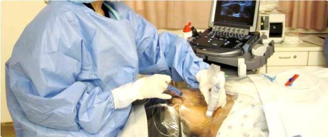
7. Guide CVC placement with ultrasound
Central line placement is performed in US hospitals more than 5 million times per year, with CVC placement complication rates as high as 15% when the needle is inserted using only the landmark method (Feller-Kopman 2007).
In a 2011 RCT study of subclavian CVC placements, patients whose lines were inserted “blindly” (using the landmark method) had a 4.8% incidence rate of pneumothorax. Compare this with the CVCs placed using real-time ultrasound guidance: these patients had a 0% rate of pneumothorax (Fragou 2011). The same rate of 0% was observed during a RCT study of 900 critical care patients receiving ultrasound-guided placements in the internal jugular vein, down from a 2.4% complication rate using landmark methods. There was also a nearly 10-fold reduction in the rate of accidental carotid artery puncture using ultrasound versus landmark method (Karakitsos 2006).
8. Remove unnecessary central lines immediately
A CVC should be continually assessed for necessity and removed immediately after it is no longer deemed necessary. These daily assessments can be planned during physician rounds or shift changes.
Interested in reducing CVC complications in your facility? Contact us regarding Sonosite Solutions.