


Costantino Balestra, Professor of Physiology at Haute Ecole Bruxelles-Brabant in Belgium, uses point-of-care ultrasound (POCUS) in environments that could not be more different from a typical hospital setting. His expertise lies in studying the effects of extreme conditions on the human body, including temperatures, altitudes, and ambient pressures found in deep oceans.
One of his areas of research has focused on evaluating the formation of vascular gas emboli (VGE) in divers after they have resurfaced from high-pressure depths. While the presence of these air bubbles in the heart is a normal physiological reaction to hyperbaric environments, VGE can block or distort blood vessels and trigger associated inflammatory responses, leading to decompression illness. VGE can be quantified by ultrasound Doppler and precordial echocardiography; the higher the number of bubbles, the more likely decompression illness may occur. Dr. Balestra explains more about his research:
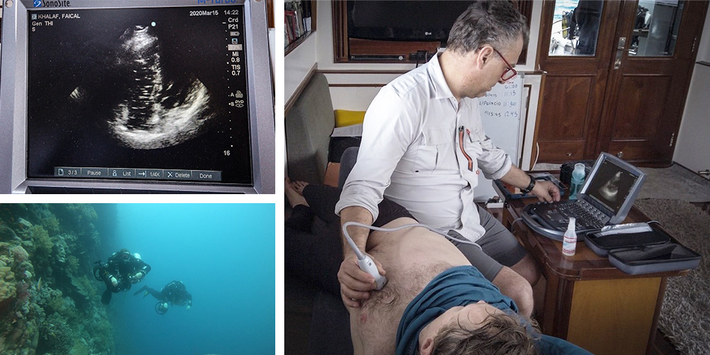
“Very little research has been carried out on VGE after deep dives, so this is where we have focused our efforts in recent years. We began by looking at the presence of gas bubbles that form in the heart when a diver begins an ascent, hoping that understanding more about why and how they form could influence the decompression algorithms used to control safety. We carried out examinations on the boat, after the diver came back on board and at regular intervals thereafter. For deeper dives of around 75-100 meters, the mix of gasses you need in your tank is different to when you only dive to 30-40 meters. At medium-depth dives, a mixture of oxygen and nitrogen is used, but for deeper dives there is the risk that this will cause neurological toxicity. Therefore a third gas, helium, is introduced as the primary gas. Very little is known about the formation of VGE when divers have been using helium in this ‘trimix’ ratio.”
On his most recent research trip, Dr. Balestra had a team of four divers carrying out a 28-day saturation dive in the Red Sea, at depths of 120-140 meters, using the trimix gas. The divers were underwater for six hours a day—the pressures at this depth are equivalent to 11 times atmospheric pressure. Underwater, both the divers and the FUJIFILM Sonosite system they had aboard had to face the same tough conditions, as divers had to face tough conditions and the same could be said for the FUJIFILM Sonosite ultrasound system used on board, as Dr. Balestra explains:
“In my field of work, I need a very tough instrument that can work well in any environment, whether that’s sandy, hot desert conditions, extremely cold environments, or on an unstable surface, such as a boat. My research depends on having a reliable ultrasound system that can do its job with no hitches. I need high-quality images to accurately see and count the VGE, as well as decent battery power that allows time to do all the necessary measurements. It has to be a compact and lightweight system for use on an overcrowded boat, and must be able to withstand frequent knocks and bumps. Now that we have seen how Sonosite devices can match up to these rigorous demands, we won’t be trying anything else.”
Dr. Balestra’s research has shed light on how complex the formation of VGE is in individual divers, and on the associated risk of decompression illness. He explains there is not a simple equation for who will and won’t experience sickness after deep diving:
“Our studies in the Red Sea support the notion that there is individual variation in the presence of VGE and great variability based on age, fitness, type, and frequency of physical activities. Our results continue to show that VGE influences the risk of decompression sickness and opens new possibilities for decompression algorithms by considering the diver's individual susceptibility, lifestyle, and recent exercise to predict the level of VGE during and after decompression.”