Blue Cross Vets Clinic is the 1st Advanced Veterinary centre located at Navi Mumbai and offers different services of diagnostic, surgical & emergency care for small pet animals. They have a dedicated team of veterinarians taking care of companion animals and we tried to find out from them the various advantages of using point of care ultrasound in their daily practice.

Since how long you have been using Ultrasound?
We have been using Ultrasound since September 2018, and have found it extremely useful as a quick non-invasive diagnostic tool for our routine veterinary practice involving different species of pets.
Which Ultrasound machine are you currently using and how do you find it useful in your daily practice?
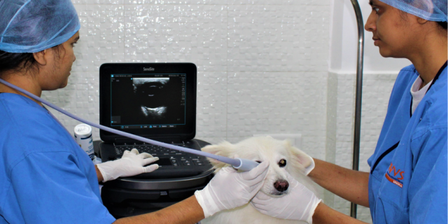
We are currently using Sonosite EDGE II color Doppler Ultrasound system which is very light-weight, compact and user-friendly. The probes are extremely light weight and doesn’t fatigue the hand even after heavy use, neither does it stress out our smallest patients like bird/cats, etc. The clarity of linear probe is mind blowing giving us exact anatomy, location and structure characteristics. Its quick boot time and processing speed is extremely useful in our practice. The machine is quite reliable, durable, gives us quality images and is easy to set up anywhere and start scanning immediately. We use it extensively for most of the pet patients to quickly rule-in or rule-out disease conditions. It is especially useful in our set up where mobility is restricted and the patient is sometimes difficult to restrain.
“Point-of-care Ultrasound
advantages are
limitless and uses are countless.”
How this Ultrasound system has changed the way you used to practice earlier?
We are thrilled, clients are happy and patients are getting better. It is complimentary to x-ray and has shortened the time to diagnose and provides better clinical results for making differential diagnosis. No blind centesis and reduced risk of major vessels puncture or other organs any more as our patients tend to move and are unpredictable, helps differentiating pericardial and pleural fluid accumulations and identifying exact location for biopsy collection. Now we can detect early pregnancies, observe foetal development within seconds. In case of opthalmic study, it is easy to scan quickly the posterior ocular structures which cannot be visible with naked eye. We also used it for FAST (Focussed Assessement for Screening in Trauma) scanning in emergency trauma patient & in all major/minor diagnostic procedures.
How efficient is this ultrasound in US guided procedure and what are the benefits?
With the help of Ultrasound, safety and efficacy has been significantly improved. Under ultrasound visualization the internal organs are safe from iatrogenic trauma during cystocentesis and FNAC biopsy of liver and spleen have become easy to perform. Also it is easy to aspirate & perform thoracentesis/ pericardiocentesis under ultrasound guidance, which earlier used to pose several risks when performed through blind techniques.
“It is easy to perform
procedures under ultrasound guidance
which earlier were risky to perform
through blind techniques.”
