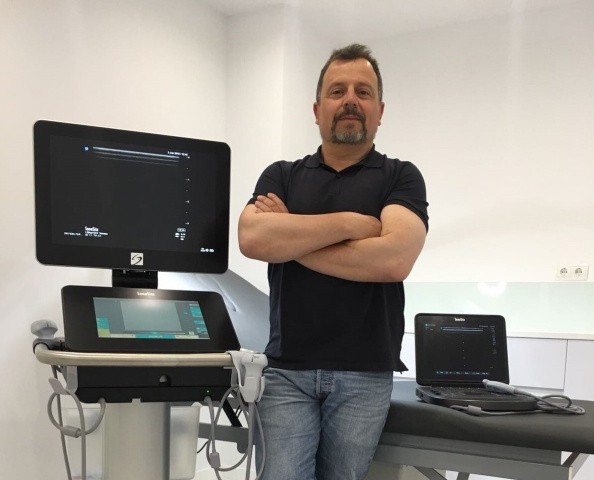
Ultrasound is a vital tool for vascular surgeons in the 21stcentury. Dr Fidel Fernández Quesada, a sixth-generation vascular surgeon and associate professor at the University of Granada, Spain, describes the difference it has made to his clinical practice and reflects on what his forefathers would have made of such technological advancement.
I have worked in the field of angiology and vascular surgery for 25 years, and currently practise both at the San Cecilio University Hospital in Granada and privately. I am also the current president of the Phlebology and Lymphology division of the Spanish Angiology and Vascular Surgery Association. Like every other specialist in vascular medicine and surgery, I use ultrasound every day in my clinical practice. I have been using FUJIFILM Sonosite systems for more than 15 years now, and have embraced each new system as it has been developed.
Every standard diagnosis of vessel pathology starts with an ultrasound examination, allowing the clinician to see the anatomy and the functionality of the artery or vein, and helping them to ‘dig deeper’ into an area to obtain more detailed, essential information on the appearance or function of the vessel. For example, it is possible to take a patient into theatre to operate on an embolic episode already knowing precisely where the obstruction is, allowing us to mark the exact entry point on the skin. Another useful diagnostic role for ultrasound is in the examination of the condition of the carotid artery; using ultrasound I can measure the degree of stenosis, the tightness of the artery based on the speed of blood flow, and can check where the plaque starts and ends, which is vital information to have prior to surgery. For all of these reasons, arteriography has been established as the first-option imaging tool.
Similarly, ultrasound has proved to be integral to modern procedures for treating diseased vessels, offering advantages over alternative treatments. For example, when treating varicose veins by sclerotherapy, we use ultrasound to determine the puncture site and to control the injection of the sclerosant. Vein cartography drawn by ultrasound guidance allows us to determine the exact point of incision, reducing the size of the cut required compared to other techniques. In endoluminal therapies, such as laser and radiofrequency therapies, ultrasound gives us control during the incision and the follow up of the whole process. Patient outcomes are improved, and the risk of complications post-surgery is reduced.
Point-of-care ultrasound has many applications in surgery, and it is used on a regular basis to place catheters for vascular access, to determine the pathologies to be corrected on dialysis fistulas, or for the placement of catheters for chemotherapy; guided puncture ensures pin-point precision and is essential for determining how the vessels are channelled. In addition, ultrasound examination is a key aid to monitoring disease effects over time. For example, following a diagnosis of ischaemia, changes to the arterial and venous pathology can be closely monitored over time to help inform further potential medical or surgical interventions.
I discovered FUJIFILM Sonosite during my time as a resident in the United States. I was hugely impressed with what the company offered and how its systems functioned, and thought ‘this is for me’. I needed a durable device and heard that the probes were resistant to impact, and when I saw the grey scale it was exceptional. My first system was a TITAN, which performed exactly as its name suggests, even in the Saharan refugee camps where I worked as part of international cooperation projects. I also liked the reassurance provided by the five-year warranty. Since then, I have upgraded as new Sonosite systems have come to market, using first the M-Turbo, then the Edge and, more recently, the Edge II and X-Porte models, keeping abreast of the latest developments in ultrasound technology and, most importantly, helping to enhance patient care.
I often visit patients at home, travelling by motorbike, and the Edge II is perfect for this situation. It is small and portable, and the image quality is excellent. The system is very robust too, and easily withstands the bumps and knocks that it regularly encounters. I once fell off my motorbike while carrying the system and there wasn’t a scratch on it! In theatre, I use both the Edge II and an X-Porte system. The X-Porte provides extreme definition, and is ideal if I am teaching, especially for its big screen. It is easy to demonstrate, and the intuitive touchscreen operation is perfect for today’s students, who have grown up using tablets and smart phones.
I am the sixth generation of doctors in my family, and consider myself fortunate to be practising angiology and vascular surgery in this exciting period of technological advancement. When I was a resident, I never thought that we would have such a tool in my lifetime; even just 40 years ago, the medical community could not imagine the revolution point-of-care ultrasound would be. In the near future, I can see general practitioners taking ultrasound machines out on home visits to aid diagnosis, reducing the need for hospital admissions. Point-of-care ultrasound has become the eyes and hands of the vascular surgeons. If my grandfather, great-grandfather and great-great-grandfather, who were also surgeons, could see the technology we have today and the way I work with my patients, they would be stunned!
Sonosite, the Sonosite logo and Edge, Edge II, M-Turbo, TITAN, and X-Porte are trademarks and registered trademarks of FUJIFILM Sonosite, Inc. in various jurisdictions. FUJIFILM is a trademark and registered trademark of FUJIFILM Corporation in various jurisdictions. All other trademarks are the property of their respective owners. Copyright (c) 2018 FUJIFILM Sonosite, Inc. All rights reserved. Subject to change.
Medical scope of practice can vary by state, country and/or local jurisdiction.

