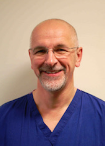
Ultrasound guidance has proven invaluable for the regional neurosurgical centre at the Salford Royal Hospital, helping to improve safety, save time and enhance the patient experience. Dr. Jim Corcoran, consultant neuroanaesthetist and clinical director for perioperative care at the hospital, explained.
When I joined the Salford Royal in [2006], my initial interest in ultrasound grew out of a need to alleviate the delays experienced by trauma patients needing a cardiac echo. Training opportunities for regional anaesthesia in the North West were very limited, but we realised that training clinicians to use point-of-care echo could significantly reduce delays in excluding aortic stenosis. At the same time, we took the opportunity to introduce training on the use of ultrasound for regional anaesthesia. Interest in this field has grown considerably since then, and for the past nine years we have worked with FUJIFILM Sonosite to run twice-yearly courses in ultrasound-guided regional anaesthesia.
Before ultrasound guidance became commonplace, regional anaesthesia was performed using nerve stimulators. Although this worked, the procedure wasn’t slick. Ultrasound offers an improved way of administering the anaesthetic, which is quicker, safer and more comfortable for the patient. It allows you to visualise the nerve, giving assurance that the needle is correctly positioned, and gives you confidence that the block will work; there is always a degree of uncertainty if ultrasound is not used.
We can also dramatically reduce the amount of anaesthetic used with ultrasound-guided procedures, decreasing the likelihood of side effects. Whereas previously 30 or 40 ml of anaesthetic would be injected – because it was hard to target a specific area with certainty – you now know exactly where the needle is, and can see the anaesthetic spreading across the area. As a result, as little as 10 to 20 ml of anaesthetic is required, which is a huge reduction.
Today, we use ultrasound-guided regional anaesthesia for both awake surgery and analgesia, for example, interscalene blocks for patients having shoulder surgery under general anaesthesia. Regional anaesthesia has totally transformed shoulder surgery, significantly reducing the length of patient stays. Ten years ago, patients were admitted for one or two days, but subacromial decompressions, for example, are now treated as day cases, and even a shoulder replacement is only an overnight stay. I also use ultrasound to place catheters in some of the more challenging shoulder replacement cases, which makes a big difference; the interscalene groove is a difficult area, and knowing the exact location of the needle is really important.
The efficiency of our hand surgery lists has improved too, as we work almost in parallel with the surgeon; while one patient is in theatre, we administer a block to the next person, reducing the impact of anaesthetic time and increasing throughput. In addition, we’ve trained our A&E colleagues to administer ultrasound-guided fascia iliaca blocks to trauma patients. This has really improved the quality of care, ensuring patients are comfortable during transfer between A&E and the X-ray department, and minimising the morphine dose required.
When ultrasound guidance first became mandatory for vascular access, we set up a training course for all doctors within the organisation who were placing central lines. Over time, as the use of ultrasound became commonplace, this knowledge started to be handed down informally, rather than through attendance at training courses.
The risk with this approach is that messages get diluted, and so we prefer trainees to undergo more formalised training for this procedure, as they do for ultrasound-guided regional anaesthesia. It is important that they realise that they are looking at a computer-generated image and are aware of the potential for artefact formation, as well as understanding how this occurs. We also run basic level training courses for senior house surgeons across the region, plus more advanced courses that are open to clinicians from outside the area.
Ultrasound guidance is now an established procedure at the Salford Royal, and we have a considerable number of Sonosite systems shared across 20 theatres – including two X-Portes®, an Edge®, two M-Turbos®, three S-Nerves™, a MicroMaxx® and three iLooks® – which are used for both regional anaesthesia and vascular access. The systems are reliable and user-friendly, and the customer service is good.
The X-Portes have become particularly popular, not only for anaesthesia, but also for central line placement. People like the big screen and wipe-clean surface, and the training features are really good. If a trainee wants to refresh their knowledge of particular anatomical landmarks, they can simply the watch relevant video, which acts as a mini tutorial.
The big advantage of ultrasound guidance is the safety and reliability it offers, even when you are treating a patient with difficult vascular access; a large lumen line, for example, can be safely inserted using the Seldinger technique. Ultrasound makes a huge difference; it allows you to do things differently, and is saving lives.


