

.png)
Point-of-care ultrasound is an essential tool for busy nephrology departments, and dialysis units in particular. Dr. Christoph C. Haufe, clinical lead at an outpatient dialysis center in Erfurt, Germany, discusses the use of the technique in daily practice and his involvement in ultrasound training.
The nephrology center in Erfurt is an outpatient dialysis unit for people living locally and a center of renal care. The associated renal department is a regional specialist nephrology hospital that treats inpatients suffering from a range of kidney complaints from across a wide area of eastern Germany. Ultrasound technology plays an important role in nephrology, and is used many times a day to aid the treatment of both in- and outpatients. It is particularly useful for dialysis patients with fistulae that are difficult to access; ultrasound imaging makes it easier to identify the correct puncture point and insert the needle. Dr. Haufe explained:
“Most patients dialyses via a fistula, but everyone’s anatomy is different, and it is harder to obtain vascular access in some people than in others. When access is proving problematic, we find there is no better way to perform the puncture than under ultrasound guidance.”
In the past, if it was necessary to use ultrasound guidance, doctors would have to move large, cart-based ultrasound systems between wards and patients. Each move entailed shutting down and rebooting the system, which was time consuming and caused delays. Due to a lack of space at the bedside, the system had to be situated to one side of the clinician – opposite the patient – which meant that it was not possible to simultaneously look at the ultrasound image and the puncture site. Dr. Haufe continued:
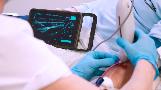
“When I was shown a small ultrasound device, I realized its potential to resolve this issue. We invested in two systems; one for the specialist nephrology department, and the other for the outpatient dialysis unit. This allows us to use ultrasound guidance for vascular access, and to quickly scan a patient’s kidney to confirm or exclude some renal conditions. The devices offer good quality imaging and are easy to handle, making them ideal to use at the bedside. They are particularly useful for dialysis patients as they can be placed close to the patient’s arm, allowing me to view the image while I perform the procedure. And with a touchscreen and smooth surface, they are easy to disinfect after use.
Traditionally, ultrasound was used only by doctors, but this does not need to be the case, and I am keen to give all our renal nurses the opportunity to learn the technique. Ultrasound scanning is quickly learned, straightforward, and does not hurt the patient, and so we began to run training courses for our renal nurses. Before ultrasound, there was a greater risk of a mispuncture occurring, which not only reduced the nurses’ confidence in their ability to perform the procedure – they would often call for a doctor rather than try a second time – but was uncomfortable for the patient. It is so much easier with ultrasound guidance. Having a good quality ultrasound image of the needle and vein reduces the potential for a mispuncture, which is invaluable for anyone learning the technique. The nurses can now be confident of success first time, which is better for them and the patients.”
Once all of the renal nurses in Erfurt had been trained in the use of ultrasound, there was a drive to establish nurse-led courses to enable this service improvement to be passed on to other hospitals in the region. As Dr. Haufe concludes,
“I encouraged one of my renal nurses to give a presentation at Erfurter Dialysefachtagung, and this generated a great deal of interest. Nurses could see the benefits it offered, and were keen to attend courses to learn how to use ultrasound. It is a really good thing for nurses, doctors and, most of all, the patients.”