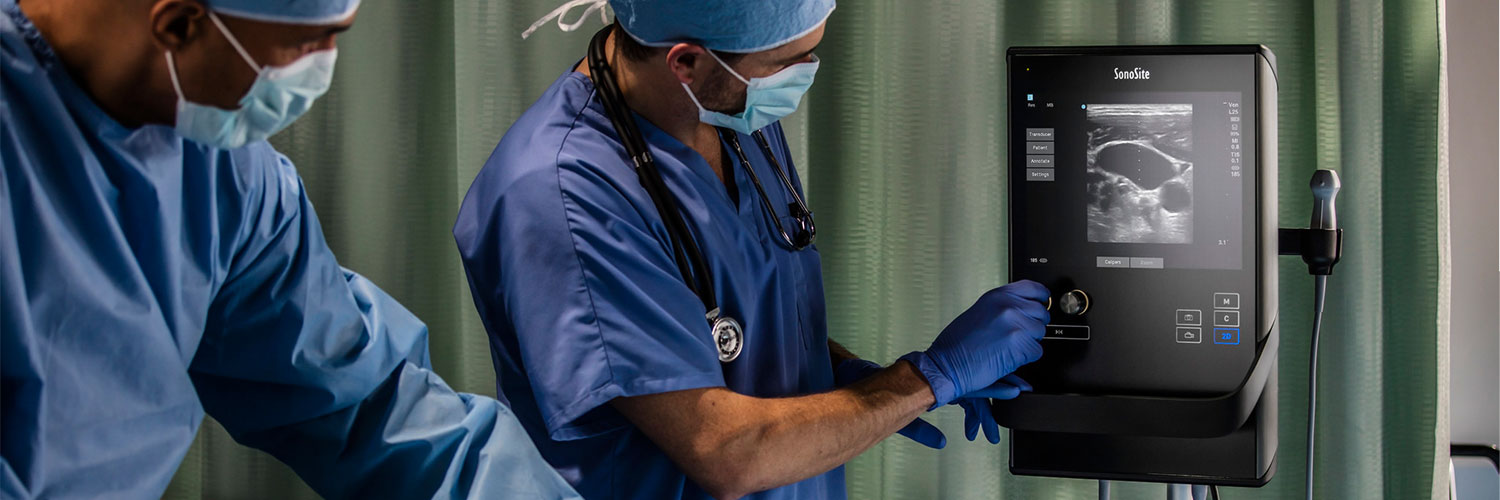
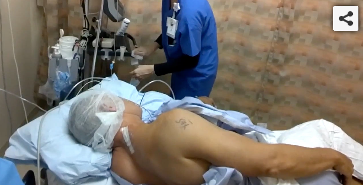
Beyond the Block: Why Would an Anaesthesiologist Use Ultrasound?

 Increasingly, anesthesiologists have been using ultrasound guidance to help visualize soft tissue anatomy and nerve location while performing regional nerve blocks. Correct placement of local anesthetics lead to long lasting pain management and enhanced

Increasingly, anaesthesiologists have been using ultrasound guidance to help visualise soft tissue anatomy and nerve location while performing regional nerve blocks. Correct placement of local anaesthetics lead to long lasting pain management and enhanced recovery times.

But beyond the block, how does ultrasound help anaesthesiologists do their jobs?

The answer has a lot to do with the changing practise of medicine.

Beyond the Block: Why Would an Anaesthesiologist Use Ultrasound?

Increasingly, anaesthesiologists have been using ultrasound guidance to help visualise soft tissue anatomy and nerve location while performing regional nerve blocks. Correct placement of local anaesthetics lead to long lasting pain management and enhanced recovery times.

But beyond the block, how does ultrasound help anaesthesiologists do their jobs?

The answer has a lot to do with the changing practise of medicine.

Ultrasound Answers Questions in Emergency Care in Spain

Ultrasound crtical to stabilizing patients and saving lives

In the world of emergency medicine, there’s nothing fun about the guessing game. With a seriously ill or injured patient, every second counts and the wrong diagnosis can actually have a significant impact on health. That’s why having all available information is absolutely critical to stabilising patients and saving lives.

That’s also what makes point-of-care ultrasound so critically important in emergency care.

Ultrasound in MSK

Sonosite was recently graced with visiting doctors from around the country, one of which was Dr Byron Patterson who is a partner in Primary Care Sports Medicine in Tarzana California.  He made a wonderful presentation to more than 160 employees about ultrasound applications in sports medicine.

Glimpse Episode 4: Dr. Francis Yamazaki, Discusses Anesthesia

Dr. Francis Yamazaki

Dr. Francis Yamazaki is a 30 year veteran in Anesthesiology. He is an Anesthesiologist with a cutting edge sports medicine group at Kerlan Jobe Surgery Center, LA. In this interview, he discusses the expectations of patients in the elite world of sports and the progression of anesthesia, particularly in the realm of nerve blocks. In particular how the progress of technology has improved safety for both patients and surgeons.

HIMSS15 Marks Major Changes in Health IT

The trend towards interoperability is palpable and what better way to underscore this positive movement than HIMSS 2015 conference which broke all time attendance records. FUJIFILM Sonosite is at the forefront of clinical workflow optimization and health IT interoperability. The need to connect modalities, especially Ultrasound, to EMRs and other Health IT systems is significant and FFSS is taking concrete steps to achieve this important goal.

Game Day Ultrasound

Courtesy of FUJIFILM Sonosite ANZ The Fremantle Dockers Football team have been using on-field Ultrasound as a way to visualize and diagnose soft tissue injuries with their players. The Sonosite Edge portable Ultrasound system provides beautiful images of the ribs and soft tissues for "match-day" diagnosis. This information allows the physician and coach to make an informed decision of whether the player should be sent back out in the field.

Ultrasound Can Diagnose and Prevent Pneumothorax

I find it ironic that one of the most efficient ways to detect pneumothorax in patients is also one of the most effective ways to prevent two of its more common clinical causes.

While scenarios that might trigger the suspicion of pneumothorax are too numerous to list, screening for it has included the standard trinity of chest-imaging procedures: x-rays, tomography, and ultrasound. In “Think Ultrasound When Evaluating for Pneumothorax” (featured in AIUM’s Journal of Ultrasound in Medicine), author Vicki E. Noble, MD, of Massachusetts General, proposes that ultrasound is the ideal screening modality due to its portability and lack of “risk associated with repeated measures as clinical scenarios change.”

Dr. Noble’s article goes on to provide instruction for performing the examination, incorporates excellent research citations, and then concludes: “Using thoracic sonography as a screening tool may lead to decreased ordering of chest radiographs, thus saving time and money and improving the efficiency of treatment.”

It may come as no surprise that my colleagues and I at Sonosite enthusiastically support Dr. Noble’s conclusion. The entire article—a part of AIUM’s “think ultrasound first” Sound Judgment Series—is available here and is worth a thorough read.