


Alexandros Mallios is a vascular surgeon at the Institut Mutualiste Montsouris (IMM) hospital in Paris. For the last four years, he has pioneered the use of ultrasound-guided percutaneous arteriovenous fistula access in patients undergoing hemodialysis. Dr. Mallios explains why he believes point-of-care ultrasound (POCUS) should be the standard of care for vascular access in all patients.
During my vascular surgical fellowship at the University of Oklahoma, it became clear to me that using ultrasound was fundamental to providing high quality care in hemodialysis patients. It helps to ensure vascular access is maintained in the long term, while reducing discomfort and complications for patients. I saw the benefits of this approach for guiding needle insertion for a wide range of procedures.
Improving Precision, Quality, and Safety
I moved to the IMM in Paris as a senior vascular surgeon in 2015. I now work predominantly in hemodialysis, and a large part of my role is creating and preserving vascular access in patients who need chronic hemodialysis. For these patients, an arteriovenous (AV) fistula—connecting a vein to an artery – is created, to allow the vein to mature and develop, make repeated punctures possible, and ensure there’s enough blood flow to allow treatment. Each time a patient comes in for hemodialysis, insertion of two needles through the skin is required to access the AV fistula and connect the patient to the dialysis machine. There are a number of challenges to successful cannulation, requiring accurate needle insertion to avoid hitting pre-existing hematomas (caused by previous cannulations or unsuccessful attempts), natural bifurcations and tortuosity of the veins, perforation of the vessel walls, or missing the fistula entirely. Essentially, improving cannulation means less suffering for the patient, less risk of complications, and increased longevity of the fistula.
Benefits for All
The use of ultrasound to guide needle placement reduces these challenges, as well as the complications that can occur afterwards. Rather than “going in blind,” the clinician can examine the vascular landscape and determine the best location for the puncture; any hematomas present can be avoided, changes in the depth or lateral position of the vein can be accounted for, and the needle can be tracked as it travels further into the vessel to avoid perforating the back wall. The accuracy gained from visualizing the vessels with POCUS not only decreases the risk of cannulation failure, but also reduces the discomfort for the patient because we are more likely to get the puncture right on the first attempt. There is also less chance of causing hematomas and bruising with ultrasound guidance, helping the fistula to remain accessible for a longer period of time.
I use POCUS with all of my patients, to map the anatomy, locate the site of incisions, and measure physiological parameters. Ultrasound images provide the anatomical and physiological information required to make the procedure as safe and effective as possible. It is also useful for all other applications in medicine where vessel puncture is required. Patients experience less pain and discomfort when ultrasound is used, and physicians have greater confidence in the success of the procedure because they know exactly where to access the vessel.
Pioneering Punctures
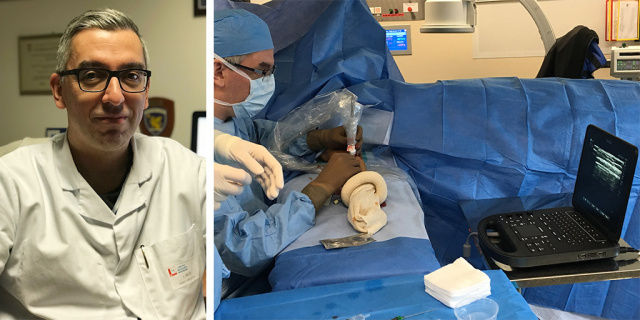
In the last few years, we have been developing an ultrasound-guided percutaneous technique to create AV fistulae without cutting through the skin with a scalpel. With POCUS, the needle puncture can be achieved with absolute accuracy, and the entire procedure of fistula creation is completed without need for X-rays or contrast injections. Starting with the transducer in the transverse position, you can clearly see the needle as it is inserted, and can ensure it is in the center of the vessel at all times. A longitudinal plane then allows you to visualize the length of the vessel as the needle advances through it, which is particularly useful for veins that drop or are tortuous.
Spreading Knowledge
I regularly share my ultrasound skills with others. I am training surgeons to use it when creating surgical fistulas and nephrologists when evaluating a fistula, and teaching nurses to know where to insert needles and helping them with cannulation. I find our FUJIFILM Sonosite ultrasound systems to be very easy to use for training, as they are intuitive, with only a few buttons, allowing first-time users to navigate the device without being overwhelmed. Sonosite machines are lightweight, easy to carry, and very robust.