

Understanding Venous Congestion
Venous congestion refers to the accumulation of blood in the venous system, which can adversely affect organ function. Conditions such as pulmonary edema, cerebral edema, and congestive nephropathy can arise from venous congestion. Traditionally, the focus has been on arterial blood flow and cardiac output, but recent studies highlight the critical role of venous return in overall perfusion.
The VExUS Exam
The VExUS exam is a Venous Excess Ultrasound Grading System designed to assess venous congestion through Doppler ultrasound. Key components include:
- Waveform Analysis: The exam evaluates waveforms from the hepatic vein, portal vein, and intrarenal veins, which change predictably with increasing congestion.
- Scoring System: The VExUS C grading system was identified as the most predictive of acute kidney injury (AKI) in patients, particularly post-cardiac surgery.
| Vessel | Normal Waveform | Congested Waveform |
| Hepatic Vein | S>D | D>S or S reversal |
| Portal Vein | Continuous Flow | Pulsability increases |
| Intrarenal Veins | Continuous Flow | Biphasic to monphasic |
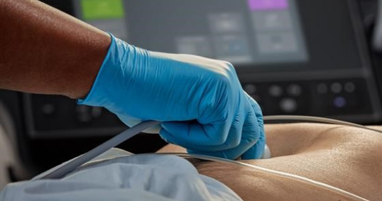
Performing the Vexus Exam
To effectively perform a VExUS exam follow these steps:
- Inferior Vena Cava (IVC) Assessment:
- Obtain both long-axis and short-axis views to confirm IVC collapse and shape.
- A small, collapsible IVC indicates no significant venous congestion.
- Hepatic Vein Evaluation:
- Use a lateral approach to obtain a Doppler waveform.
- Normal waveforms show S and D waves, while congestion alters this pattern.
- Portal Vein Examination:
- Identify the portal vein using color Doppler.
- Normal flow is continuous; increased pulsatility indicates congestion.
- Intrarenal Vein Analysis:
- Locate the intrarenal veins and assess for continuous low-grade flow.
- Changes in waveform patterns indicate varying degrees of congestion.
Interpretation of Findings
How to interpret the findings from the VExUS exam:
- IVC: A plethoric, non-collapsible IVC suggests elevated right atrial pressure.
- Hepatic Vein: A reversal of the S wave indicates severe congestion, often associated with tricuspid regurgitation.
- Portal Vein: Increased pulsatility and flow reversal below the baseline indicate severe congestion.
- Intrarenal Veins: A biphasic pattern suggests mild congestion, while monophasic flow indicates severe congestion.
Caveats and Pitfalls
- Technical Difficulties: The hepatic vein can be challenging to interpret without ECG gating.
- Respiratory Variability: Respiratory motion can affect Doppler waveforms, making it essential to have patients hold their breath during the exam.
- Clinical Context: It is crucial to interpret ultrasound findings in conjunction with the overall clinical picture, avoiding over-reliance on single data points.
Clinical Integration
The VExUS exam can significantly influence clinical decision-making, particularly in patients with heart failure or acute kidney injury. VExUS has been proven to guide treatment decisions, such as diuresis in patients with suspected congestive nephropathy.
The VExUS exam represents a promising advancement in the assessment of venous congestion, offering a more nuanced understanding of fluid status in patients. As the evidence base continues to evolve, the integration of VExUS findings into clinical practice may enhance decision-making and patient care in various medical settings.
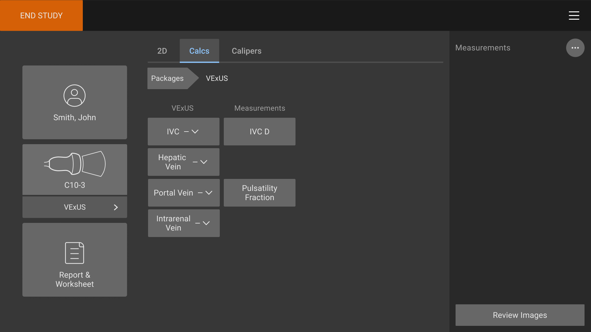
Sonosite’s dedicated exam type
Until now the calculations required to evaluate venous congestion using the VExUS score were performed off-modality.
Sonosite’s solution to support VExUS is a dedicated exam type and analysis package that provides users a protocol style workflow to evaluate and score the severity of venous congestion.
The VExUS exam type has been specially optimized for obtaining the four views required for a VExUS score and combined it with a dedicated measurements package for maximum efficiency.
The VExUS exam type with ECG compatibility is available on Sonosite LX and Sonosite PX with the P5-1, C5-1 , and C10-3 transducers.
- Complete the VExUS scoring exam with a single transducer and dedicated exam type for maximum efficiency
- VExUS exam type includes a VExUS score calculation package to help simplify the process of assessing fluid status
To get started with VExUS, download our quick reference guide today.
Click to download the guide today.
1. Beaubien-Souligny, W., Rola, P., Haycock, K., et al. (2020). "The VExUS Score Predicts Acute Kidney Injury in Critical Illness". Journal of Clinical Ultrasound.
2. Ciozda, W., Kedan, I., Kehl, D. W., et al. (2016). "The IVC assessment with point-of-care ultrasound: what is the best view and best mode?". Journal of Cardiac Failure.
3. Danforth, W. H., & Tissot, C. (2019). "Echocardiographic approach to fluid management in critical illness". Critical Care Clinics.
4. Jardin, F., Vieillard-Baron, A., et al. (2019). "ARDS and venous return: let's go back to physiology". Intensive Care Medicine.
5. Neumann, A., Hoyer, D., Bendrat, K., et al. (2021). "Quantifying intrarenal venous flow patterns in patients with acute kidney injury". Intensive Care Medicine Experimental.
6. Stawicki, S. P., Singh, S., et al. (2018). "Basic Concepts in Hemodynamics: As Applied in the Intensive Care Unit". Operational Medicine.