

In the Emergency Department, it is important to get answers quickly in patients presenting with acute traumatic injury, shock, bleeding, or respiratory distress. In these life-threatening scenarios, ultrasound can provide answers quickly with one transducer in less than five minutes. The eFAST exam (Extended Focused Assessment with Sonography in Trauma) is performed to evaluate the heart, pelvis, abdomen, and lung with one scan and can guide treatment for the unstable patient. The goal of this protocol is to quickly assess the patient for free fluid. The eFAST exam includes five-to-six views; (1) Right Upper Quadrant (RUQ), (2) Cardiac (subcostal) view, (3) Left Upper Quadrant (LUQ), (4) Pelvic view, (5) Right Lung, and (6) Left Lung. The diagram below represents the locations of these 6 views. In the abdominal views, including RUQ and LUQ, free fluid can be easily seen with ultrasound.
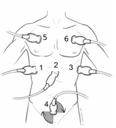
1. RUQ (Right Upper Quadrant)
The RUQ view includes the liver and kidney, the space between these two organs is called Morrison’s pouch and is where fluid is likely to accumulate. Imaging in the region superior to the liver and diaphragm is also important in evaluating for pleural effusion. For the RUQ, the caudal tip of the liver is where fluid is likely to accumulate.
 – eFAST")

2. Cardiac
The cardiac view uses the liver as an imaging window to evaluate for fluid in the pericardium. The pericardium is the sac around the heart. Fluid surrounding the heart may increase pressure on the heart. When the heart is unable to pump effectively due to this pressure, cardiac tamponade may occur. Cardiac tamponade is an emergent condition and requires a procedure to promptly drain the fluid.

3. LUQ (Left Upper Quadrant)
The LUQ includes the spleen and kidney. Fluid commonly accumulates in the perisplenic space, or around the spleen, and at the spleen tip. Less commonly, fluid can accumulate between the spleen and left kidney, or the splenorenal recess. As in the RUQ, imaging in the region superior to the spleen can rule out pleural effusion.
 – eFAST")
4. Pelvic view
For the pelvic view, it is helpful for the patient to have a full bladder to enhance visualisation of the pelvic structures. It is also important to scan in two imaging planes (sagittal and transverse) and sweep the transducer left and right to not miss any pockets of fluid. In males, the rectovesical pouch is where fluid is likely to accumulate. In females, the Cul de Sac or Pouch of Douglas is where fluid will accumulate.
")

5–6. Lung view(s)
As part of the eFAST, imaging of the lungs is performed to evaluate for pneumothorax. If lung sliding is present, pneumothorax is ruled out. If lung sliding is absent, further investigation must be done to evaluate for pneumothorax because more than one condition can cause this. A lung point is the point at which lung sliding and no lung sliding is seen. This confirms pneumothorax. M-mode may be used to better visualise this. On M-mode, normal lung sliding is described as the “seashore sign” represented on the image below. When lung sliding is absent it is described as a “barcode sign”. eFAST is useful to assess the critical patient for fluid, especially when the patient is too unstable for other imaging modalities such as CT. Ultrasound is repeatable and non-invasive. The information obtained from eFAST can direct a surgeon as to the course of surgery needed for treatment and can be lifesaving.

- Waves
- Sandy beach
The eFAST ultrasound exam is a safe and easy way to identify abnormal fluid collections within the abdominal, cardiac/chest, and retroperitoneal spaces. By incorporating lung views health care providers can also determine the presence of pneumothorax and other plural conditions. It is recommended that when performing the various views outlined above that the practitioner use the appropriate (Abdomen, Cardiac, Lung) exam type suitable for each view.