ASA “Practice Guidelines for Central Venous Access” Published
It may be an exaggeration to suggest that every other month a new guideline or requirement is released by a professional medical body recommending or directing that ultrasound—especially bedside (a.k.a., point-of-care) ultrasound—be incorporated into clinical best practices, but often it feels like it.
Case in point: In my last CMO Corner, I commented on the AIUM recognizing the American College of Emergency Physician’s Policy Statement “Emergency Ultrasound Guidelines,” which acknowledged point-of-care ultrasound as part of emergency care’s best practices.
Not long ago, new ACGME Program Requirements for Graduate Medical Education in Pulmonary Disease and Critical Care Medicine directed that instructors include the teaching of specific bedside ultrasound techniques to graduate students. (By the way, Sonosite is offering equipment and training to help educators meet the requirements that go into effect July 1, 2012.) …
A Big Thank-You From Tenwek, Kenya
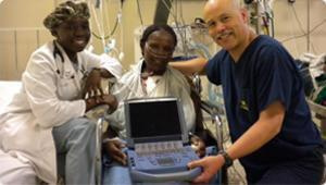
This story reminds us yet again how lives can be saved when dedicated teamwork combines with progressive medical technology.
…
AIUM Recognizes ACEP's Emergency Ultrasound Guidelines
While not all portable ultrasound examinations are of an emergency nature, its predominant use originated within the “we-need-it-STAT” category. Hence, the significant role of emergency medicine in contributing to point-of-care ultrasound best practices and insight into the valuable role ultrasonography can play at the bedside.
In mid-November, the contributions of emergency physicians to the proper use of “focused emergency ultrasound examinations” (a.k.a. point-of-care ultrasound) was acknowledged by the American Institute of Ultrasound in Medicine (AIUM). According to their November 17, 2011, press release, the AIUM now recognizes the American College of Emergency Physician’s (ACEP) Policy Statement Emergency Ultrasound Guidelines as “meeting the qualifications for performing ultrasound in the emergency settings. These guidelines describe the education and training required by emergency physicians to achieve competency for the performance of focused emergency ultrasound applications in clinical practice.” …
EAU Recommends Ultrasound First for Renal Stone Disease
I’ve seen many patients present to the E.D. in severe pain from kidney stones. Renal colic is a common and recurrent condition; it affects over a million people each year in the U.S. and accounts for approximately 1 percent of admissions.1
Diagnosing kidney stones in patients who present with renal colic is often performed with tomography (CT). In the past, Intravenous Urography (IVU) was used. While CT and IVU are accurate diagnostic tests and define clearly the size, shape, and position of uric acid stones, they also present a number of factors that would discourage use, including the potential risks of exposing patients to repeated doses of ionizing radiation.
In its 2011 Guidelines on Urolithiasis, the European Association of Urology (EAU) calls for ultrasound to be used as the primary imaging procedure. The guidelines state, “it is a safe (no risk of radiation), reproducible and inexpensive method of urinary stone detection.”2 The EAU notes that ultrasonography can detect stones in the calices, pelvis, pyelo-ureteric junction and vesicoureteric junction, and dilatation of the upper urinary tract. For renal stones greater than 5 mm, ultrasound offers a sensitivity of 96% and a specificity of 100%. For location of all stones, ultrasound’s sensitivity and specificity was 78% and 31% respectively.3, 4 …
Joint Commission and the Risk of Medical Imaging Radiation
The Joint Commission is sending a powerful notice about the radiation risks of diagnostic imaging. Its August 24, 2011, Sentinel Event Alert clearly states that diagnostic radiation can save lives. That is not in dispute. What is at issue is the amount of radiation patients receive over time and the risk for long-term damage.
Over the last 20 years, Americans’ exposure to ionizing radiation has nearly doubled. As the Joint Commission states, “…any physician can order tests involving exposure to radiation at any frequency, with no knowledge of when the last of when the patient was irradiated or how much radiation the patient received.” The World Health Organization’s International Agency for Research on Cancer, the Agency for Toxic Substances and Disease Registry of the Centers for Disease Control and Prevention and the National institute of Environmental Health Sciences all classify x-rays as a carcinogen.
As an Emergency Physician, I recognize the value of x-rays and CT scans and use them when they are medically appropriate. In its Sentinel Event Alert, one of the Joint Commission’s recommended actions is use other imaging modalities when appropriate: “In order to reduce the expose of the patient to ionizing radiation, use other imaging techniques, such as ultrasound or MRI, whenever these tests will produce the required diagnostic information at a similar quality level.” …
Dr. Jeff Gonzales' Story

Catching a bus shouldn't be life-threatening. But for one 23-year-old woman, running for the bus could have cost her everything.After rushing to hop on board, she fell unconscious, leaving her arm outstretched beyond the door. Not knowing this, the driver closed the door and drove to the next stop, about two-tenths of a mile away.The passenger was shocked awake by her AICD, which she wore because of a history of hypertrophic obstructive cardiomyopathy. As she regained consciousness, she realized her forearm was still outside of the bus. …
Training Standards for Critical Care Ultrasonography
Ultrasound within critical care is growing rapidly and has a large role for multiple diagnostic applications and for guidance of invasive procedures.
A recent international roundtable, composed of 29 experts from five continents, just published recommendations on the need for developing training standards for intensive care medical students. Twelve Critical Care societies from around the world have endorsed the framework. They state, “There was 100% agreement among participants that general critical care ultrasound and ‘basic’ critical care echocardiography should be mandatory in the curriculum of intensive care unit (ICU) physicians.”
The first step to proficiency is good education, and I am sure that I am among many who work in acute care who applaud the move to make critical care ultrasound training mandatory in ICU curriculums around the world. …
Improving Care while Reducing Costs in Health Care
Would you let your family fly in an airplane at night if the pilot didn’t have radar? I certainly wouldn’t. That pilot would be flying blind. It’s the same for ultrasound. When a physician performs an invasive procure without ultrasound guidance, it’s akin to a flying blind. …
Training Standards for Critical Care Ultrasonography
Ultrasound within critical care is growing rapidly and has a large role for multiple diagnostic applications and for guidance of invasive procedures. A recent international roundtable, composed of 29 experts from five continents, just published recommendations on the need for developing training standards for intensive care medical students. Twelve Critical Care societies from around the world have endorsed the framework. …
Welcome to the CMO Corner
As an emergency physician, I have taken care of thousands of patients who have benefitted from bedside ultrasound and I consider it to be an indispensible tool. Many across the medical profession share the same view. In fact, the value of ultrasound is of such importance that the sheer volume of published information is overwhelming. That’s why we’ve created CMO Corner. It is meant distill relevant information about portable ultrasound so you can access it without spending hours online. …