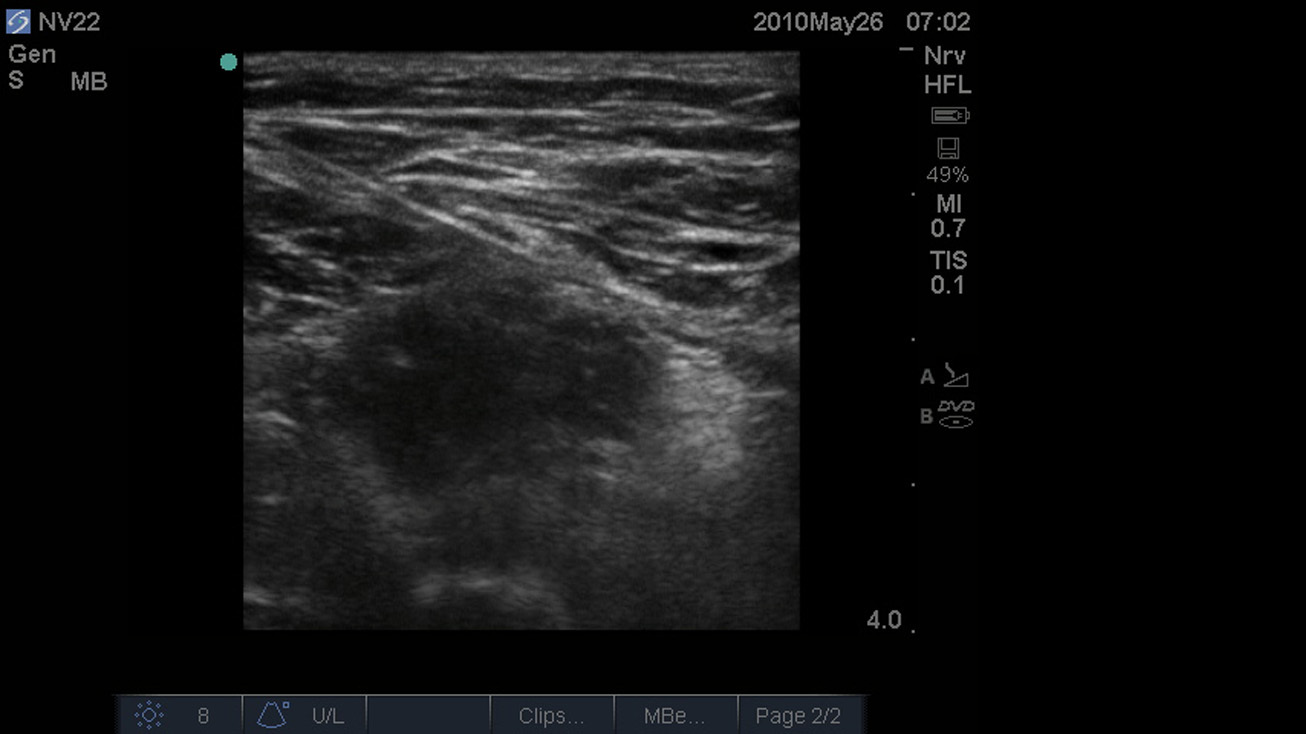
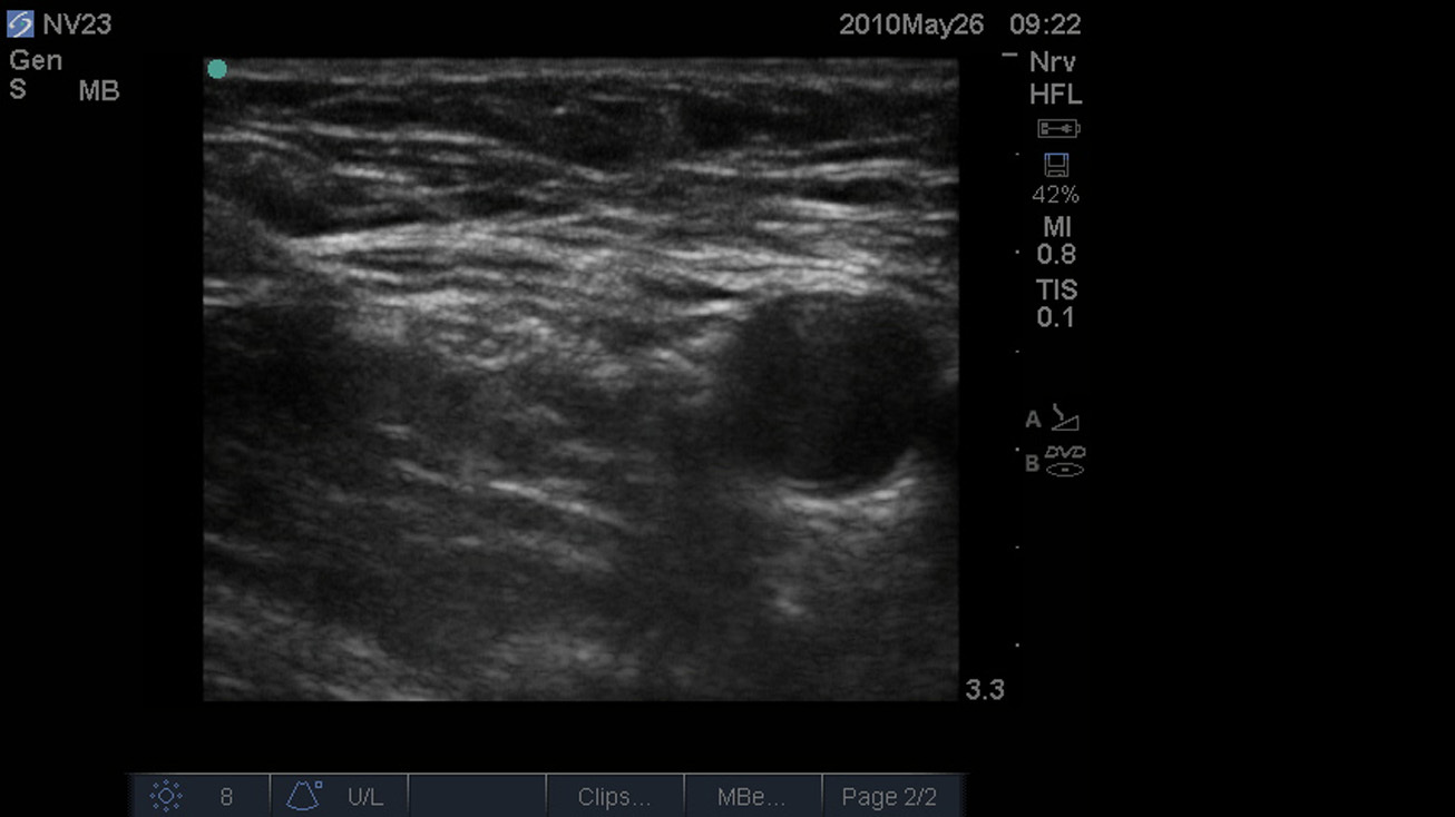
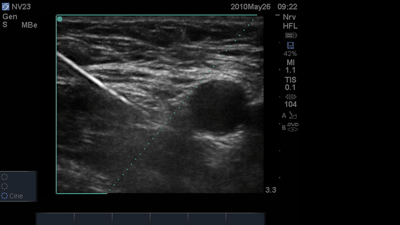
3D How To: Lumbar Puncture Procedure

3D How To: Lumbar Puncture Procedure

/sites/default/files/LumbarPuncture_edu00480_thumbnail.jpg
3D animation demonstrating an ultrasound guided lumbar puncture procedure.
Clinical Specialties
Media Library Type
Subtitles
<p begin="00:00:07.227" end="00:00:08.945" style="s2">- [Voiceover] A linear array transducer</p>
<p begin="00:00:08.945" end="00:00:11.605" style="s2">with a superficial exam<br />type is used to perform</p>
<p begin="00:00:11.605" end="00:00:14.437" style="s2">an ultrasound guided lumbar puncture.</p>
<p begin="00:00:14.437" end="00:00:16.616" style="s2">The patient is placed<br />in a sitting position</p>
<p begin="00:00:16.616" end="00:00:18.991" style="s2">with the spine maximally flexed.</p>
<p begin="00:00:18.991" end="00:00:22.142" style="s2">The transducer is placed<br />in a transverse direction</p>
<p begin="00:00:22.142" end="00:00:24.333" style="s2">with the orientation<br />marker directed toward</p>
<p begin="00:00:24.333" end="00:00:26.758" style="s2">the patient's left side<br />in the middle of the back</p>
<p begin="00:00:26.758" end="00:00:29.416" style="s2">at the level of the iliac crests.</p>
<p begin="00:00:29.416" end="00:00:32.008" style="s2">The spinous processes<br />will appear as distinct</p>
<p begin="00:00:32.008" end="00:00:35.181" style="s2">hyperechoic peaks with<br />acoustic shadowing below</p>
<p begin="00:00:35.181" end="00:00:37.632" style="s2">and define the midline of the spine.</p>
<p begin="00:00:37.632" end="00:00:40.818" style="s2">The transducer is slowly moved superiorly</p>
<p begin="00:00:40.818" end="00:00:43.706" style="s2">until the next spinous<br />process is identified.</p>
<p begin="00:00:43.706" end="00:00:46.199" style="s2">The transducer is rotated 90 degrees</p>
<p begin="00:00:46.199" end="00:00:48.242" style="s2">with the orientation<br />marker directed toward</p>
<p begin="00:00:48.242" end="00:00:49.707" style="s2">the patient's head.</p>
<p begin="00:00:49.707" end="00:00:52.646" style="s2">The two previously<br />identified spinous processes</p>
<p begin="00:00:52.646" end="00:00:55.552" style="s2">will now appear as<br />crescent-shaped, concave,</p>
<p begin="00:00:55.552" end="00:00:57.792" style="s2">hyperechoic structures.</p>
<p begin="00:00:57.792" end="00:01:00.857" style="s2">The gap between the bright<br />hyperechoic convexities</p>
<p begin="00:01:00.857" end="00:01:03.796" style="s2">is the inner spinous<br />space and defines the area</p>
<p begin="00:01:03.796" end="00:01:07.879" style="s2">in which the lumbar puncture<br />should be performed.</p>
Brightcove ID
5508123506001
https://youtube.com/watch?v=ndnZxAcNjdg

3D How To: Interscalene Nerve Block

3D How To: Interscalene Nerve Block

/sites/default/files/Interscalene_edu00493_thumbnail.jpg
3D animation demonstrating an ultrasound guided interscalene nerve block.
Clinical Specialties
Media Library Type
Subtitles
<p begin="00:00:07.292" end="00:00:09.121" style="s2">- [Voiceover] A linear array transducer</p>
<p begin="00:00:09.121" end="00:00:11.513" style="s2">with a nerve exam type is used to perform</p>
<p begin="00:00:11.513" end="00:00:14.888" style="s2">an ultrasound-guided,<br />interscalene nerve block.</p>
<p begin="00:00:14.888" end="00:00:18.497" style="s2">The target depth is approximately<br />one to three centimeters</p>
<p begin="00:00:18.497" end="00:00:20.715" style="s2">in an 80-kilogram adult.</p>
<p begin="00:00:20.715" end="00:00:24.654" style="s2">The patient is positioned in<br />a 45-degree reclining position</p>
<p begin="00:00:24.654" end="00:00:26.373" style="s2">with a pillow under their head</p>
<p begin="00:00:26.373" end="00:00:29.536" style="s2">and the head turned toward<br />the contralateral side.</p>
<p begin="00:00:29.536" end="00:00:31.900" style="s2">The examination begins by finding</p>
<p begin="00:00:31.900" end="00:00:34.916" style="s2">the supraclavicular region<br />of the brachial plexus</p>
<p begin="00:00:34.916" end="00:00:36.709" style="s2">as a landmark technique.</p>
<p begin="00:00:36.709" end="00:00:39.039" style="s2">The transducer is placed posterior</p>
<p begin="00:00:39.039" end="00:00:41.013" style="s2">to the midpoint of the clavicle</p>
<p begin="00:00:41.013" end="00:00:42.373" style="s2">with the orientation marker</p>
<p begin="00:00:42.373" end="00:00:46.540" style="s2">directed to the patient's right<br />at a three-o'clock position.</p>
<p begin="00:00:49.085" end="00:00:51.056" style="s2">The subclavian artery is seen</p>
<p begin="00:00:51.056" end="00:00:53.399" style="s2">as a round, pulsatile structure</p>
<p begin="00:00:53.399" end="00:00:56.778" style="s2">superior to the bright<br />reflection of the first rib.</p>
<p begin="00:00:56.778" end="00:01:00.337" style="s2">The pleura is seen as a<br />bright, hyperechoic reflection</p>
<p begin="00:01:00.337" end="00:01:03.590" style="s2">deep to or at the same<br />depth as the first rib.</p>
<p begin="00:01:03.590" end="00:01:07.432" style="s2">The nerves of the brachial<br />plexus are posterior or superior</p>
<p begin="00:01:07.432" end="00:01:09.681" style="s2">to the subclavian artery.</p>
<p begin="00:01:09.681" end="00:01:12.728" style="s2">The nerves appear as<br />hypoechoic dark circles</p>
<p begin="00:01:12.728" end="00:01:14.814" style="s2">within the bright hyperechoic fascia</p>
<p begin="00:01:14.814" end="00:01:17.115" style="s2">surrounding the brachial plexus.</p>
<p begin="00:01:17.115" end="00:01:21.158" style="s2">To identify the interscalene<br />region of the brachial plexus,</p>
<p begin="00:01:21.158" end="00:01:23.987" style="s2">slowly slide the transducer up the neck.</p>
<p begin="00:01:23.987" end="00:01:26.568" style="s2">The interscalene muscles<br />will frame the nerves</p>
<p begin="00:01:26.568" end="00:01:29.020" style="s2">within the interscalene groove.</p>
<p begin="00:01:29.020" end="00:01:30.848" style="s2">The nerves will change in shape</p>
<p begin="00:01:30.848" end="00:01:34.236" style="s2">from a grape-like cluster in<br />the supraclavicular region</p>
<p begin="00:01:34.236" end="00:01:37.710" style="s2">to a chain-link appearance<br />in the interscalene groove.</p>
<p begin="00:01:37.710" end="00:01:42.445" style="s2">The C5 and C6 nerve roots<br />appear as three circles.</p>
<p begin="00:01:42.445" end="00:01:45.348" style="s2">The hypoechoic pulsatile carotid artery</p>
<p begin="00:01:45.348" end="00:01:48.969" style="s2">will appear medial to the<br />anterior scalene muscle.</p>
<p begin="00:01:48.969" end="00:01:50.345" style="s2">The needle can be advanced</p>
<p begin="00:01:50.345" end="00:01:53.771" style="s2">using an in-plane or<br />out-of-plane technique.</p>
<p begin="00:01:53.771" end="00:01:55.547" style="s2">For an in-plane technique,</p>
<p begin="00:01:55.547" end="00:01:58.220" style="s2">the needle is positioned<br />one to two centimeters</p>
<p begin="00:01:58.220" end="00:02:02.344" style="s2">lateral to the transducer and<br />advanced under the transducer</p>
<p begin="00:02:02.344" end="00:02:05.776" style="s2">until the tip is just<br />posterior to or between</p>
<p begin="00:02:05.776" end="00:02:07.943" style="s2">the C5 and C6 nerve roots.</p>
<p begin="00:02:08.909" end="00:02:11.627" style="s2">The local anesthetic is<br />injected incrementally</p>
<p begin="00:02:11.627" end="00:02:13.691" style="s2">close to the nerve roots.</p>
<p begin="00:02:13.691" end="00:02:15.098" style="s2">The needle can be redirected</p>
<p begin="00:02:15.098" end="00:02:18.749" style="s2">if the spread of local anesthetic<br />is not deemed adequate.</p>
<p begin="00:02:18.749" end="00:02:21.425" style="s2">It is not necessary to<br />have local anesthetic</p>
<p begin="00:02:21.425" end="00:02:24.665" style="s2">anterior and posterior to the nerve roots.</p>
<p begin="00:02:24.665" end="00:02:28.832" style="s2">Posterior spread only of<br />local anesthetic is effective.</p>
Brightcove ID
5508123555001
https://youtube.com/watch?v=wAABZF1aSO8

3D How To: Infraclavicular Nerve Block

3D How To: Infraclavicular Nerve Block

/sites/default/files/Infraclavicular_edu00493_thumbnail.jpg
3D animation demonstrating an ultrasound guided infraclavicular nerve block.
Clinical Specialties
Media Library Type
Subtitles
<p begin="00:00:07.354" end="00:00:09.128" style="s2">- [Voiceover] A linear array transducer</p>
<p begin="00:00:09.128" end="00:00:11.643" style="s2">with a nerve exam type is used to perform</p>
<p begin="00:00:11.643" end="00:00:15.694" style="s2">an ultrasound-guided,<br />infraclavicular regional nerve block.</p>
<p begin="00:00:15.694" end="00:00:19.134" style="s2">The target depth is approximately<br />three to four centmeters</p>
<p begin="00:00:19.134" end="00:00:21.341" style="s2">in an 80 kilogram adult.</p>
<p begin="00:00:21.341" end="00:00:23.542" style="s2">The patient is positioned supine</p>
<p begin="00:00:23.542" end="00:00:25.523" style="s2">with the arm abducted 90 degrees</p>
<p begin="00:00:25.523" end="00:00:27.616" style="s2">and the elbow bent 90 degrees</p>
<p begin="00:00:27.616" end="00:00:29.525" style="s2">to move the clavicle posterior,</p>
<p begin="00:00:29.525" end="00:00:32.434" style="s2">and permit a shallow<br />angle of needle insertion.</p>
<p begin="00:00:32.434" end="00:00:34.126" style="s2">The patient's head should be rotated</p>
<p begin="00:00:34.126" end="00:00:36.427" style="s2">toward the contralateral side.</p>
<p begin="00:00:36.427" end="00:00:40.380" style="s2">The transducer is placed on the<br />chest in a paramedian plane,</p>
<p begin="00:00:40.380" end="00:00:42.936" style="s2">inferior to the midpoint of the clavicle</p>
<p begin="00:00:42.936" end="00:00:46.372" style="s2">with the orientation marker<br />directed to the patient's head.</p>
<p begin="00:00:46.372" end="00:00:48.368" style="s2">The axillary artery is seen as</p>
<p begin="00:00:48.368" end="00:00:52.247" style="s2">a dark, round pulsatile<br />structure with a hyperechoic wall</p>
<p begin="00:00:52.247" end="00:00:56.144" style="s2">inferior to the pectoralis<br />major and minor muscles.</p>
<p begin="00:00:56.144" end="00:00:59.668" style="s2">The axillary vein lies<br />beside the axillary artery</p>
<p begin="00:00:59.668" end="00:01:01.751" style="s2">and collapses with compression.</p>
<p begin="00:01:01.751" end="00:01:04.801" style="s2">The nerve cords appear<br />as hyperechoic circles</p>
<p begin="00:01:04.801" end="00:01:06.193" style="s2">with a honeycomb appearance</p>
<p begin="00:01:06.193" end="00:01:10.370" style="s2">within the bright hyperechoic<br />fascia of the brachial plexus.</p>
<p begin="00:01:10.370" end="00:01:13.618" style="s2">The transducer is slowly<br />moved in a lateral direction,</p>
<p begin="00:01:13.618" end="00:01:16.454" style="s2">following the course<br />of the axillary artery.</p>
<p begin="00:01:16.454" end="00:01:18.722" style="s2">The nerves will split into a lateral,</p>
<p begin="00:01:18.722" end="00:01:21.793" style="s2">medial, and posterior cord.</p>
<p begin="00:01:21.793" end="00:01:24.069" style="s2">The optimum position of the transducer</p>
<p begin="00:01:24.069" end="00:01:26.136" style="s2">for the infraclavicular nerve block</p>
<p begin="00:01:26.136" end="00:01:28.306" style="s2">is as lateral as possible.</p>
<p begin="00:01:28.306" end="00:01:31.280" style="s2">The needle is positioned<br />two centimeters cephalad</p>
<p begin="00:01:31.280" end="00:01:35.432" style="s2">to the transducer and advanced<br />using an in-plane technique.</p>
<p begin="00:01:35.432" end="00:01:37.913" style="s2">The needle path is<br />directed over the clavicle</p>
<p begin="00:01:37.913" end="00:01:40.181" style="s2">and through the pectoral muscles.</p>
<p begin="00:01:40.181" end="00:01:42.143" style="s2">The initial endpoint for the needle is</p>
<p begin="00:01:42.143" end="00:01:45.627" style="s2">immediately posterior to<br />the midpoint of the artery.</p>
<p begin="00:01:45.627" end="00:01:48.770" style="s2">The spread of local<br />anesthetic should be observed.</p>
<p begin="00:01:48.770" end="00:01:52.364" style="s2">Supplemental injections can<br />be made anterior to the artery</p>
<p begin="00:01:52.364" end="00:01:53.954" style="s2">to deposit local anesthetic</p>
<p begin="00:01:53.954" end="00:01:57.954" style="s2">around the lateral and<br />medial cords if required.</p>
Brightcove ID
5745561363001
https://youtube.com/watch?v=8xRDVLKb5BY

3D How To: Ultrasound Guided TAP Block

3D How To: Ultrasound Guided TAP Block

/sites/default/files/Ilioinquinal_Iliohypogastric_edu00498_thumnail.jpg
3D animation demonstrating an ultrasound guided Ilioinguinal and Iliohypogastric nerve blocks.
Clinical Specialties
Media Library Type
Subtitles
<p begin="00:00:07.549" end="00:00:09.196" style="s2">- [Voiceover] A linear array transducer</p>
<p begin="00:00:09.196" end="00:00:11.475" style="s2">with a nerve exam type is used to perform</p>
<p begin="00:00:11.475" end="00:00:13.828" style="s2">an ultrasound guided ilioinguinal</p>
<p begin="00:00:13.828" end="00:00:16.351" style="s2">and iliohypogastric nerve block.</p>
<p begin="00:00:16.351" end="00:00:19.067" style="s2">The target depth is<br />approximately three centimeters</p>
<p begin="00:00:19.067" end="00:00:21.384" style="s2">in an 80 kilogram adult.</p>
<p begin="00:00:21.384" end="00:00:24.528" style="s2">The patient is positioned<br />in a supine position.</p>
<p begin="00:00:24.528" end="00:00:27.639" style="s2">The transducer is placed<br />over the bony prominence</p>
<p begin="00:00:27.639" end="00:00:30.408" style="s2">of the anterior superior iliac spine</p>
<p begin="00:00:30.408" end="00:00:33.991" style="s2">with the orientation marker<br />directed to the patient's right.</p>
<p begin="00:00:33.991" end="00:00:37.495" style="s2">The transducer is rotated<br />slightly counterclockwise</p>
<p begin="00:00:37.495" end="00:00:41.199" style="s2">so one side rests on the<br />anterior superior iliac spine</p>
<p begin="00:00:41.199" end="00:00:43.968" style="s2">and the other side<br />points at the umbilicus.</p>
<p begin="00:00:43.968" end="00:00:47.696" style="s2">The medial side of the transducer<br />is slowly swept superiorly</p>
<p begin="00:00:47.696" end="00:00:51.231" style="s2">and inferiorly while<br />maintaining the bony iliac crest</p>
<p begin="00:00:51.231" end="00:00:53.956" style="s2">in the lateral part of<br />the ultrasound image.</p>
<p begin="00:00:53.956" end="00:00:56.495" style="s2">The structures of the<br />anterior abdominal wall</p>
<p begin="00:00:56.495" end="00:00:59.425" style="s2">should be identified<br />from superficial to deep,</p>
<p begin="00:00:59.425" end="00:01:03.104" style="s2">including adipose tissue,<br />external oblique muscle,</p>
<p begin="00:01:03.104" end="00:01:04.727" style="s2">internal oblique muscle,</p>
<p begin="00:01:04.727" end="00:01:07.759" style="s2">and the transversus abdominis muscle.</p>
<p begin="00:01:07.759" end="00:01:10.688" style="s2">A fourth muscle, the<br />iliacus, may occasionally</p>
<p begin="00:01:10.688" end="00:01:14.527" style="s2">be visualized underneath<br />the transversus abdominis.</p>
<p begin="00:01:14.527" end="00:01:17.271" style="s2">The nerves are located<br />between the internal oblique</p>
<p begin="00:01:17.271" end="00:01:20.056" style="s2">and the transversus<br />abdominis muscle layers.</p>
<p begin="00:01:20.056" end="00:01:22.816" style="s2">They may appear only as<br />hyperechoic thickness</p>
<p begin="00:01:22.816" end="00:01:25.738" style="s2">between these two muscles<br />or as dark hypoechoic</p>
<p begin="00:01:25.738" end="00:01:29.166" style="s2">oval structures with the<br />bright hyperechoic covering.</p>
<p begin="00:01:29.166" end="00:01:32.752" style="s2">The nerves are very closely<br />related to the iliac crest.</p>
<p begin="00:01:32.752" end="00:01:35.150" style="s2">The needle is advanced using an in-plane</p>
<p begin="00:01:35.150" end="00:01:37.031" style="s2">or out-of-plane technique.</p>
<p begin="00:01:37.031" end="00:01:39.007" style="s2">If using an in-plane technique,</p>
<p begin="00:01:39.007" end="00:01:41.607" style="s2">the needle is positioned<br />one to two centimeters</p>
<p begin="00:01:41.607" end="00:01:45.855" style="s2">medial to the transducer and<br />advanced under the transducer.</p>
<p begin="00:01:45.855" end="00:01:47.343" style="s2">The end point for the needle is</p>
<p begin="00:01:47.343" end="00:01:50.999" style="s2">between internal oblique<br />and transversus abdominis.</p>
<p begin="00:01:50.999" end="00:01:53.689" style="s2">The local anesthetic is<br />injected incrementally</p>
<p begin="00:01:53.689" end="00:01:55.439" style="s2">close to the nerves.</p>
Brightcove ID
5508136009001
https://youtube.com/watch?v=uurxBFRT_EE

3D How To: Gluteal Sciatic Nerve Block

3D How To: Gluteal Sciatic Nerve Block

/sites/default/files/GlutealSciatic_edu00500_thumbnail.jpg
3D animation demonstrating an ultrasound guided Gluteal Sciatic nerve block.
Clinical Specialties
Media Library Type
Subtitles
<p begin="00:00:07.596" end="00:00:09.143" style="s2">- [Voiceover] A curved array transducer</p>
<p begin="00:00:09.143" end="00:00:11.456" style="s2">with a nerve exam type is used to perform</p>
<p begin="00:00:11.456" end="00:00:14.407" style="s2">a ultrasound guided sciatic nerve block.</p>
<p begin="00:00:14.407" end="00:00:17.696" style="s2">The target depth is approximately<br />four to six centimeters</p>
<p begin="00:00:17.696" end="00:00:20.063" style="s2">in an 80 kilogram adult.</p>
<p begin="00:00:20.063" end="00:00:23.396" style="s2">The patient is positioned in<br />a lateral decubitus position</p>
<p begin="00:00:23.396" end="00:00:25.591" style="s2">with the hip and knee flexed.</p>
<p begin="00:00:25.591" end="00:00:28.711" style="s2">The transducer is placed<br />between the greater trochanter</p>
<p begin="00:00:28.711" end="00:00:31.763" style="s2">and the ischial tuberosity<br />in a transverse position</p>
<p begin="00:00:31.763" end="00:00:34.303" style="s2">just proximal to the gluteal fold</p>
<p begin="00:00:34.303" end="00:00:37.479" style="s2">with the orientation<br />marker directed laterally.</p>
<p begin="00:00:37.479" end="00:00:39.056" style="s2">The bony landmarks will appear</p>
<p begin="00:00:39.056" end="00:00:43.374" style="s2">as bright hyperechoic crescents<br />with posterior shadowing.</p>
<p begin="00:00:43.374" end="00:00:44.886" style="s2">There are several layers of tissue</p>
<p begin="00:00:44.886" end="00:00:47.454" style="s2">between the bony landmarks,<br />which should be identified</p>
<p begin="00:00:47.454" end="00:00:51.249" style="s2">from superficial to deep,<br />including adipose tissue,</p>
<p begin="00:00:51.249" end="00:00:55.303" style="s2">the gluteus maximus muscle<br />and quadratus femoris muscle.</p>
<p begin="00:00:55.303" end="00:00:58.156" style="s2">The sciatic nerve lies<br />deep to the gluteus maximus</p>
<p begin="00:00:58.156" end="00:01:01.744" style="s2">and superficial to the<br />quadratus femoris muscle.</p>
<p begin="00:01:01.744" end="00:01:04.321" style="s2">It appears as a bright hyperechoic oval</p>
<p begin="00:01:04.321" end="00:01:06.888" style="s2">or triangular shaped density.</p>
<p begin="00:01:06.888" end="00:01:10.176" style="s2">The transducer should be<br />moved slightly cranially</p>
<p begin="00:01:10.176" end="00:01:12.296" style="s2">or caudally from the initial position</p>
<p begin="00:01:12.296" end="00:01:14.357" style="s2">by slightly rocking the transducer</p>
<p begin="00:01:14.357" end="00:01:16.596" style="s2">so the beam is perpendicular to the nerve</p>
<p begin="00:01:16.596" end="00:01:20.739" style="s2">for the best echo reflection<br />if it is difficult to identify.</p>
<p begin="00:01:20.739" end="00:01:23.307" style="s2">When the transducer is<br />moved distally in the thigh,</p>
<p begin="00:01:23.307" end="00:01:26.138" style="s2">the sciatic nerve becomes<br />more oval in shape</p>
<p begin="00:01:26.138" end="00:01:29.267" style="s2">and is found more superficially<br />between the biceps femoris</p>
<p begin="00:01:29.267" end="00:01:31.675" style="s2">and abductor magnus muscles.</p>
<p begin="00:01:31.675" end="00:01:34.922" style="s2">The needle is advanced<br />using an in plane technique.</p>
<p begin="00:01:34.922" end="00:01:38.036" style="s2">The needle is positioned one<br />to two centimeters lateral</p>
<p begin="00:01:38.036" end="00:01:42.219" style="s2">to the transducer and advanced<br />slowly under the transducer.</p>
<p begin="00:01:42.219" end="00:01:43.828" style="s2">The initial end point for the needle</p>
<p begin="00:01:43.828" end="00:01:46.907" style="s2">is just lateral and deep to the nerve.</p>
<p begin="00:01:46.907" end="00:01:49.538" style="s2">The local anesthetic is<br />injected incrementally</p>
<p begin="00:01:49.538" end="00:01:51.011" style="s2">close to the nerve.</p>
<p begin="00:01:51.011" end="00:01:53.355" style="s2">For a successful block, a spread of locals</p>
<p begin="00:01:53.355" end="00:01:55.500" style="s2">should be observed around the medial side</p>
<p begin="00:01:55.500" end="00:01:58.250" style="s2">or tibial component of the nerve.</p>
Brightcove ID
5508134308001
https://youtube.com/watch?v=xvAY_bu_S7A

3D How To: Femoral Nerve Block

3D How To: Femoral Nerve Block

/sites/default/files/Femoral_edu00499_thumbnail.jpg
3D animation demonstrating an ultrasound guided femoral nerve block.
Clinical Specialties
Media Library Type
Subtitles
<p begin="00:00:07.378" end="00:00:09.000" style="s2">- [Voiceover] A linear array transducer</p>
<p begin="00:00:09.000" end="00:00:11.275" style="s2">with a nerve exam type is used to perform</p>
<p begin="00:00:11.275" end="00:00:14.181" style="s2">an ultrasound-guided femoral nerve block.</p>
<p begin="00:00:14.181" end="00:00:17.382" style="s2">The target depth is approximately<br />one to three centimeters</p>
<p begin="00:00:17.382" end="00:00:19.631" style="s2">in an 80 kilogram adult.</p>
<p begin="00:00:19.631" end="00:00:22.367" style="s2">The patient is placed<br />in a supine position.</p>
<p begin="00:00:22.367" end="00:00:24.579" style="s2">The transducer is placed just superior</p>
<p begin="00:00:24.579" end="00:00:26.294" style="s2">to the inguinal skin crease</p>
<p begin="00:00:26.294" end="00:00:30.461" style="s2">with the orientation marker<br />directed to the patient's right.</p>
<p begin="00:00:31.318" end="00:00:33.157" style="s2">The common femoral artery is seen</p>
<p begin="00:00:33.157" end="00:00:35.893" style="s2">as a round, pulsatile structure.</p>
<p begin="00:00:35.893" end="00:00:38.069" style="s2">The transducer should be moved laterally</p>
<p begin="00:00:38.069" end="00:00:41.966" style="s2">one to two centimeters to<br />identify the femoral nerve.</p>
<p begin="00:00:41.966" end="00:00:44.927" style="s2">The nerve lies deep to the fascia iliaca</p>
<p begin="00:00:44.927" end="00:00:48.332" style="s2">and appears as an oblong,<br />bright hyperechoic structure</p>
<p begin="00:00:48.332" end="00:00:50.561" style="s2">lying on the iliacus muscle.</p>
<p begin="00:00:50.561" end="00:00:53.344" style="s2">It may contain dark hyperechoic circles</p>
<p begin="00:00:53.344" end="00:00:55.686" style="s2">that represent the nerve fascicles.</p>
<p begin="00:00:55.686" end="00:00:58.920" style="s2">The nerve always lies<br />deep to the fascia iliaca</p>
<p begin="00:00:58.920" end="00:01:00.135" style="s2">and should not be confused</p>
<p begin="00:01:00.135" end="00:01:03.340" style="s2">with the bright hyperechoic<br />tissue lying above the fascia,</p>
<p begin="00:01:03.340" end="00:01:05.481" style="s2">which is lymphatic tissue.</p>
<p begin="00:01:05.481" end="00:01:08.793" style="s2">The needle is advanced<br />using an in-plane technique.</p>
<p begin="00:01:08.793" end="00:01:11.734" style="s2">The needle is positioned one<br />to two centimeters lateral</p>
<p begin="00:01:11.734" end="00:01:15.563" style="s2">to the transducer and<br />advanced under the transducer.</p>
<p begin="00:01:15.563" end="00:01:17.219" style="s2">The initial end point for the needle</p>
<p begin="00:01:17.219" end="00:01:21.762" style="s2">is just lateral to the nerve<br />and deep to the fascia iliaca.</p>
<p begin="00:01:21.762" end="00:01:24.475" style="s2">The local anesthetic is<br />injected incrementally,</p>
<p begin="00:01:24.475" end="00:01:25.860" style="s2">close to the nerve.</p>
<p begin="00:01:25.860" end="00:01:28.643" style="s2">For a successful block, a spread of local</p>
<p begin="00:01:28.643" end="00:01:31.367" style="s2">should be observed behind<br />the femoral artery.</p>
<p begin="00:01:31.367" end="00:01:34.771" style="s2">If local anesthetic is identified<br />anterior to the artery,</p>
<p begin="00:01:34.771" end="00:01:38.097" style="s2">then the needle is superficial<br />to the fascia iliaca</p>
<p begin="00:01:38.097" end="00:01:42.014" style="s2">and the needle needs to<br />be repositioned deeper.</p>
Brightcove ID
5508117977001
https://youtube.com/watch?v=pCkjioc-EmQ

3D How To: Subclavian Vein Catheter Insertion

3D How To: Subclavian Vein Catheter Insertion

/sites/default/files/CentralVenous_SubclavianVein_edu00473_thumbnail.jpg
3D animation demonstrating an ultrasound guided insertion of a Subclavian Vein Catheter.
Applications
Clinical Specialties
Media Library Type
Subtitles
<p begin="00:00:07.010" end="00:00:09.026" style="s2">- [Voiceover] A linear array transducer</p>
<p begin="00:00:09.026" end="00:00:11.485" style="s2">with a venous exam<br />type, is used to perform</p>
<p begin="00:00:11.485" end="00:00:14.424" style="s2">an ultrasound guided<br />insertion of a subclavian</p>
<p begin="00:00:14.424" end="00:00:17.250" style="s2">vein catheter via an transverse approach.</p>
<p begin="00:00:17.250" end="00:00:19.487" style="s2">The patient is in a supine position</p>
<p begin="00:00:19.487" end="00:00:21.144" style="s2">with the head neutral.</p>
<p begin="00:00:21.144" end="00:00:24.198" style="s2">The operator should stand<br />to the patient's right side.</p>
<p begin="00:00:24.198" end="00:00:26.531" style="s2">The transducer is placed transversely</p>
<p begin="00:00:26.531" end="00:00:29.380" style="s2">just inferior to the mid<br />portion of the clavicle,</p>
<p begin="00:00:29.380" end="00:00:31.338" style="s2">with the orientation marker directed</p>
<p begin="00:00:31.338" end="00:00:34.428" style="s2">to the patient's head at<br />a 12 o'clock position.</p>
<p begin="00:00:34.428" end="00:00:36.407" style="s2">The hyperechoic clavicle can be seen</p>
<p begin="00:00:36.407" end="00:00:39.304" style="s2">in the superior portion<br />of the ultrasound image.</p>
<p begin="00:00:39.304" end="00:00:41.387" style="s2">The vein is dark, and anechoic,</p>
<p begin="00:00:41.387" end="00:00:43.977" style="s2">just inferior and deep to the clavicle.</p>
<p begin="00:00:43.977" end="00:00:47.219" style="s2">The transducer should be<br />slowly moved one to two inches</p>
<p begin="00:00:47.219" end="00:00:49.877" style="s2">toward the shoulder, with<br />the face of the transducer</p>
<p begin="00:00:49.877" end="00:00:52.685" style="s2">staying below the clavicle<br />to obtain the best view</p>
<p begin="00:00:52.685" end="00:00:55.397" style="s2">of the subclavian or axillary vein.</p>
<p begin="00:00:55.397" end="00:00:58.063" style="s2">It is important to note<br />that the lung lies directly</p>
<p begin="00:00:58.063" end="00:00:59.861" style="s2">posterior to the vessel.</p>
<p begin="00:00:59.861" end="00:01:02.691" style="s2">So, posterior wall puncture<br />of the axillary vein</p>
<p begin="00:01:02.691" end="00:01:04.214" style="s2">should be avoided.</p>
<p begin="00:01:04.214" end="00:01:07.472" style="s2">Adjust the transducer so it<br />is centered over the vein.</p>
<p begin="00:01:07.472" end="00:01:10.469" style="s2">Follow the needle entry by<br />slowly sliding the transducer</p>
<p begin="00:01:10.469" end="00:01:12.943" style="s2">in the direction of needle advancement.</p>
<p begin="00:01:12.943" end="00:01:16.832" style="s2">The needle will appear as a<br />small, bright, hyperechoic dot.</p>
<p begin="00:01:16.832" end="00:01:19.415" style="s2">When the needle tip<br />appears, the transducer</p>
<p begin="00:01:19.415" end="00:01:21.964" style="s2">should be advanced a<br />short distance distally</p>
<p begin="00:01:21.964" end="00:01:24.265" style="s2">to follow the tip of<br />the needle trajectory,</p>
<p begin="00:01:24.265" end="00:01:26.819" style="s2">and stay in advance of the needle entry.</p>
<p begin="00:01:26.819" end="00:01:28.455" style="s2">The needle is slowly advanced</p>
<p begin="00:01:28.455" end="00:01:30.590" style="s2">under direct ultrasound visualization</p>
<p begin="00:01:30.590" end="00:01:34.266" style="s2">until the tip is seen to<br />puncture the subclavian vein.</p>
<p begin="00:01:34.266" end="00:01:37.399" style="s2">The probe should be moved<br />slightly proximally and distally</p>
<p begin="00:01:37.399" end="00:01:39.357" style="s2">to confirm that the needle tip lies</p>
<p begin="00:01:39.357" end="00:01:41.940" style="s2">in the mid portion of the vein.</p>
Brightcove ID
5508120188001
https://youtube.com/watch?v=IBmbc1ak5fY

3D How To: IJV Catheter Insertion (Transverse)

3D How To: IJV Catheter Insertion (Transverse)

/sites/default/files/CentralVenous_IJ_Transverse_edu00471_thumbnail.jpg
3D animation demonstrating an ultrasound guided insertion of an internal jugular catheter (transverse).
Applications
Clinical Specialties
Media Library Type
Subtitles
<p begin="00:00:07.397" end="00:00:09.087" style="s2">- [Voiceover] A linear array transducer</p>
<p begin="00:00:09.087" end="00:00:10.710" style="s2">with a venous exam type</p>
<p begin="00:00:10.710" end="00:00:13.822" style="s2">is used to perform an<br />ultrasound-guided insertion</p>
<p begin="00:00:13.822" end="00:00:17.827" style="s2">of an internal jugular catheter<br />via a transverse approach.</p>
<p begin="00:00:17.827" end="00:00:19.996" style="s2">The patient is in a supine position</p>
<p begin="00:00:19.996" end="00:00:21.420" style="s2">with the head slightly turned</p>
<p begin="00:00:21.420" end="00:00:23.431" style="s2">toward the contralateral side.</p>
<p begin="00:00:23.431" end="00:00:26.331" style="s2">The operator is positioned<br />at the head of the bed.</p>
<p begin="00:00:26.331" end="00:00:28.712" style="s2">The transducer is placed transversely</p>
<p begin="00:00:28.712" end="00:00:29.970" style="s2">just below the apex</p>
<p begin="00:00:29.970" end="00:00:32.663" style="s2">of the sternocleidomastoid<br />muscle triangle,</p>
<p begin="00:00:32.663" end="00:00:35.671" style="s2">with the orientation marker<br />directed to the patient's left</p>
<p begin="00:00:35.671" end="00:00:37.655" style="s2">at a 9:00 position.</p>
<p begin="00:00:37.655" end="00:00:39.098" style="s2">The most superficial structure</p>
<p begin="00:00:39.098" end="00:00:41.086" style="s2">identified in the ultrasound image</p>
<p begin="00:00:41.086" end="00:00:43.548" style="s2">is the sternocleidomastoid muscle.</p>
<p begin="00:00:43.548" end="00:00:46.077" style="s2">Deeper to this, the internal jugular vein</p>
<p begin="00:00:46.077" end="00:00:49.080" style="s2">will appear as a dark<br />anechoic elliptical shape,</p>
<p begin="00:00:49.080" end="00:00:51.990" style="s2">and is compressible with<br />transducer pressure.</p>
<p begin="00:00:51.990" end="00:00:55.354" style="s2">The artery will be circular and pulsatile.</p>
<p begin="00:00:55.354" end="00:00:58.421" style="s2">The thyroid gland lies<br />medial to these structures,</p>
<p begin="00:00:58.421" end="00:01:01.367" style="s2">and has a light gray echo signature.</p>
<p begin="00:01:01.367" end="00:01:03.420" style="s2">Adjust the transducer so it is centered</p>
<p begin="00:01:03.420" end="00:01:05.804" style="s2">over the internal jugular vein.</p>
<p begin="00:01:05.804" end="00:01:08.867" style="s2">Follow the needle entry by<br />slowly sliding the transducer</p>
<p begin="00:01:08.867" end="00:01:11.272" style="s2">in the direction of needle advancement.</p>
<p begin="00:01:11.272" end="00:01:15.230" style="s2">The needle will appear as a<br />small bright hyperechoic dot.</p>
<p begin="00:01:15.230" end="00:01:16.923" style="s2">When the needle tip appears,</p>
<p begin="00:01:16.923" end="00:01:20.067" style="s2">the transducer should be advanced<br />a short distance distally</p>
<p begin="00:01:20.067" end="00:01:22.537" style="s2">to follow the tip of<br />the needle trajectory,</p>
<p begin="00:01:22.537" end="00:01:25.144" style="s2">and stay in advance of the needle entry.</p>
<p begin="00:01:25.144" end="00:01:26.523" style="s2">The needle is slowly advanced</p>
<p begin="00:01:26.523" end="00:01:28.724" style="s2">under direct ultrasound visualization</p>
<p begin="00:01:28.724" end="00:01:30.616" style="s2">until the tip is seen to indent,</p>
<p begin="00:01:30.616" end="00:01:33.391" style="s2">and then puncture the<br />internal jugular vein.</p>
<p begin="00:01:33.391" end="00:01:34.837" style="s2">The transducer should be moved</p>
<p begin="00:01:34.837" end="00:01:36.859" style="s2">slightly proximally and distally</p>
<p begin="00:01:36.859" end="00:01:38.740" style="s2">to confirm that the needle tip lies</p>
<p begin="00:01:38.740" end="00:01:41.990" style="s2">in the mid-portion of the jugular vein.</p>
Brightcove ID
5742626540001
https://youtube.com/watch?v=eesN9rGoXFM

3D How To: IJV Catheter Insertion (Long)

3D How To: IJV Catheter Insertion (Long)

/sites/default/files/CentralVenous_IJ_Long_edu00472_thumbnail.jpg
3D animation demonstrating an ultrasound guided insertion of an Internal Jugular Catheter (Longitudinal).
Applications
Clinical Specialties
Media Library Type
Subtitles
<p begin="00:00:07.010" end="00:00:09.026" style="s2">- [Voiceover] A linear array transducer</p>
<p begin="00:00:09.026" end="00:00:11.485" style="s2">with a venous exam<br />type, is used to perform</p>
<p begin="00:00:11.485" end="00:00:14.424" style="s2">an ultrasound guided<br />insertion of a subclavian</p>
<p begin="00:00:14.424" end="00:00:17.250" style="s2">vein catheter via an transverse approach.</p>
<p begin="00:00:17.250" end="00:00:19.487" style="s2">The patient is in a supine position</p>
<p begin="00:00:19.487" end="00:00:21.144" style="s2">with the head neutral.</p>
<p begin="00:00:21.144" end="00:00:24.198" style="s2">The operator should stand<br />to the patient's right side.</p>
<p begin="00:00:24.198" end="00:00:26.531" style="s2">The transducer is placed transversely</p>
<p begin="00:00:26.531" end="00:00:29.380" style="s2">just inferior to the mid<br />portion of the clavicle,</p>
<p begin="00:00:29.380" end="00:00:31.338" style="s2">with the orientation marker directed</p>
<p begin="00:00:31.338" end="00:00:34.428" style="s2">to the patient's head at<br />a 12 o'clock position.</p>
<p begin="00:00:34.428" end="00:00:36.407" style="s2">The hyperechoic clavicle can be seen</p>
<p begin="00:00:36.407" end="00:00:39.304" style="s2">in the superior portion<br />of the ultrasound image.</p>
<p begin="00:00:39.304" end="00:00:41.387" style="s2">The vein is dark, and anechoic,</p>
<p begin="00:00:41.387" end="00:00:43.977" style="s2">just inferior and deep to the clavicle.</p>
<p begin="00:00:43.977" end="00:00:47.219" style="s2">The transducer should be<br />slowly moved one to two inches</p>
<p begin="00:00:47.219" end="00:00:49.877" style="s2">toward the shoulder, with<br />the face of the transducer</p>
<p begin="00:00:49.877" end="00:00:52.685" style="s2">staying below the clavicle<br />to obtain the best view</p>
<p begin="00:00:52.685" end="00:00:55.397" style="s2">of the subclavian or axillary vein.</p>
<p begin="00:00:55.397" end="00:00:58.063" style="s2">It is important to note<br />that the lung lies directly</p>
<p begin="00:00:58.063" end="00:00:59.861" style="s2">posterior to the vessel.</p>
<p begin="00:00:59.861" end="00:01:02.691" style="s2">So, posterior wall puncture<br />of the axillary vein</p>
<p begin="00:01:02.691" end="00:01:04.214" style="s2">should be avoided.</p>
<p begin="00:01:04.214" end="00:01:07.472" style="s2">Adjust the transducer so it<br />is centered over the vein.</p>
<p begin="00:01:07.472" end="00:01:10.469" style="s2">Follow the needle entry by<br />slowly sliding the transducer</p>
<p begin="00:01:10.469" end="00:01:12.943" style="s2">in the direction of needle advancement.</p>
<p begin="00:01:12.943" end="00:01:16.832" style="s2">The needle will appear as a<br />small, bright, hyperechoic dot.</p>
<p begin="00:01:16.832" end="00:01:19.415" style="s2">When the needle tip<br />appears, the transducer</p>
<p begin="00:01:19.415" end="00:01:21.964" style="s2">should be advanced a<br />short distance distally</p>
<p begin="00:01:21.964" end="00:01:24.265" style="s2">to follow the tip of<br />the needle trajectory,</p>
<p begin="00:01:24.265" end="00:01:26.819" style="s2">and stay in advance of the needle entry.</p>
<p begin="00:01:26.819" end="00:01:28.455" style="s2">The needle is slowly advanced</p>
<p begin="00:01:28.455" end="00:01:30.590" style="s2">under direct ultrasound visualization</p>
<p begin="00:01:30.590" end="00:01:34.266" style="s2">until the tip is seen to<br />puncture the subclavian vein.</p>
<p begin="00:01:34.266" end="00:01:37.399" style="s2">The probe should be moved<br />slightly proximally and distally</p>
<p begin="00:01:37.399" end="00:01:39.357" style="s2">to confirm that the needle tip lies</p>
<p begin="00:01:39.357" end="00:01:41.940" style="s2">in the mid portion of the vein.</p>
Brightcove ID
5508120188001
https://youtube.com/watch?v=IBmbc1ak5fY

3D How To: Femoral Vein Catheter Insertion

3D How To: Femoral Vein Catheter Insertion

/sites/default/files/CentralVenous_FemoralVein_edu00503_thumbnail.jpg
3D animation demonstrating an ultrasound guided insertion of a Femoral Vein Catheter.
Applications
Clinical Specialties
Media Library Type
Subtitles
<p begin="00:00:07.545" end="00:00:09.217" style="s2">- [Voiceover] A linear array transducer</p>
<p begin="00:00:09.217" end="00:00:11.689" style="s2">with a venous exam type is used to perform</p>
<p begin="00:00:11.689" end="00:00:15.584" style="s2">an ultrasound guided insertion<br />of a femoral vein catheter</p>
<p begin="00:00:15.584" end="00:00:17.953" style="s2">via a transverse approach.</p>
<p begin="00:00:17.953" end="00:00:20.153" style="s2">The patient is in a supine position</p>
<p begin="00:00:20.153" end="00:00:22.616" style="s2">with the operator at the right side.</p>
<p begin="00:00:22.616" end="00:00:24.913" style="s2">The transducer is placed transversely,</p>
<p begin="00:00:24.913" end="00:00:27.362" style="s2">just inferior to the inguinal crease</p>
<p begin="00:00:27.362" end="00:00:30.502" style="s2">with the orientation marker<br />directed to the patient's right</p>
<p begin="00:00:30.502" end="00:00:32.815" style="s2">at a 9 o'clock position.</p>
<p begin="00:00:32.815" end="00:00:35.934" style="s2">The superficial femoral<br />artery is seen as a dark</p>
<p begin="00:00:35.934" end="00:00:38.806" style="s2">anechoic pulsatile circular structure</p>
<p begin="00:00:38.806" end="00:00:41.503" style="s2">in the mid portion of<br />the ultrasound image.</p>
<p begin="00:00:41.503" end="00:00:44.022" style="s2">Deeper and slightly to<br />the right of the image,</p>
<p begin="00:00:44.022" end="00:00:45.838" style="s2">the femoral vein will appear as a</p>
<p begin="00:00:45.838" end="00:00:48.119" style="s2">dark anechoic elliptical shape</p>
<p begin="00:00:48.119" end="00:00:51.470" style="s2">that is compressible<br />with transducer pressure.</p>
<p begin="00:00:51.470" end="00:00:53.632" style="s2">Adjust the transducer so it is centered</p>
<p begin="00:00:53.632" end="00:00:57.199" style="s2">over the femoral vein<br />medial to the artery.</p>
<p begin="00:00:57.199" end="00:01:00.510" style="s2">Follow the needle entry by<br />slowly sliding the transducer</p>
<p begin="00:01:00.510" end="00:01:02.902" style="s2">in the direction of needle advancement.</p>
<p begin="00:01:02.902" end="00:01:06.317" style="s2">The needle will appear<br />as a small bright dot.</p>
<p begin="00:01:06.317" end="00:01:07.849" style="s2">When the needle tip appears,</p>
<p begin="00:01:07.849" end="00:01:11.318" style="s2">the transducer should be advanced<br />a short distance distally</p>
<p begin="00:01:11.318" end="00:01:14.038" style="s2">to follow the tip of<br />the needle trajectory.</p>
<p begin="00:01:14.038" end="00:01:15.838" style="s2">The needle is slowly advanced under</p>
<p begin="00:01:15.838" end="00:01:17.990" style="s2">direct ultrasound visualization</p>
<p begin="00:01:17.990" end="00:01:19.837" style="s2">until the tip is seen to indent</p>
<p begin="00:01:19.837" end="00:01:22.070" style="s2">and then puncture the femoral vein.</p>
<p begin="00:01:22.070" end="00:01:24.942" style="s2">The transducer should be<br />moved slightly proximally</p>
<p begin="00:01:24.942" end="00:01:27.510" style="s2">and distally to confirm<br />that the needle tip</p>
<p begin="00:01:27.510" end="00:01:31.177" style="s2">lies in the mid portion<br />of the femoral vein.</p>
Brightcove ID
5508120181001
https://youtube.com/watch?v=gNvbxeoUMlc