

Biceps Tendon - Longitudinal
Biceps Tendon - Longitudinal

/sites/default/files/M-Turbo_MSK_Longitudinal_Biceps_Tendon-2_SA.jpg
M-Turbo - Longitudinal View Biceps Tendon.
Clinical Specialties
Media Library Type
Media Library Tag
Biceps Tendon - Transverse View
Biceps Tendon - Transverse View
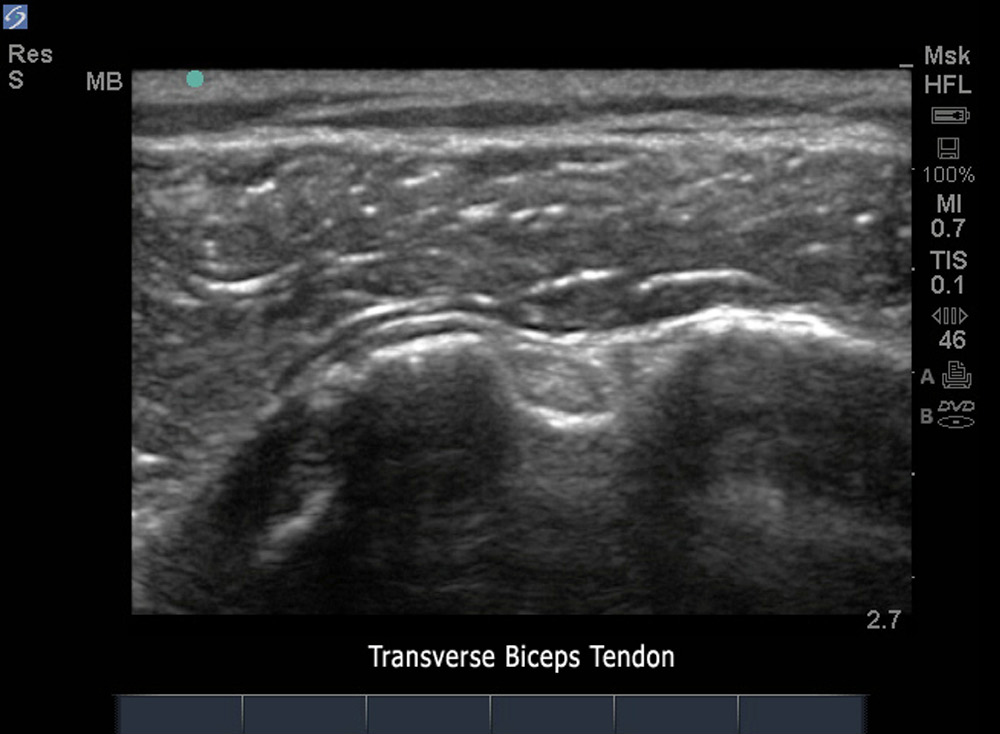
/sites/default/files/M-Turbo_MSK_Biceps_Tendon-2_SA.jpg
M-Turbo - Transverse View Biceps Tendon.
Clinical Specialties
Media Library Type
Media Library Tag
Biceps Tendon - Transverse View Abnormal
Biceps Tendon - Transverse View Abnormal
/sites/default/files/M-Turbo_MSK_Transverse_View_Abnormal_Biceps_Tendon-2_SA.jpg
M-Turbo - Transverse View Abnormal Biceps Tendon.
Clinical Specialties
Media Library Type
Media Library Tag