

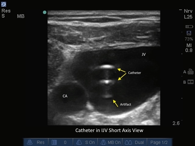
M-Turbo: Catheter in IJV Short Axis.
M-Turbo: Catheter in IJV Short Axis.
/sites/default/files/201410_Image_M-Turbo_Catheter_in_IJV_Short_Axis.jpg
M-Turbo: Catheter in IJV Short Axis.
Clinical Specialties
Media Library Type
Media Library Tag
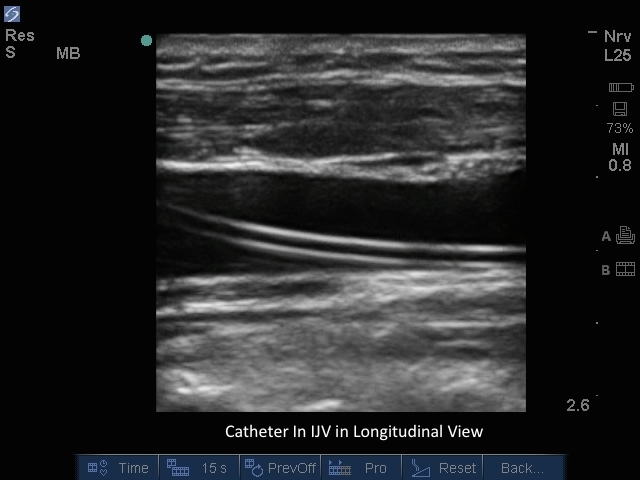
M-Turbo: Catheter in IJV Long Axis
M-Turbo: Catheter in IJV Long Axis
/sites/default/files/201410_Image_M-Turbo_Catheter_in_IJV_Long_Axis.jpg
M-Turbo: Catheter in IJV Long Axis.
Media Library Type
Media Library Tag