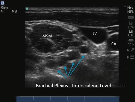
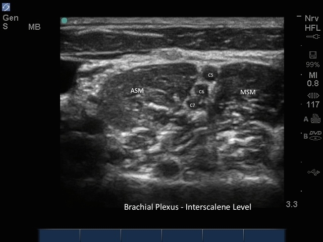
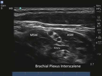
3D How To: Saphenous Nerve Block

3D How To: Saphenous Nerve Block

/sites/default/files/Saphenous_edu00504_thumbnail.jpg

3D animation demonstrating an ultrasound guided saphenous nerve block.

Clinical Specialties
Media Library Type
Subtitles
<p begin="00:00:07.382" end="00:00:09.372" style="s2">- [Voiceover] A linear array transducer</p>
<p begin="00:00:09.372" end="00:00:11.583" style="s2">with a nerve exam type is used to perform</p>
<p begin="00:00:11.583" end="00:00:14.256" style="s2">an ultrasound-guided<br />saphenous nerve block.</p>
<p begin="00:00:14.256" end="00:00:16.488" style="s2">The target depth is approximately</p>
<p begin="00:00:16.488" end="00:00:19.895" style="s2">one to three centimeters<br />in an 80 kilogram adult.</p>
<p begin="00:00:19.895" end="00:00:22.767" style="s2">The patient is positioned<br />in a supine position</p>
<p begin="00:00:22.767" end="00:00:26.181" style="s2">with the leg slightly abducted<br />and externally rotated.</p>
<p begin="00:00:26.181" end="00:00:29.113" style="s2">The transducer is placed<br />in the middle of the thigh</p>
<p begin="00:00:29.113" end="00:00:32.156" style="s2">in a transverse plane,<br />with the orientation marker</p>
<p begin="00:00:32.156" end="00:00:34.315" style="s2">directed to the patient's right.</p>
<p begin="00:00:34.315" end="00:00:36.942" style="s2">The leg is scanned medially to laterally</p>
<p begin="00:00:36.942" end="00:00:39.876" style="s2">to identify the<br />superficial femoral artery,</p>
<p begin="00:00:39.876" end="00:00:42.604" style="s2">which lies underneath<br />the sartorius muscle.</p>
<p begin="00:00:42.604" end="00:00:44.704" style="s2">The saphenous nerve can lie either</p>
<p begin="00:00:44.704" end="00:00:47.489" style="s2">anterior or posterior to the artery.</p>
<p begin="00:00:47.489" end="00:00:50.222" style="s2">The saphenous nerve may not be visible.</p>
<p begin="00:00:50.222" end="00:00:52.648" style="s2">If it is, it will appear as a bright,</p>
<p begin="00:00:52.648" end="00:00:55.461" style="s2">hyperechoic oval or triangular structure.</p>
<p begin="00:00:55.461" end="00:00:58.688" style="s2">The needle is positioned one<br />to two centimeters lateral</p>
<p begin="00:00:58.688" end="00:01:02.121" style="s2">to the transducer, and<br />advanced under the transducer.</p>
<p begin="00:01:02.121" end="00:01:04.989" style="s2">Local anesthetic is<br />injected incrementally,</p>
<p begin="00:01:04.989" end="00:01:07.315" style="s2">superficial and deep to the artery</p>
<p begin="00:01:07.315" end="00:01:10.482" style="s2">to complete the saphenous nerve block.</p>
Brightcove ID
5508114714001
https://youtube.com/watch?v=54VG2GhJ3w4
Body

3D animation demonstrating an ultrasound guided saphenous nerve block.

3D How To: Supraclavicular Nerve Block

3D How To: Supraclavicular Nerve Block

/sites/default/files/Supraclavicular_edu00495_thumbnail_.jpg

3D animation demonstrating an ultrasound guided Supraclavicular nerve block.

Clinical Specialties
Media Library Type
Subtitles
<p begin="00:00:07.580" end="00:00:09.407" style="s2">- [Voiceover] A linear<br />array transducer with</p>
<p begin="00:00:09.407" end="00:00:13.087" style="s2">a nerve exam type is used to<br />perform an ultrasound guided</p>
<p begin="00:00:13.087" end="00:00:15.690" style="s2">supraclavicular regional nerve block.</p>
<p begin="00:00:15.690" end="00:00:18.986" style="s2">The target depth is approximately<br />one to three centimeters</p>
<p begin="00:00:18.986" end="00:00:21.352" style="s2">in an 80 kilogram adult.</p>
<p begin="00:00:21.352" end="00:00:24.233" style="s2">The patient is positioned in a 45 degree</p>
<p begin="00:00:24.233" end="00:00:27.042" style="s2">reclining position with<br />a pillow under their head</p>
<p begin="00:00:27.042" end="00:00:29.627" style="s2">and the neck exposed<br />on the operative side.</p>
<p begin="00:00:29.627" end="00:00:33.564" style="s2">The patient's head is rotated<br />toward the contralateral side.</p>
<p begin="00:00:33.564" end="00:00:37.068" style="s2">The examination begins by finding<br />the supraclavicular region</p>
<p begin="00:00:37.068" end="00:00:40.404" style="s2">of the brachial plexus<br />as a landmark technique.</p>
<p begin="00:00:40.404" end="00:00:42.977" style="s2">The transducer is placed posterior</p>
<p begin="00:00:42.977" end="00:00:46.300" style="s2">to the midpoint of the<br />clavicle at an acute angle</p>
<p begin="00:00:46.300" end="00:00:47.878" style="s2">with the orientation marker directed</p>
<p begin="00:00:47.878" end="00:00:50.750" style="s2">to the patient's right<br />at a ten o'clock position</p>
<p begin="00:00:50.750" end="00:00:54.250" style="s2">with the transducer aimed into the thorax.</p>
<p begin="00:00:56.045" end="00:00:59.684" style="s2">The subclavian artery is<br />seen as a round pulsal tile</p>
<p begin="00:00:59.684" end="00:01:01.824" style="s2">structure superior to<br />the bright reflection</p>
<p begin="00:01:01.824" end="00:01:03.662" style="s2">of the first rib.</p>
<p begin="00:01:03.662" end="00:01:06.728" style="s2">The plura is seen as a<br />bright hyperechoic reflection</p>
<p begin="00:01:06.728" end="00:01:10.679" style="s2">deep two, or at the same<br />depth as the first rib.</p>
<p begin="00:01:10.679" end="00:01:13.352" style="s2">The nerves of the brachial<br />plexus are posterior</p>
<p begin="00:01:13.352" end="00:01:16.608" style="s2">or superior to the subclavian artery.</p>
<p begin="00:01:16.608" end="00:01:19.723" style="s2">The nerve trunks appear as<br />hypoechoic dark circles</p>
<p begin="00:01:19.723" end="00:01:24.288" style="s2">within the bright hyperechoic<br />fascia of the brachial plexus.</p>
<p begin="00:01:24.288" end="00:01:25.915" style="s2">Colored doppler imaging may be used</p>
<p begin="00:01:25.915" end="00:01:28.343" style="s2">to identify smaller arterial branches</p>
<p begin="00:01:28.343" end="00:01:30.056" style="s2">running through the brachial plexus</p>
<p begin="00:01:30.056" end="00:01:33.394" style="s2">or lying in the path<br />of needle advancement.</p>
<p begin="00:01:33.394" end="00:01:35.861" style="s2">The needle is positioned<br />one to two centimeters</p>
<p begin="00:01:35.861" end="00:01:38.295" style="s2">lateral to the transducer, and advanced</p>
<p begin="00:01:38.295" end="00:01:40.770" style="s2">using an in plane technique.</p>
<p begin="00:01:40.770" end="00:01:42.600" style="s2">The initial end point of the needle</p>
<p begin="00:01:42.600" end="00:01:44.320" style="s2">is just posterior to the artery</p>
<p begin="00:01:44.320" end="00:01:46.747" style="s2">immediately above the first rib.</p>
<p begin="00:01:46.747" end="00:01:48.937" style="s2">It is important to distinguish the plura</p>
<p begin="00:01:48.937" end="00:01:52.676" style="s2">from the first rib to<br />avoid a pneumothorax.</p>
<p begin="00:01:52.676" end="00:01:54.378" style="s2">The local anesthetic is injected</p>
<p begin="00:01:54.378" end="00:01:56.994" style="s2">incrementally close to the nerves.</p>
<p begin="00:01:56.994" end="00:01:58.640" style="s2">The needle can be redirected towards</p>
<p begin="00:01:58.640" end="00:02:00.757" style="s2">the upper trunks of the brachial plexus</p>
<p begin="00:02:00.757" end="00:02:04.924" style="s2">if the spread of local anesthetic<br />is not deemed adequate.</p>
Brightcove ID
5750031878001
https://youtube.com/watch?v=9vW1uo7mKDc
Body

3D animation demonstrating an ultrasound guided Supraclavicular nerve block.

How to: Stellate Ganglion Block

How to: Stellate Ganglion Block

/sites/default/files/Stellate_Ganglion_Block_edu00310.jpg
This videos discusses some of the scanning techniques involved when performing a stellate ganglion nerve block under ultrasound guidance.
Clinical Specialties
Media Library Type
Subtitles
<p begin="00:00:09.402" end="00:00:11.659" style="s2">- Today I'm going to demonstrate<br />the stellate ganglion block</p>
<p begin="00:00:11.659" end="00:00:13.495" style="s2">performed under ultrasound guidance.</p>
<p begin="00:00:13.495" end="00:00:15.962" style="s2">Traditionally this block<br />was performed blindly,</p>
<p begin="00:00:15.962" end="00:00:18.270" style="s2">without the use of any imaging modality.</p>
<p begin="00:00:18.270" end="00:00:20.964" style="s2">Currently thoracoscopy<br />is a preferred method,</p>
<p begin="00:00:20.964" end="00:00:24.667" style="s2">however utilizing ultrasound<br />such as this M Turbo system</p>
<p begin="00:00:24.667" end="00:00:28.071" style="s2">utilizing the HFL 50 linear probe,</p>
<p begin="00:00:28.071" end="00:00:30.052" style="s2">this can be done with less radiation</p>
<p begin="00:00:30.052" end="00:00:31.672" style="s2">and avoidance of vascular structures</p>
<p begin="00:00:31.672" end="00:00:33.446" style="s2">that might not otherwise be seen</p>
<p begin="00:00:33.446" end="00:00:35.143" style="s2">under thoracoscopic guidance.</p>
<p begin="00:00:35.143" end="00:00:39.310" style="s2">The C6 transverse process,<br />Chassaignac's tubercle,</p>
<p begin="00:00:40.457" end="00:00:43.574" style="s2">immediately inferior is the C7 body</p>
<p begin="00:00:43.574" end="00:00:45.491" style="s2">and transverse process.</p>
<p begin="00:00:46.380" end="00:00:49.120" style="s2">The probe is placed in a<br />transverse orientation,</p>
<p begin="00:00:49.120" end="00:00:53.287" style="s2">clearly identifying and<br />avoiding the vertebral artery.</p>
<p begin="00:00:54.506" end="00:00:57.084" style="s2">And the needle coming in contact with</p>
<p begin="00:00:57.084" end="00:00:59.251" style="s2">the C7 transverse process.</p>
<p begin="00:01:01.288" end="00:01:03.217" style="s2">To perform this particular block,</p>
<p begin="00:01:03.217" end="00:01:05.682" style="s2">the following equipment is necessary.</p>
<p begin="00:01:05.682" end="00:01:08.398" style="s2">The chlorhexidine prep,</p>
<p begin="00:01:08.398" end="00:01:09.957" style="s2">one percent buffered lidocaine</p>
<p begin="00:01:09.957" end="00:01:13.862" style="s2">with a one and one half<br />inch 27 gauge needle,</p>
<p begin="00:01:13.862" end="00:01:18.297" style="s2">a 25 gauge three and one<br />half inch spinal needle,</p>
<p begin="00:01:18.297" end="00:01:21.172" style="s2">a 10 cc syringe that<br />will contain seven ccs</p>
<p begin="00:01:21.172" end="00:01:23.851" style="s2">of one percent lidocaine with epinephrine,</p>
<p begin="00:01:23.851" end="00:01:27.719" style="s2">or quarter percent<br />bupivacaine with epinephrine.</p>
<p begin="00:01:27.719" end="00:01:29.722" style="s2">The procedure is<br />performed with the patient</p>
<p begin="00:01:29.722" end="00:01:32.412" style="s2">in the supine position, with the chin up</p>
<p begin="00:01:32.412" end="00:01:35.728" style="s2">and the following anatomic<br />structures are identified.</p>
<p begin="00:01:35.728" end="00:01:39.358" style="s2">The thyroid cartilage, the<br />cricoid cartilage, and then,</p>
<p begin="00:01:39.358" end="00:01:42.176" style="s2">the finger is placed in the<br />groove next to the trachea,</p>
<p begin="00:01:42.176" end="00:01:46.480" style="s2">and pressure applied until<br />a bony protuberance is felt.</p>
<p begin="00:01:46.480" end="00:01:48.317" style="s2">This is Chassaignac's tubercle or</p>
<p begin="00:01:48.317" end="00:01:51.128" style="s2">the transverse process of C6.</p>
<p begin="00:01:51.128" end="00:01:53.099" style="s2">Currently, the use of thoracoscopy</p>
<p begin="00:01:53.099" end="00:01:55.903" style="s2">indicates use at the C7 level.</p>
<p begin="00:01:55.903" end="00:01:57.658" style="s2">The reason for this is to provide</p>
<p begin="00:01:57.658" end="00:02:00.853" style="s2">better sympathetic blockade<br />while avoiding some</p>
<p begin="00:02:00.853" end="00:02:03.716" style="s2">of the common side effects<br />such as Horner's syndrome,</p>
<p begin="00:02:03.716" end="00:02:07.382" style="s2">as well as recurrent<br />pharyngeal nerve paralysis.</p>
<p begin="00:02:07.382" end="00:02:10.301" style="s2">In this case, the HFL 50<br />probe will be utilized.</p>
<p begin="00:02:10.301" end="00:02:14.213" style="s2">It provides a very high<br />definition, high resolution view</p>
<p begin="00:02:14.213" end="00:02:16.796" style="s2">of the subcutaneous structures.</p>
<p begin="00:02:19.814" end="00:02:23.647" style="s2">Gel is placed at the<br />formerly marked C6 level.</p>
<p begin="00:02:25.278" end="00:02:29.549" style="s2">And the HFL 50 probe is placed on the skin</p>
<p begin="00:02:29.549" end="00:02:31.882" style="s2">in a transverse arrangement.</p>
<p begin="00:02:34.594" end="00:02:37.150" style="s2">The initial view is of<br />the cricoid cartilage,</p>
<p begin="00:02:37.150" end="00:02:40.861" style="s2">and then the probe is<br />moved slightly cephalad</p>
<p begin="00:02:40.861" end="00:02:44.116" style="s2">and then rotated slightly outward.</p>
<p begin="00:02:44.116" end="00:02:48.060" style="s2">At this point the thyroid as<br />well as the carotid artery</p>
<p begin="00:02:48.060" end="00:02:50.833" style="s2">and Chassaignac's tubercle<br />are clearly visible</p>
<p begin="00:02:50.833" end="00:02:54.786" style="s2">as the hump located immediately<br />in the center of the screen.</p>
<p begin="00:02:54.786" end="00:02:57.203" style="s2">Since this is the C6 tubercle,</p>
<p begin="00:02:57.203" end="00:03:00.856" style="s2">we will move slightly<br />interior until it flattens out</p>
<p begin="00:03:00.856" end="00:03:04.439" style="s2">and this becomes the<br />C7 transverse process.</p>
<p begin="00:03:05.932" end="00:03:10.667" style="s2">It's important to note<br />at this point to utilize</p>
<p begin="00:03:10.667" end="00:03:13.426" style="s2">the color flow Doppler which will indicate</p>
<p begin="00:03:13.426" end="00:03:16.941" style="s2">vascular structures such as<br />perforating thyroidal arteries</p>
<p begin="00:03:16.941" end="00:03:19.859" style="s2">that are not to be violated,</p>
<p begin="00:03:19.859" end="00:03:21.930" style="s2">as well as the carotid artery,</p>
<p begin="00:03:21.930" end="00:03:24.533" style="s2">and the vertebral artery more laterally.</p>
<p begin="00:03:24.533" end="00:03:28.700" style="s2">The structure of interest<br />is the longus colli muscle.</p>
<p begin="00:03:29.590" end="00:03:31.727" style="s2">At this point, local anesthetic</p>
<p begin="00:03:31.727" end="00:03:35.144" style="s2">one percent lidocaine buffer is injected,</p>
<p begin="00:03:36.173" end="00:03:40.233" style="s2">and a 25 gauge three<br />and a half inch needle</p>
<p begin="00:03:40.233" end="00:03:44.400" style="s2">is advanced through this track<br />to contact the oss at C7.</p>
<p begin="00:03:48.349" end="00:03:51.806" style="s2">The needle is backed off<br />slightly and then the injection</p>
<p begin="00:03:51.806" end="00:03:55.787" style="s2">of local anesthetic,<br />preferably epinephrine</p>
<p begin="00:03:55.787" end="00:03:58.541" style="s2">containing one percent lidocaine,</p>
<p begin="00:03:58.541" end="00:04:02.229" style="s2">six to seven ccs is gently<br />and slowly injected.</p>
<p begin="00:04:02.229" end="00:04:06.922" style="s2">At this point, the probe<br />is removed, the skin wiped</p>
<p begin="00:04:06.922" end="00:04:10.755" style="s2">and cleaned, and if<br />needed, a band aid placed</p>
<p begin="00:04:10.755" end="00:04:12.591" style="s2">over the small puncture site.</p>
<p begin="00:04:12.591" end="00:04:15.199" style="s2">This successfully concludes<br />a stellate ganglion block</p>
<p begin="00:04:15.199" end="00:04:18.199" style="s2">performed under ultrasound guidance.</p>
Brightcove ID
5508120224001
https://youtube.com/watch?v=RrDOsfoOSuw

How to: Piriformis Injection

How to: Piriformis Injection

/sites/default/files/Piriformis_injection_edu00307.jpg
This videos discusses some of the scanning techniques involved while performing a piriformis injection.
Clinical Specialties
Media Library Type
Subtitles
<p begin="00:00:09.683" end="00:00:12.658" style="s2">- Today I'm going to demonstrate<br />the piriformis injection.</p>
<p begin="00:00:12.658" end="00:00:15.497" style="s2">Piriformis injection under<br />ultrasound guidance is useful</p>
<p begin="00:00:15.497" end="00:00:17.249" style="s2">in diagnostic maneuvers,</p>
<p begin="00:00:17.249" end="00:00:18.672" style="s2">specifically for patients</p>
<p begin="00:00:18.672" end="00:00:20.710" style="s2">that may have sciatic-like symptoms</p>
<p begin="00:00:20.710" end="00:00:24.529" style="s2">as the sciatic nerve passes<br />close to the piriformis muscle.</p>
<p begin="00:00:24.529" end="00:00:25.709" style="s2">In piriformis syndrome</p>
<p begin="00:00:25.709" end="00:00:28.103" style="s2">the piriformis muscle is hypertrophied</p>
<p begin="00:00:28.103" end="00:00:30.632" style="s2">and can mimic sciatic symptoms</p>
<p begin="00:00:30.632" end="00:00:33.389" style="s2">caused by lumbar herniated disks.</p>
<p begin="00:00:33.389" end="00:00:34.722" style="s2">If this has been excluded,</p>
<p begin="00:00:34.722" end="00:00:36.467" style="s2">this particular procedure can be done</p>
<p begin="00:00:36.467" end="00:00:38.598" style="s2">under ultrasonographic guidance.</p>
<p begin="00:00:38.598" end="00:00:40.381" style="s2">The patient is placed<br />in the prone position</p>
<p begin="00:00:40.381" end="00:00:44.048" style="s2">with a pillow underneath<br />to provide bolster.</p>
<p begin="00:00:48.281" end="00:00:51.638" style="s2">The side in question is<br />identified and prepped</p>
<p begin="00:00:51.638" end="00:00:53.467" style="s2">in this particular fashion.</p>
<p begin="00:00:53.467" end="00:00:55.414" style="s2">The gluteus is exposed.</p>
<p begin="00:00:55.414" end="00:00:58.256" style="s2">For facilitation of visualization</p>
<p begin="00:00:58.256" end="00:01:01.351" style="s2">I utilize the Sonosite M Turbo</p>
<p begin="00:01:01.351" end="00:01:06.008" style="s2">with the musculoskeletal<br />setting to help enhance</p>
<p begin="00:01:06.008" end="00:01:09.299" style="s2">the muscular fibers of<br />the piriformis muscle,</p>
<p begin="00:01:09.299" end="00:01:11.143" style="s2">the gluteus minimus and maximus,</p>
<p begin="00:01:11.143" end="00:01:14.489" style="s2">and the curvilinear shape<br />facilitates entry of the needle</p>
<p begin="00:01:14.489" end="00:01:16.744" style="s2">into the targeted site.</p>
<p begin="00:01:16.744" end="00:01:18.415" style="s2">To perform this particular block</p>
<p begin="00:01:18.415" end="00:01:21.556" style="s2">the following equipment is necessary.</p>
<p begin="00:01:21.556" end="00:01:24.336" style="s2">Chlorhexidine for sterile technique,</p>
<p begin="00:01:24.336" end="00:01:28.503" style="s2">local anesthetic syringe<br />containing 1% buffered lidocaine,</p>
<p begin="00:01:30.111" end="00:01:34.278" style="s2">and then a syringe containing<br />five cc of bupivicaine 1/2%</p>
<p begin="00:01:35.932" end="00:01:39.878" style="s2">mixed with five cc of 1% lidocaine,</p>
<p begin="00:01:39.878" end="00:01:43.843" style="s2">and then 40 to 80 milligrams<br />of triamcinolone kenalog,</p>
<p begin="00:01:43.843" end="00:01:46.704" style="s2">which is a particulate steroid.</p>
<p begin="00:01:46.704" end="00:01:48.943" style="s2">A 3-1/2-inch 25-gauge spinal needle</p>
<p begin="00:01:48.943" end="00:01:52.173" style="s2">will also be utilized for the procedure.</p>
<p begin="00:01:52.173" end="00:01:53.482" style="s2">There will be sterile four by fours</p>
<p begin="00:01:53.482" end="00:01:56.627" style="s2">necessary to clean the<br />area at the conclusion</p>
<p begin="00:01:56.627" end="00:02:00.634" style="s2">and a simple BAND-AID will<br />suffice to cover the wound.</p>
<p begin="00:02:00.634" end="00:02:02.384" style="s2">Copious amounts of gel are required</p>
<p begin="00:02:02.384" end="00:02:05.677" style="s2">for this particular procedure.</p>
<p begin="00:02:05.677" end="00:02:08.878" style="s2">The middle portion of<br />the gluteus is identified</p>
<p begin="00:02:08.878" end="00:02:12.559" style="s2">and then in a transverse or<br />left-to-right positioning</p>
<p begin="00:02:12.559" end="00:02:16.309" style="s2">the probe is placed in<br />contact with the skin.</p>
<p begin="00:02:24.660" end="00:02:27.660" style="s2">The leg in question can be mobilized</p>
<p begin="00:02:29.333" end="00:02:32.083" style="s2">in a abduction-adduction maneuver</p>
<p begin="00:02:34.757" end="00:02:38.329" style="s2">demonstrating motion of<br />the piriformis muscle</p>
<p begin="00:02:38.329" end="00:02:40.985" style="s2">as it inserts out towards<br />the greater trochanter</p>
<p begin="00:02:40.985" end="00:02:44.042" style="s2">and towards the ischium centrally.</p>
<p begin="00:02:44.042" end="00:02:45.208" style="s2">Clearly in the middle of the screen</p>
<p begin="00:02:45.208" end="00:02:49.141" style="s2">you can also identify the sciatic nerve</p>
<p begin="00:02:49.141" end="00:02:50.858" style="s2">as it passes very close</p>
<p begin="00:02:50.858" end="00:02:54.108" style="s2">to the fibers of the piriformis muscle.</p>
<p begin="00:02:55.482" end="00:02:58.353" style="s2">A central point is identified,</p>
<p begin="00:02:58.353" end="00:03:01.180" style="s2">making sure to stay away<br />from the investing fascia</p>
<p begin="00:03:01.180" end="00:03:03.263" style="s2">around the sciatic nerve.</p>
<p begin="00:03:04.531" end="00:03:08.462" style="s2">The skin is marked and then 1%<br />local anesthetic is injected</p>
<p begin="00:03:08.462" end="00:03:10.712" style="s2">directly in short axis view</p>
<p begin="00:03:14.359" end="00:03:16.971" style="s2">and then a 25-gauge 3-1/2-inch needle</p>
<p begin="00:03:16.971" end="00:03:20.121" style="s2">is passed through this anesthetized track</p>
<p begin="00:03:20.121" end="00:03:23.460" style="s2">and approximately six<br />cc of local anesthetic</p>
<p begin="00:03:23.460" end="00:03:26.903" style="s2">containing bupivicaine and<br />lidocaine in a one to one mixture</p>
<p begin="00:03:26.903" end="00:03:31.070" style="s2">with a small amount of steroid<br />if desired can be injected.</p>
<p begin="00:03:31.926" end="00:03:33.186" style="s2">- [Voiceover] This is a short axis view</p>
<p begin="00:03:33.186" end="00:03:35.477" style="s2">and the curvilinear probe is applied</p>
<p begin="00:03:35.477" end="00:03:38.224" style="s2">in the transverse orientation.</p>
<p begin="00:03:38.224" end="00:03:40.699" style="s2">The leg is being adducted and abducted</p>
<p begin="00:03:40.699" end="00:03:42.866" style="s2">to show the muscle moving.</p>
<p begin="00:03:46.159" end="00:03:47.673" style="s2">The sciatic nerve is clearly visible</p>
<p begin="00:03:47.673" end="00:03:50.345" style="s2">in the substance of the muscle.</p>
<p begin="00:03:50.345" end="00:03:53.845" style="s2">It is the band-like hyperechoic structure.</p>
<p begin="00:03:57.117" end="00:03:59.101" style="s2">The injectate is the hypoechoic structure</p>
<p begin="00:03:59.101" end="00:04:02.934" style="s2">that grows within the<br />substance of the muscle.</p>
<p begin="00:04:22.176" end="00:04:24.259" style="s2">- The probe is withdrawn,</p>
<p begin="00:04:25.810" end="00:04:28.727" style="s2">the area wiped with a four by four,</p>
<p begin="00:04:33.066" end="00:04:36.566" style="s2">and then a BAND-AID placed over the wound.</p>
<p begin="00:04:38.385" end="00:04:40.235" style="s2">This is the successful completion</p>
<p begin="00:04:40.235" end="00:04:44.402" style="s2">of a piriformis injection under<br />ultrasonographic guidance.</p>
Brightcove ID
5508136045001
https://youtube.com/watch?v=ZGcNFTHlFeQ

How to: Cervical Spine Injection

How to: Cervical Spine Injection

/sites/default/files/Cervical_Spine_edu00308.jpg
This videos discusses some of the scanning techniques involved while performing the cervical spine injection.
Clinical Specialties
Media Library Type
Subtitles
<p begin="00:00:09.277" end="00:00:12.473" style="s2">- Today I will perform<br />an anatomic demonstration</p>
<p begin="00:00:12.473" end="00:00:14.176" style="s2">of the cervical spine utilizing</p>
<p begin="00:00:14.176" end="00:00:16.894" style="s2">the M-Turbo Ultrasound System,</p>
<p begin="00:00:16.894" end="00:00:20.272" style="s2">utilizing the C60 curvilinear probe.</p>
<p begin="00:00:20.272" end="00:00:22.765" style="s2">The point of this<br />exercise is to demonstrate</p>
<p begin="00:00:22.765" end="00:00:25.246" style="s2">the anatomic considerations<br />when performing</p>
<p begin="00:00:25.246" end="00:00:28.005" style="s2">cervical epidural steroid injections</p>
<p begin="00:00:28.005" end="00:00:29.905" style="s2">and cervical facet injections.</p>
<p begin="00:00:29.905" end="00:00:32.402" style="s2">Typical patients may have have<br />a variance in their anatomy</p>
<p begin="00:00:32.402" end="00:00:35.125" style="s2">and the traditional approaches utilizing</p>
<p begin="00:00:35.125" end="00:00:36.739" style="s2">loss of resistance techniques for</p>
<p begin="00:00:36.739" end="00:00:40.204" style="s2">cervical epidural steroids<br />can present come challenges.</p>
<p begin="00:00:40.204" end="00:00:43.271" style="s2">The purpose of utilizing<br />the ultrasound system</p>
<p begin="00:00:43.271" end="00:00:46.105" style="s2">for an anatomic survey<br />is that the patient,</p>
<p begin="00:00:46.105" end="00:00:49.308" style="s2">who normally has a cervical<br />epidural steroid injection</p>
<p begin="00:00:49.308" end="00:00:51.419" style="s2">performed under fluoroscopic technique</p>
<p begin="00:00:51.419" end="00:00:54.799" style="s2">may occasionally encounter<br />false losses of resistance.</p>
<p begin="00:00:54.799" end="00:00:57.909" style="s2">Utilizing this particular<br />tool as a sounding device</p>
<p begin="00:00:57.909" end="00:01:01.742" style="s2">may perhaps improve the<br />accuracy of injection.</p>
<p begin="00:01:02.726" end="00:01:06.584" style="s2">The way this is performed is<br />utilizing again the C60 probe,</p>
<p begin="00:01:06.584" end="00:01:11.234" style="s2">setting the depth to<br />approximately 9.2 centimeters,</p>
<p begin="00:01:11.234" end="00:01:14.283" style="s2">remembering that the average<br />depth to the epidural space</p>
<p begin="00:01:14.283" end="00:01:16.584" style="s2">is appx six centimeters.</p>
<p begin="00:01:16.584" end="00:01:19.283" style="s2">The nerve setting will also be used</p>
<p begin="00:01:19.283" end="00:01:22.646" style="s2">to highlight any neural structures<br />that may be of interest.</p>
<p begin="00:01:22.646" end="00:01:25.938" style="s2">On the patient, initially, I like to mark</p>
<p begin="00:01:25.938" end="00:01:29.605" style="s2">utilizing a Sharpie,<br />the C7 spinous process.</p>
<p begin="00:01:32.137" end="00:01:35.181" style="s2">This is the most easily<br />palpable spinous process,</p>
<p begin="00:01:35.181" end="00:01:37.149" style="s2">and this is facilitated with the patient</p>
<p begin="00:01:37.149" end="00:01:40.496" style="s2">in the prone position, a<br />bolster under the thorax,</p>
<p begin="00:01:40.496" end="00:01:42.913" style="s2">and the head slightly flexed.</p>
<p begin="00:01:43.954" end="00:01:45.925" style="s2">I place a copious amount of gel</p>
<p begin="00:01:45.925" end="00:01:49.821" style="s2">across the patient's C7 spinous process,</p>
<p begin="00:01:49.821" end="00:01:53.738" style="s2">and then utilizing the<br />probe in the transverse,</p>
<p begin="00:01:54.573" end="00:01:56.570" style="s2">that is the left-to-right positioning,</p>
<p begin="00:01:56.570" end="00:02:00.366" style="s2">I place contact on the<br />skin, and clearly visible</p>
<p begin="00:02:00.366" end="00:02:03.366" style="s2">is the patient's C7 spinous process.</p>
<p begin="00:02:04.461" end="00:02:07.097" style="s2">In this particular view you<br />can also see the lamina,</p>
<p begin="00:02:07.097" end="00:02:10.930" style="s2">and the transverse<br />processes of T1 just below.</p>
<p begin="00:02:12.973" end="00:02:14.856" style="s2">And in the center of the screen,</p>
<p begin="00:02:14.856" end="00:02:18.064" style="s2">under the shadow of the spinous<br />process is a white line.</p>
<p begin="00:02:18.064" end="00:02:21.897" style="s2">This white line, and I<br />will highlight it here,</p>
<p begin="00:02:24.556" end="00:02:29.129" style="s2">demonstrates the actual<br />location of the epidural space.</p>
<p begin="00:02:29.129" end="00:02:32.904" style="s2">So looking off to the right of the screen,</p>
<p begin="00:02:32.904" end="00:02:35.199" style="s2">we can see that the depth is approximately</p>
<p begin="00:02:35.199" end="00:02:38.866" style="s2">five centimeters in<br />this particular patient.</p>
<p begin="00:02:40.790" end="00:02:43.256" style="s2">Other potential views include</p>
<p begin="00:02:43.256" end="00:02:48.086" style="s2">the cranial carotid<br />orientation, or sagittal plane</p>
<p begin="00:02:48.086" end="00:02:51.910" style="s2">of the probe, and placed along the neck,</p>
<p begin="00:02:51.910" end="00:02:53.993" style="s2">and slightly off midline,</p>
<p begin="00:02:54.938" end="00:02:57.649" style="s2">again now can be seen newer structures.</p>
<p begin="00:02:57.649" end="00:03:01.164" style="s2">In this particular case<br />the facet joint line</p>
<p begin="00:03:01.164" end="00:03:05.238" style="s2">is very clearly indicated<br />at these locations.</p>
<p begin="00:03:05.238" end="00:03:08.513" style="s2">The actual location for<br />a medial branch block</p>
<p begin="00:03:08.513" end="00:03:11.513" style="s2">would be on these superior surfaces.</p>
<p begin="00:03:12.556" end="00:03:14.499" style="s2">On the lateral mass.</p>
<p begin="00:03:14.499" end="00:03:18.690" style="s2">If the probe is then rotated further out,</p>
<p begin="00:03:18.690" end="00:03:21.440" style="s2">and a more oblique view obtained,</p>
<p begin="00:03:22.365" end="00:03:25.371" style="s2">the actual foramen of the nerve roots</p>
<p begin="00:03:25.371" end="00:03:27.451" style="s2">can thus be discerned.</p>
<p begin="00:03:27.451" end="00:03:30.118" style="s2">And as we move further cephalad,</p>
<p begin="00:03:33.346" end="00:03:37.513" style="s2">it is possible to view the<br />vertebral artery pulsations.</p>
<p begin="00:03:43.765" end="00:03:44.930" style="s2">And this obviously is a structure</p>
<p begin="00:03:44.930" end="00:03:48.905" style="s2">that would want to be<br />avoided at all costs.</p>
<p begin="00:03:48.905" end="00:03:51.318" style="s2">This concludes the<br />successful anatomical survey</p>
<p begin="00:03:51.318" end="00:03:55.401" style="s2">utilizing ultrasonography<br />of the cervical spine.</p>
Brightcove ID
5508121212001
https://youtube.com/watch?v=qlLfe5CE454

How to: Caudal Epidural Steroid Injection

How to: Caudal Epidural Steroid Injection

/sites/default/files/Caudal_Epidural_Steroid_Injection_edu00311.jpg
This video discusses some of the scanning techniques involved when performing a caudal epidural steroid injection under ultrasound guidance.
Clinical Specialties
Media Library Type
Subtitles
<p begin="00:00:09.298" end="00:00:10.544" style="s2">- Today I'm going to demonstrate a</p>
<p begin="00:00:10.544" end="00:00:13.071" style="s2">caudal epidural steroid injection.</p>
<p begin="00:00:13.071" end="00:00:14.521" style="s2">This is a variation on the technique</p>
<p begin="00:00:14.521" end="00:00:17.296" style="s2">of a lumbar epidural steroid injection.</p>
<p begin="00:00:17.296" end="00:00:19.276" style="s2">With the probe in this location we can see</p>
<p begin="00:00:19.276" end="00:00:21.828" style="s2">the following anatomic structures.</p>
<p begin="00:00:21.828" end="00:00:25.568" style="s2">The coccyx, the sacrococcygeal ligament,</p>
<p begin="00:00:25.568" end="00:00:27.843" style="s2">and the sacral cornua.</p>
<p begin="00:00:27.843" end="00:00:31.650" style="s2">This indicates the entry<br />at the sacral hiatus.</p>
<p begin="00:00:31.650" end="00:00:35.293" style="s2">The needle is placed thusly,<br />performing the block.</p>
<p begin="00:00:35.293" end="00:00:37.433" style="s2">Indications for this procedure include</p>
<p begin="00:00:37.433" end="00:00:40.066" style="s2">patients with lumbar spinal stenosis,</p>
<p begin="00:00:40.066" end="00:00:42.637" style="s2">lumbar radiculopathy, and any other cause</p>
<p begin="00:00:42.637" end="00:00:45.680" style="s2">of radiating and low back leg pain.</p>
<p begin="00:00:45.680" end="00:00:47.718" style="s2">In this particular case,<br />the caudal epidural</p>
<p begin="00:00:47.718" end="00:00:50.724" style="s2">steroid approach is utilized<br />to reduce the need for</p>
<p begin="00:00:50.724" end="00:00:54.471" style="s2">loss of resistance syringes,<br />fluoroscope radiation</p>
<p begin="00:00:54.471" end="00:00:56.392" style="s2">exposure to the patient, and removal</p>
<p begin="00:00:56.392" end="00:00:58.734" style="s2">of the need for ionic contrast.</p>
<p begin="00:00:58.734" end="00:01:02.438" style="s2">The same benefit can be<br />achieved utilizing ultrasound.</p>
<p begin="00:01:02.438" end="00:01:05.012" style="s2">In this particular case I'm<br />going to be using a Sonosite</p>
<p begin="00:01:05.012" end="00:01:08.664" style="s2">C60 probe because of its curvilinear shape</p>
<p begin="00:01:08.664" end="00:01:11.531" style="s2">which will facilitate viewing<br />of the sacral anatomy.</p>
<p begin="00:01:11.531" end="00:01:15.596" style="s2">This is key for the demonstration<br />of the relevant anatomy</p>
<p begin="00:01:15.596" end="00:01:17.231" style="s2">and ultimate placement of the needle</p>
<p begin="00:01:17.231" end="00:01:19.674" style="s2">for successful completion<br />of the procedure.</p>
<p begin="00:01:19.674" end="00:01:22.999" style="s2">The settings are set for<br />5.6 centimeter depth.</p>
<p begin="00:01:22.999" end="00:01:24.802" style="s2">Obviously this is patient specific.</p>
<p begin="00:01:24.802" end="00:01:27.361" style="s2">A larger patient may<br />require a deeper depth.</p>
<p begin="00:01:27.361" end="00:01:29.832" style="s2">The other setting is the<br />musculoskeletal setting</p>
<p begin="00:01:29.832" end="00:01:31.464" style="s2">or the MSK setting.</p>
<p begin="00:01:31.464" end="00:01:33.689" style="s2">This provides the best bony enhancement,</p>
<p begin="00:01:33.689" end="00:01:34.814" style="s2">and as we'll see we'll need to be</p>
<p begin="00:01:34.814" end="00:01:37.156" style="s2">passing a needle between two bony plates,</p>
<p begin="00:01:37.156" end="00:01:40.491" style="s2">and this setting facilitates<br />the best view of this.</p>
<p begin="00:01:40.491" end="00:01:43.038" style="s2">In order to facilitate<br />placement of the patient</p>
<p begin="00:01:43.038" end="00:01:45.533" style="s2">for the procedure, it is necessary to have</p>
<p begin="00:01:45.533" end="00:01:47.832" style="s2">the patient in the prone<br />position as seen here.</p>
<p begin="00:01:47.832" end="00:01:49.615" style="s2">This is a typical position<br />that would normally</p>
<p begin="00:01:49.615" end="00:01:52.710" style="s2">be used for a lumbar<br />epidural steroid approach.</p>
<p begin="00:01:52.710" end="00:01:55.119" style="s2">One key difference,<br />however, is the need to</p>
<p begin="00:01:55.119" end="00:01:58.087" style="s2">move the gluteal folds so that access</p>
<p begin="00:01:58.087" end="00:02:00.549" style="s2">to the sacral hiatus, which is the</p>
<p begin="00:02:00.549" end="00:02:02.929" style="s2">key target, is facilitated.</p>
<p begin="00:02:02.929" end="00:02:05.503" style="s2">In this particular case you<br />can see that the gluteal folds</p>
<p begin="00:02:05.503" end="00:02:08.223" style="s2">have been taped down and away in</p>
<p begin="00:02:08.223" end="00:02:10.739" style="s2">a caudal fashion to the patient.</p>
<p begin="00:02:10.739" end="00:02:13.369" style="s2">We now have a clean access point</p>
<p begin="00:02:13.369" end="00:02:15.605" style="s2">within the two gluteal folds.</p>
<p begin="00:02:15.605" end="00:02:17.921" style="s2">In order to facilitate a proper block,</p>
<p begin="00:02:17.921" end="00:02:19.836" style="s2">the following equipment is required.</p>
<p begin="00:02:19.836" end="00:02:23.276" style="s2">A chlorhexidine prep<br />for sterile technique,</p>
<p begin="00:02:23.276" end="00:02:25.750" style="s2">a local anesthetic syringe with a 27 gauge</p>
<p begin="00:02:25.750" end="00:02:29.260" style="s2">one and a half inch needle<br />for local anesthetic placement</p>
<p begin="00:02:29.260" end="00:02:31.244" style="s2">preferably one percent bicarbonated</p>
<p begin="00:02:31.244" end="00:02:33.051" style="s2">lidocaine, buffered lidocaine.</p>
<p begin="00:02:33.051" end="00:02:36.631" style="s2">A syringe containing<br />the epidural steroid mix</p>
<p begin="00:02:36.631" end="00:02:40.798" style="s2">which is composed of one<br />percent, one CC lidocaine,</p>
<p begin="00:02:42.246" end="00:02:45.367" style="s2">six CCs of bacteriostatic saline,</p>
<p begin="00:02:45.367" end="00:02:49.255" style="s2">and then 40 to 80 milligrams<br />of triamcinolone kenalog</p>
<p begin="00:02:49.255" end="00:02:52.003" style="s2">which is a particulate steroid.</p>
<p begin="00:02:52.003" end="00:02:53.662" style="s2">The needle utilized to enter the space</p>
<p begin="00:02:53.662" end="00:02:57.583" style="s2">is a three and a half inch spinal needle.</p>
<p begin="00:02:57.583" end="00:02:58.711" style="s2">For the conclusion of the procedure</p>
<p begin="00:02:58.711" end="00:03:00.652" style="s2">it will be necessary to have</p>
<p begin="00:03:00.652" end="00:03:03.043" style="s2">sterile four by four for cleanup</p>
<p begin="00:03:03.043" end="00:03:05.130" style="s2">and then a simple Band-Aid will suffice</p>
<p begin="00:03:05.130" end="00:03:07.130" style="s2">to cover the entry site.</p>
<p begin="00:03:08.659" end="00:03:11.710" style="s2">Placement of a copious amount of gel</p>
<p begin="00:03:11.710" end="00:03:13.996" style="s2">for coupling, aqueous<br />coupling is important</p>
<p begin="00:03:13.996" end="00:03:16.996" style="s2">to enhance resolution on the screen.</p>
<p begin="00:03:18.078" end="00:03:21.411" style="s2">And then contact is made in the midline.</p>
<p begin="00:03:22.951" end="00:03:26.238" style="s2">Because there's anatomic<br />variation in the midline,</p>
<p begin="00:03:26.238" end="00:03:29.690" style="s2">physiologically, a sagittal view</p>
<p begin="00:03:29.690" end="00:03:33.139" style="s2">and then gently rocking the probe</p>
<p begin="00:03:33.139" end="00:03:37.713" style="s2">in the oblique direction can<br />help find the spinous processes</p>
<p begin="00:03:37.713" end="00:03:40.130" style="s2">of the sacral space, and then</p>
<p begin="00:03:42.469" end="00:03:46.741" style="s2">the probe is moved<br />caudally and very clearly</p>
<p begin="00:03:46.741" end="00:03:48.840" style="s2">the sacral hiatus comes into view.</p>
<p begin="00:03:48.840" end="00:03:51.796" style="s2">There is a superior plate<br />and an inferior plate</p>
<p begin="00:03:51.796" end="00:03:55.038" style="s2">and in between them is the sacral hiatus.</p>
<p begin="00:03:55.038" end="00:03:58.693" style="s2">I'll demonstrate where<br />that access point is.</p>
<p begin="00:03:58.693" end="00:04:01.026" style="s2">The needle is angled thusly.</p>
<p begin="00:04:02.072" end="00:04:03.435" style="s2">At this point, I would utilize</p>
<p begin="00:04:03.435" end="00:04:07.953" style="s2">the local anesthetic to perform<br />a local anesthetic track</p>
<p begin="00:04:07.953" end="00:04:09.876" style="s2">down to this level.</p>
<p begin="00:04:09.876" end="00:04:11.394" style="s2">Removing that, I would then place</p>
<p begin="00:04:11.394" end="00:04:14.997" style="s2">the three and a half inch<br />25 gauge spinal needle</p>
<p begin="00:04:14.997" end="00:04:18.008" style="s2">along with the same anesthetized track</p>
<p begin="00:04:18.008" end="00:04:20.102" style="s2">and then once between the two plates,</p>
<p begin="00:04:20.102" end="00:04:24.269" style="s2">I would attach my local<br />anesthetic and steroids mix.</p>
<p begin="00:04:26.572" end="00:04:27.472" style="s2">- [Voiceover] The needle is visible</p>
<p begin="00:04:27.472" end="00:04:30.436" style="s2">as this view is in the long axis.</p>
<p begin="00:04:30.436" end="00:04:32.544" style="s2">The sacral plate is clearly visible,</p>
<p begin="00:04:32.544" end="00:04:34.961" style="s2">diving anterior and cephelad.</p>
<p begin="00:04:36.684" end="00:04:38.886" style="s2">Once the injectate enters the canal,</p>
<p begin="00:04:38.886" end="00:04:41.629" style="s2">the colored doppler shows<br />the fluid perturbation</p>
<p begin="00:04:41.629" end="00:04:44.393" style="s2">confined within the sacral epidural space,</p>
<p begin="00:04:44.393" end="00:04:46.650" style="s2">indicating a successful injection</p>
<p begin="00:04:46.650" end="00:04:49.567" style="s2">without need for contrast or X-ray.</p>
<p begin="00:04:51.366" end="00:04:54.324" style="s2">- Once the injectate is done<br />and the image is recorded</p>
<p begin="00:04:54.324" end="00:04:56.978" style="s2">as necessary for the medical record,</p>
<p begin="00:04:56.978" end="00:04:58.728" style="s2">the probe is removed.</p>
<p begin="00:04:59.783" end="00:05:02.578" style="s2">The needles are removed.</p>
<p begin="00:05:02.578" end="00:05:05.495" style="s2">The area is cleaned, and then place</p>
<p begin="00:05:07.427" end="00:05:09.976" style="s2">a Band-Aid over the wound.</p>
<p begin="00:05:09.976" end="00:05:12.671" style="s2">And thus this concludes<br />a successfully placed</p>
<p begin="00:05:12.671" end="00:05:15.504" style="s2">caudal epidural steroid injection.</p>
Brightcove ID
5508128556001
https://youtube.com/watch?v=yD6L4aSbSZA

How To Perform An Interscalene Nerve Block

How To Perform An Interscalene Nerve Block

/sites/default/files/youtube_0Cboqf1Qnhc.jpg
Dr. David Auyong reviews scanning techniques and sonographic landmarks for an interscalene brachial plexus nerve block.
Media Library Type
Subtitles
<p begin="00:00:13.460" end="00:00:14.935" style="s2">- The interscalene block is used</p>
<p begin="00:00:14.935" end="00:00:18.092" style="s2">for shoulder surgery and clavicle surgery.</p>
<p begin="00:00:18.092" end="00:00:19.843" style="s2">So, to start the interscalene block,</p>
<p begin="00:00:19.843" end="00:00:22.290" style="s2">proper positioning is very important.</p>
<p begin="00:00:22.290" end="00:00:25.260" style="s2">The best way to get to<br />the interscalene block</p>
<p begin="00:00:25.260" end="00:00:28.713" style="s2">is to have the patient sitting up</p>
<p begin="00:00:28.713" end="00:00:30.970" style="s2">about 30 or 45 degrees.</p>
<p begin="00:00:30.970" end="00:00:32.721" style="s2">Next, we turn the patients head away</p>
<p begin="00:00:32.721" end="00:00:35.731" style="s2">from the shoulder to the opposite side.</p>
<p begin="00:00:35.731" end="00:00:38.395" style="s2">This gives us a lot of<br />room to put the probe</p>
<p begin="00:00:38.395" end="00:00:42.012" style="s2">and have our hands come<br />from the posterior side.</p>
<p begin="00:00:42.012" end="00:00:44.829" style="s2">The proper approach to<br />the interscalene block</p>
<p begin="00:00:44.829" end="00:00:48.244" style="s2">is to have the needle approach<br />from the posterior side.</p>
<p begin="00:00:48.244" end="00:00:52.424" style="s2">This avoids the phrenic<br />nerve and allows us</p>
<p begin="00:00:52.424" end="00:00:56.019" style="s2">to not injure the phrenic<br />nerve with our needle approach.</p>
<p begin="00:00:56.019" end="00:00:57.452" style="s2">So, for the interscalene block,</p>
<p begin="00:00:57.452" end="00:01:01.022" style="s2">we usually use a high<br />frequency linear probe.</p>
<p begin="00:01:01.022" end="00:01:02.989" style="s2">The high frequency linear probe is best</p>
<p begin="00:01:02.989" end="00:01:05.550" style="s2">for structures that are superficial.</p>
<p begin="00:01:05.550" end="00:01:08.058" style="s2">Usually, in the interscalene groove,</p>
<p begin="00:01:08.058" end="00:01:10.303" style="s2">the interscalene nerves or the roots</p>
<p begin="00:01:10.303" end="00:01:13.271" style="s2">of the brachial plexus lie very shallow.</p>
<p begin="00:01:13.271" end="00:01:15.581" style="s2">Usually, two centimeters or less</p>
<p begin="00:01:15.581" end="00:01:17.684" style="s2">even in large patients.</p>
<p begin="00:01:17.684" end="00:01:20.607" style="s2">So, to start, I usually<br />set my ultrasound depth</p>
<p begin="00:01:20.607" end="00:01:22.642" style="s2">to approximately three centimeters</p>
<p begin="00:01:22.642" end="00:01:24.873" style="s2">in an average sized patient.</p>
<p begin="00:01:24.873" end="00:01:28.953" style="s2">I also set the frequency<br />to general setting</p>
<p begin="00:01:28.953" end="00:01:32.163" style="s2">or resolution setting,<br />in skinnier patients.</p>
<p begin="00:01:32.163" end="00:01:34.483" style="s2">To get to the interscalene groove</p>
<p begin="00:01:34.483" end="00:01:38.742" style="s2">the best place to start is in<br />the supraclavicular region.</p>
<p begin="00:01:38.742" end="00:01:41.557" style="s2">The reason we start in<br />the supraclavicular region</p>
<p begin="00:01:41.557" end="00:01:44.021" style="s2">is that it allows us to use</p>
<p begin="00:01:44.021" end="00:01:47.354" style="s2">a vascular structure to find the nerves.</p>
<p begin="00:01:49.333" end="00:01:51.564" style="s2">So, when I start, I put the probe on</p>
<p begin="00:01:51.564" end="00:01:53.312" style="s2">just posterior to the clavicle</p>
<p begin="00:01:53.312" end="00:01:55.812" style="s2">aiming straight down the body.</p>
<p begin="00:01:56.948" end="00:02:00.847" style="s2">In this area we will<br />see a pulsating artery</p>
<p begin="00:02:00.847" end="00:02:02.538" style="s2">sitting on the first rib,</p>
<p begin="00:02:02.538" end="00:02:05.288" style="s2">as well as some pleura, possibly.</p>
<p begin="00:02:06.139" end="00:02:09.506" style="s2">Posterior to the pulsating<br />subclavian artery</p>
<p begin="00:02:09.506" end="00:02:10.422" style="s2">are your nerves.</p>
<p begin="00:02:10.422" end="00:02:13.804" style="s2">Your nerves in this setting<br />are hyperechoic, or bright,</p>
<p begin="00:02:13.804" end="00:02:18.530" style="s2">and have many fascicles, or<br />dark circles, within 'em.</p>
<p begin="00:02:18.530" end="00:02:21.060" style="s2">These are the nerves that are gonna become</p>
<p begin="00:02:21.060" end="00:02:22.984" style="s2">the roots of the brachial plexus</p>
<p begin="00:02:22.984" end="00:02:25.615" style="s2">as we trace backwards up the neck.</p>
<p begin="00:02:25.615" end="00:02:28.367" style="s2">Now, to find the interscalene groove</p>
<p begin="00:02:28.367" end="00:02:30.283" style="s2">we take our pulsating artery,</p>
<p begin="00:02:30.283" end="00:02:32.066" style="s2">look for the nerves posterior,</p>
<p begin="00:02:32.066" end="00:02:35.037" style="s2">and we're gonna slide the<br />probe back up the neck.</p>
<p begin="00:02:35.037" end="00:02:37.820" style="s2">The probe slides up the<br />neck as well as tilts</p>
<p begin="00:02:37.820" end="00:02:39.900" style="s2">as we move the probe up the neck.</p>
<p begin="00:02:39.900" end="00:02:41.830" style="s2">Here, we are moving up the neck</p>
<p begin="00:02:41.830" end="00:02:45.690" style="s2">following the upper trunk,<br />this most superior nerve,</p>
<p begin="00:02:45.690" end="00:02:49.440" style="s2">as we go up the neck<br />those nerves will become</p>
<p begin="00:02:50.338" end="00:02:55.060" style="s2">more dark and larger<br />fascicles, or dark circles.</p>
<p begin="00:02:55.060" end="00:02:58.031" style="s2">Now, we are up at the interscalene groove.</p>
<p begin="00:02:58.031" end="00:03:01.368" style="s2">The interscalene groove<br />is found by identifying</p>
<p begin="00:03:01.368" end="00:03:02.935" style="s2">the anterior scalene muscle,</p>
<p begin="00:03:02.935" end="00:03:05.591" style="s2">anterior here is to the left of the screen</p>
<p begin="00:03:05.591" end="00:03:07.531" style="s2">and the middle scalene muscle</p>
<p begin="00:03:07.531" end="00:03:10.183" style="s2">posterior to the right of the screen.</p>
<p begin="00:03:10.183" end="00:03:13.603" style="s2">The nerves are hypoechoic,<br />or dark, surrounded by</p>
<p begin="00:03:13.603" end="00:03:17.020" style="s2">hyperechoic, or bright, fascial covering.</p>
<p begin="00:03:18.088" end="00:03:21.938" style="s2">Here, we are looking at<br />the C5 and C6 nerve roots</p>
<p begin="00:03:21.938" end="00:03:23.780" style="s2">in the interscalene groove.</p>
<p begin="00:03:23.780" end="00:03:25.470" style="s2">If I slide the probe anterior,</p>
<p begin="00:03:25.470" end="00:03:27.383" style="s2">we get a carotid artery</p>
<p begin="00:03:27.383" end="00:03:30.368" style="s2">with a internal jugular vein on top of it.</p>
<p begin="00:03:30.368" end="00:03:34.350" style="s2">The sternocleidomastoid<br />is above these structures.</p>
<p begin="00:03:34.350" end="00:03:38.094" style="s2">As I slide posterior, we<br />have out anterior scalene,</p>
<p begin="00:03:38.094" end="00:03:39.941" style="s2">our interscalene groove,</p>
<p begin="00:03:39.941" end="00:03:42.427" style="s2">and posterior is our middle scalene.</p>
<p begin="00:03:42.427" end="00:03:44.003" style="s2">Here is a very good picture</p>
<p begin="00:03:44.003" end="00:03:47.276" style="s2">of the nerve roots here and they are</p>
<p begin="00:03:47.276" end="00:03:50.560" style="s2">sandwiched between the<br />anterior scalene on the left</p>
<p begin="00:03:50.560" end="00:03:54.137" style="s2">and the middle scalene on the right.</p>
<p begin="00:03:54.137" end="00:03:56.007" style="s2">So, now, we are looking specifically</p>
<p begin="00:03:56.007" end="00:03:58.424" style="s2">at the C5 and C6 nerve roots.</p>
<p begin="00:04:00.034" end="00:04:02.877" style="s2">Our needle approach comes from posterior.</p>
<p begin="00:04:02.877" end="00:04:04.716" style="s2">Usually, I start the needle</p>
<p begin="00:04:04.716" end="00:04:07.939" style="s2">approximately one centimeter<br />away from the probe.</p>
<p begin="00:04:07.939" end="00:04:10.555" style="s2">In this image we see<br />the interscalene groove</p>
<p begin="00:04:10.555" end="00:04:12.888" style="s2">with the C5, C6 nerve roots.</p>
<p begin="00:04:14.372" end="00:04:17.978" style="s2">The needle is passing through<br />the middle scalene muscle.</p>
<p begin="00:04:17.978" end="00:04:20.975" style="s2">You'll see an injection<br />on the posterior side</p>
<p begin="00:04:20.975" end="00:04:22.892" style="s2">of the brachial plexus.</p>
<p begin="00:04:24.778" end="00:04:26.268" style="s2">The needle will be then moved</p>
<p begin="00:04:26.268" end="00:04:28.685" style="s2">underneath the C6 nerve root.</p>
<p begin="00:04:30.696" end="00:04:33.279" style="s2">An injection will be given now.</p>
<p begin="00:04:36.229" end="00:04:38.259" style="s2">You can see the local anesthetic spreading</p>
<p begin="00:04:38.259" end="00:04:41.089" style="s2">on the anterior side<br />of the brachial plexus,</p>
<p begin="00:04:41.089" end="00:04:44.933" style="s2">between the brachial plexus and<br />the anterior scalene muscle.</p>
<p begin="00:04:44.933" end="00:04:49.436" style="s2">And the needle is positioned<br />below the C6 nerve roots.</p>
<p begin="00:04:49.436" end="00:04:50.971" style="s2">I usually deposit about</p>
<p begin="00:04:50.971" end="00:04:54.133" style="s2">20 to 30 milliliters of local anesthetic.</p>
<p begin="00:04:54.133" end="00:04:57.042" style="s2">Some people use less to avoid</p>
<p begin="00:04:57.042" end="00:04:59.407" style="s2">paralysis of the phrenic<br />nerve, temporarily,</p>
<p begin="00:04:59.407" end="00:05:01.574" style="s2">from the local anesthetic.</p>
Brightcove ID
5508105692001
https://youtube.com/watch?v=0Cboqf1Qnhc
Body

Dr. David Auyong reviews scanning techniques and sonographic landmarks for an interscalene brachial plexus nerve block.

How to: Infraclavicular Brachial Plexus Nerve Block

How to: Infraclavicular Brachial Plexus Nerve Block

/sites/default/files/ST_BPB_Infraclavicular_EDU00163.jpg

Dr. David Auyong reviews scanning techniques and sonographic landmarks for an ultrasound guided nerve block .

Clinical Specialties
Media Library Type
Subtitles
<p begin="00:00:13.602" end="00:00:16.030" style="s2">- The infraclavicular<br />block is used for surgery</p>
<p begin="00:00:16.030" end="00:00:17.898" style="s2">below the mid-humerus.</p>
<p begin="00:00:17.898" end="00:00:21.592" style="s2">Any surgery of the elbow,<br />forearm, wrist or hand</p>
<p begin="00:00:21.592" end="00:00:24.759" style="s2">can be performed under a properly executed</p>
<p begin="00:00:24.759" end="00:00:27.369" style="s2">infraclavicular block.</p>
<p begin="00:00:27.369" end="00:00:30.945" style="s2">Many people use curvilinear, low-frequency</p>
<p begin="00:00:30.945" end="00:00:35.112" style="s2">or mid-frequency probe to do<br />the infraclavicular block.</p>
<p begin="00:00:36.067" end="00:00:38.153" style="s2">With proper positioning</p>
<p begin="00:00:38.153" end="00:00:40.450" style="s2">you can do a infraclavicular block</p>
<p begin="00:00:40.450" end="00:00:42.617" style="s2">with a basic linear probe.</p>
<p begin="00:00:43.712" end="00:00:46.126" style="s2">I'm gonna demonstrate<br />the infraclavicular block</p>
<p begin="00:00:46.126" end="00:00:47.742" style="s2">with a basic linear probe</p>
<p begin="00:00:47.742" end="00:00:49.990" style="s2">because most people have a linear probe</p>
<p begin="00:00:49.990" end="00:00:52.573" style="s2">in their ultrasound repertoire.</p>
<p begin="00:00:53.558" end="00:00:54.622" style="s2">Proper positioning for</p>
<p begin="00:00:54.622" end="00:00:58.278" style="s2">the infraclavicular block is important.</p>
<p begin="00:00:58.278" end="00:01:00.373" style="s2">We usually keep the patient supine</p>
<p begin="00:01:00.373" end="00:01:02.502" style="s2">for infraclavicular block.</p>
<p begin="00:01:02.502" end="00:01:05.405" style="s2">We also move the patient<br />completely to the other side</p>
<p begin="00:01:05.405" end="00:01:08.680" style="s2">of the bed of the site to be blocked.</p>
<p begin="00:01:08.680" end="00:01:12.118" style="s2">Abduction of the arm<br />moves the clavicle down</p>
<p begin="00:01:12.118" end="00:01:14.694" style="s2">and out of the way of your needle.</p>
<p begin="00:01:14.694" end="00:01:16.806" style="s2">If the arm is down by the side</p>
<p begin="00:01:16.806" end="00:01:19.862" style="s2">our needle approach is gonna<br />bump into the clavicle.</p>
<p begin="00:01:19.862" end="00:01:21.485" style="s2">Usual depth settings</p>
<p begin="00:01:21.485" end="00:01:24.503" style="s2">for infraclavicular<br />block in a normal patient</p>
<p begin="00:01:24.503" end="00:01:28.799" style="s2">usually range between four to<br />six centimeters total depth.</p>
<p begin="00:01:28.799" end="00:01:32.639" style="s2">Ultrasound probe positioning<br />in the infraclavicular region</p>
<p begin="00:01:32.639" end="00:01:37.598" style="s2">is done in the parasagittal<br />plane below the clavicle.</p>
<p begin="00:01:37.598" end="00:01:38.918" style="s2">I will orient the probe</p>
<p begin="00:01:38.918" end="00:01:42.005" style="s2">so the left side of the screen is caudal</p>
<p begin="00:01:42.005" end="00:01:45.470" style="s2">and the right side of<br />the screen is cranial.</p>
<p begin="00:01:45.470" end="00:01:47.510" style="s2">This makes sense because if I bring</p>
<p begin="00:01:47.510" end="00:01:50.334" style="s2">the needle from the<br />cranial side on the screen</p>
<p begin="00:01:50.334" end="00:01:52.110" style="s2">it will also come from the right side.</p>
<p begin="00:01:52.110" end="00:01:54.055" style="s2">The first thing we see here</p>
<p begin="00:01:54.055" end="00:01:55.972" style="s2">is the pectoralis major</p>
<p begin="00:01:57.447" end="00:02:00.143" style="s2">and we also will see a pectoralis minor</p>
<p begin="00:02:00.143" end="00:02:02.718" style="s2">if I move slightly lateral.</p>
<p begin="00:02:02.718" end="00:02:06.965" style="s2">Here we now have identified<br />both the axillary vein</p>
<p begin="00:02:06.965" end="00:02:09.118" style="s2">and the axillary artery.</p>
<p begin="00:02:09.118" end="00:02:13.336" style="s2">The vein is found more<br />caudal than the artery.</p>
<p begin="00:02:13.336" end="00:02:16.076" style="s2">The artery is found cranial.</p>
<p begin="00:02:16.076" end="00:02:20.280" style="s2">Around the artery we<br />now identify our nerves.</p>
<p begin="00:02:20.280" end="00:02:21.905" style="s2">The nerves at this level</p>
<p begin="00:02:21.905" end="00:02:24.049" style="s2">are the cords of the brachial plexus.</p>
<p begin="00:02:24.049" end="00:02:26.881" style="s2">Traditionally the medial<br />cord is described as being</p>
<p begin="00:02:26.881" end="00:02:29.697" style="s2">approximately seven to ten o'clock</p>
<p begin="00:02:29.697" end="00:02:32.114" style="s2">on the artery in this picture.</p>
<p begin="00:02:32.114" end="00:02:34.537" style="s2">The posterior cord is described around</p>
<p begin="00:02:34.537" end="00:02:36.608" style="s2">six o'clock on the artery</p>
<p begin="00:02:36.608" end="00:02:38.945" style="s2">and the lateral cord is<br />described between three</p>
<p begin="00:02:38.945" end="00:02:41.945" style="s2">and six o'clock on this picture.</p>
<p begin="00:02:41.945" end="00:02:45.126" style="s2">It's difficult to see individual nerves</p>
<p begin="00:02:45.126" end="00:02:47.679" style="s2">because this is a deep block.</p>
<p begin="00:02:47.679" end="00:02:50.358" style="s2">So the important thing<br />is to surround the artery</p>
<p begin="00:02:50.358" end="00:02:52.518" style="s2">with local anesthetic.</p>
<p begin="00:02:52.518" end="00:02:54.758" style="s2">Now if we move more medially</p>
<p begin="00:02:54.758" end="00:02:58.751" style="s2">we see some lung on the bottom<br />left side of the screen here.</p>
<p begin="00:02:58.751" end="00:03:02.545" style="s2">Lateral approaches to the<br />infraclavicular block are safer</p>
<p begin="00:03:02.545" end="00:03:04.830" style="s2">because the more lateral you are</p>
<p begin="00:03:04.830" end="00:03:08.997" style="s2">the less likely you are to<br />enter the lung with your needle.</p>
<p begin="00:03:09.962" end="00:03:13.095" style="s2">Typically we use about<br />20 to 30 milliliters</p>
<p begin="00:03:13.095" end="00:03:16.582" style="s2">of local anesthetic for<br />infraclavicular block.</p>
<p begin="00:03:16.582" end="00:03:18.985" style="s2">Our first injection of the artery</p>
<p begin="00:03:18.985" end="00:03:20.905" style="s2">will be below the artery.</p>
<p begin="00:03:20.905" end="00:03:23.492" style="s2">Some studies have described<br />a single injection</p>
<p begin="00:03:23.492" end="00:03:26.586" style="s2">resulting in a complete<br />brachial plexus block</p>
<p begin="00:03:26.586" end="00:03:31.260" style="s2">by depositing our entire local<br />anesthetic below the artery.</p>
<p begin="00:03:31.260" end="00:03:34.721" style="s2">Usually I do my first<br />injection below the artery</p>
<p begin="00:03:34.721" end="00:03:36.253" style="s2">and look at the spread.</p>
<p begin="00:03:36.253" end="00:03:38.906" style="s2">If the spread is adequate I'll stop there.</p>
<p begin="00:03:38.906" end="00:03:42.010" style="s2">If I need to position the<br />needle in other places</p>
<p begin="00:03:42.010" end="00:03:44.017" style="s2">I'll go either to the lateral cord</p>
<p begin="00:03:44.017" end="00:03:45.937" style="s2">or approximately three o'clock</p>
<p begin="00:03:45.937" end="00:03:48.242" style="s2">and then lastly at the medial cord</p>
<p begin="00:03:48.242" end="00:03:51.882" style="s2">which would be about ten<br />o'clock on the artery.</p>
<p begin="00:03:51.882" end="00:03:54.481" style="s2">Complete spread of local<br />anesthetic around the artery</p>
<p begin="00:03:54.481" end="00:03:58.385" style="s2">will result in a good<br />brachial plexus block.</p>
<p begin="00:03:58.385" end="00:04:01.497" style="s2">In this image of the infraclavicular block</p>
<p begin="00:04:01.497" end="00:04:04.474" style="s2">we see the local anesthetic being injected</p>
<p begin="00:04:04.474" end="00:04:07.433" style="s2">cranial to the axillary artery.</p>
<p begin="00:04:07.433" end="00:04:10.766" style="s2">Superficial we see the pectoralis major.</p>
<p begin="00:04:12.256" end="00:04:16.053" style="s2">The pectoralis minor's not<br />very visible on this picture.</p>
<p begin="00:04:16.053" end="00:04:20.173" style="s2">Deep to the artery we<br />see the subscapularis.</p>
<p begin="00:04:20.173" end="00:04:23.237" style="s2">The needle has now injected<br />on the cranial side</p>
<p begin="00:04:23.237" end="00:04:26.893" style="s2">and is being advanced deep to the artery.</p>
<p begin="00:04:26.893" end="00:04:30.989" style="s2">And you can see the injection<br />there below the artery</p>
<p begin="00:04:30.989" end="00:04:34.710" style="s2">getting local anesthetic<br />around the posterior cord.</p>
<p begin="00:04:34.710" end="00:04:38.037" style="s2">We continued to advance the needle</p>
<p begin="00:04:38.037" end="00:04:41.037" style="s2">so it injects around the medial cord</p>
<p begin="00:04:43.006" end="00:04:46.173" style="s2">on the more caudal side of the artery.</p>
Brightcove ID
5508104662001
https://youtube.com/watch?v=1xTsXuiUNiw
Body

Dr. David Auyong reviews scanning techniques and sonographic landmarks for an ultrasound guided nerve block .

How to: Axillary Nerve Block

How to: Axillary Nerve Block

/sites/default/files/ST_Axillary_Musculocutaneous_EDU00165.jpg

Dr. David Auyong reviews scanning techniques and sonographic landmarks for an ultrasound guided nerve block .

Clinical Specialties
Media Library Type
Subtitles
<p begin="00:00:13.632" end="00:00:17.077" style="s2">- Axillary nerve blocks<br />are used for surgery,</p>
<p begin="00:00:17.077" end="00:00:19.123" style="s2">usually below the elbow.</p>
<p begin="00:00:19.123" end="00:00:21.487" style="s2">If properly executed, axillary nerve block</p>
<p begin="00:00:21.487" end="00:00:24.326" style="s2">can be performed by<br />identifying individual nerves</p>
<p begin="00:00:24.326" end="00:00:26.740" style="s2">or just in depositing local anesthetic</p>
<p begin="00:00:26.740" end="00:00:30.393" style="s2">below the artery and<br />above the axillary artery.</p>
<p begin="00:00:30.393" end="00:00:32.991" style="s2">Axillary nerve blocks under ultrasound</p>
<p begin="00:00:32.991" end="00:00:35.375" style="s2">can improve safety because you can view</p>
<p begin="00:00:35.375" end="00:00:39.223" style="s2">many of the small arteries<br />and veins in the axilla,</p>
<p begin="00:00:39.223" end="00:00:42.056" style="s2">and avoid intravascular injection.</p>
<p begin="00:00:43.035" end="00:00:46.036" style="s2">To properly position for<br />the axillary nerve block,</p>
<p begin="00:00:46.036" end="00:00:49.332" style="s2">we have moved our patient to<br />the opposite side of the bed,</p>
<p begin="00:00:49.332" end="00:00:52.994" style="s2">and we will now abduct the arm 90 degrees.</p>
<p begin="00:00:52.994" end="00:00:54.594" style="s2">For the axillary nerve block,</p>
<p begin="00:00:54.594" end="00:00:56.826" style="s2">we usually use a linear probe.</p>
<p begin="00:00:56.826" end="00:00:59.911" style="s2">Usually axillary nerve<br />blocks are very shallow,</p>
<p begin="00:00:59.911" end="00:01:02.733" style="s2">so I've put my initial depth setting</p>
<p begin="00:01:02.733" end="00:01:05.900" style="s2">to about two and a half<br />to three centimeters.</p>
<p begin="00:01:05.900" end="00:01:09.949" style="s2">Usually, I also set the frequency settings</p>
<p begin="00:01:09.949" end="00:01:14.572" style="s2">to general or resolution for<br />the axillary nerve block.</p>
<p begin="00:01:14.572" end="00:01:18.489" style="s2">To do a properly executed<br />axillary nerve block,</p>
<p begin="00:01:19.912" end="00:01:23.424" style="s2">identification of the artery<br />and vein is important.</p>
<p begin="00:01:23.424" end="00:01:25.378" style="s2">If you find the artery,</p>
<p begin="00:01:25.378" end="00:01:29.048" style="s2">injection below and<br />above the axillary artery</p>
<p begin="00:01:29.048" end="00:01:32.613" style="s2">usually results in a good nerve block.</p>
<p begin="00:01:32.613" end="00:01:36.127" style="s2">We initially place the<br />probe in the axilla,</p>
<p begin="00:01:36.127" end="00:01:39.670" style="s2">and identify a pulsating<br />artery in the axilla.</p>
<p begin="00:01:39.670" end="00:01:42.087" style="s2">This is your axillary artery.</p>
<p begin="00:01:42.940" end="00:01:46.593" style="s2">Now, as you can see, the pulsating artery,</p>
<p begin="00:01:46.593" end="00:01:49.040" style="s2">there is no vein in my initial picture.</p>
<p begin="00:01:49.040" end="00:01:51.630" style="s2">This is because the vein is collapsed</p>
<p begin="00:01:51.630" end="00:01:53.572" style="s2">with light pressure of the probe.</p>
<p begin="00:01:53.572" end="00:01:56.632" style="s2">It is very important to<br />identify the axillary vein,</p>
<p begin="00:01:56.632" end="00:01:59.914" style="s2">so you do not inject<br />into the axillary vein.</p>
<p begin="00:01:59.914" end="00:02:01.419" style="s2">As I let up some pressure,</p>
<p begin="00:02:01.419" end="00:02:03.718" style="s2">you can now see the axillary vein</p>
<p begin="00:02:03.718" end="00:02:06.635" style="s2">superficial to my pulsating artery.</p>
<p begin="00:02:07.485" end="00:02:10.818" style="s2">Other structures visualized in this shot</p>
<p begin="00:02:11.714" end="00:02:14.892" style="s2">include the biceps and coracobrachialis</p>
<p begin="00:02:14.892" end="00:02:17.217" style="s2">on the right side of the screen,</p>
<p begin="00:02:17.217" end="00:02:20.040" style="s2">and either the latissimus dorsi,</p>
<p begin="00:02:20.040" end="00:02:23.288" style="s2">or the triceps, on the<br />left side of the screen,</p>
<p begin="00:02:23.288" end="00:02:26.560" style="s2">depending on what level I am at.</p>
<p begin="00:02:26.560" end="00:02:29.474" style="s2">Our needle approach to the axillary block</p>
<p begin="00:02:29.474" end="00:02:33.068" style="s2">is always cranial to<br />caudal in this direction.</p>
<p begin="00:02:33.068" end="00:02:35.184" style="s2">The reason we come cranial to caudal</p>
<p begin="00:02:35.184" end="00:02:36.519" style="s2">is for two reasons:</p>
<p begin="00:02:36.519" end="00:02:39.339" style="s2">the axillary vein, as<br />you see on the picture,</p>
<p begin="00:02:39.339" end="00:02:40.714" style="s2">usually lies caudal,</p>
<p begin="00:02:40.714" end="00:02:43.176" style="s2">and we do not wanna<br />puncture the axillary vein</p>
<p begin="00:02:43.176" end="00:02:46.425" style="s2">with a needle approach<br />from the caudal side.</p>
<p begin="00:02:46.425" end="00:02:50.089" style="s2">Also, it's much cleaner<br />to go through the deltoid</p>
<p begin="00:02:50.089" end="00:02:53.256" style="s2">or the biceps, rather than the axilla.</p>
<p begin="00:02:54.454" end="00:02:57.204" style="s2">My initial needle insertion point</p>
<p begin="00:02:58.069" end="00:03:01.401" style="s2">will direct the needle below the artery.</p>
<p begin="00:03:01.401" end="00:03:03.322" style="s2">If you inject below the artery,</p>
<p begin="00:03:03.322" end="00:03:05.838" style="s2">local anesthetic can spread backwards</p>
<p begin="00:03:05.838" end="00:03:08.996" style="s2">along the latissimus<br />dorsi, or triceps muscle,</p>
<p begin="00:03:08.996" end="00:03:12.349" style="s2">to get to the radial and ulnar nerves.</p>
<p begin="00:03:12.349" end="00:03:13.908" style="s2">Here, we can see the needle,</p>
<p begin="00:03:13.908" end="00:03:17.175" style="s2">advancing through the biceps muscle.</p>
<p begin="00:03:17.175" end="00:03:20.519" style="s2">Our first injection is<br />gonna be below the artery,</p>
<p begin="00:03:20.519" end="00:03:23.712" style="s2">and you can see the needle<br />advancing to that area.</p>
<p begin="00:03:23.712" end="00:03:28.164" style="s2">You can see the axillary<br />artery, and the axillary vein.</p>
<p begin="00:03:28.164" end="00:03:32.376" style="s2">The radial nerve is located<br />deep to the axillary artery.</p>
<p begin="00:03:32.376" end="00:03:36.500" style="s2">The ulnar nerve is located<br />between the artery and vein,</p>
<p begin="00:03:36.500" end="00:03:41.022" style="s2">and the median nerve is<br />located at nine o'clock</p>
<p begin="00:03:41.022" end="00:03:42.962" style="s2">on the axillary artery.</p>
<p begin="00:03:42.962" end="00:03:46.867" style="s2">Now we see the needle being<br />advanced above the artery.</p>
<p begin="00:03:46.867" end="00:03:50.416" style="s2">You can see the local anesthetic<br />has already been injected</p>
<p begin="00:03:50.416" end="00:03:51.761" style="s2">deep to the artery,</p>
<p begin="00:03:51.761" end="00:03:55.085" style="s2">and now the median nerve is<br />sitting on top of the artery,</p>
<p begin="00:03:55.085" end="00:03:56.879" style="s2">at twelve o'clock.</p>
<p begin="00:03:56.879" end="00:03:59.426" style="s2">The needle is now pushing the artery down</p>
<p begin="00:03:59.426" end="00:04:02.861" style="s2">and injecting local anesthetic<br />all around the artery</p>
<p begin="00:04:02.861" end="00:04:04.611" style="s2">and the median nerve.</p>
<p begin="00:04:08.096" end="00:04:11.580" style="s2">We then advance the needle<br />towards the ulnar nerve,</p>
<p begin="00:04:11.580" end="00:04:15.330" style="s2">which is now directly<br />in front of the needle.</p>
<p begin="00:04:16.246" end="00:04:18.139" style="s2">Our goal is to get local anesthetic</p>
<p begin="00:04:18.139" end="00:04:20.472" style="s2">around the ulnar nerve here.</p>
<p begin="00:04:22.140" end="00:04:26.054" style="s2">Total volume injected appears to be large,</p>
<p begin="00:04:26.054" end="00:04:29.137" style="s2">but it is only 20 milliliters so far.</p>
<p begin="00:04:33.597" end="00:04:35.309" style="s2">Now the ulnar nerve is visible,</p>
<p begin="00:04:35.309" end="00:04:37.273" style="s2">floating in the local anesthetic,</p>
<p begin="00:04:37.273" end="00:04:39.640" style="s2">in the median on top of the artery.</p>
<p begin="00:04:39.640" end="00:04:44.395" style="s2">Next, I would like to identify<br />the musculocutaneous nerve.</p>
<p begin="00:04:44.395" end="00:04:47.514" style="s2">The musculocutaneous<br />nerve is the fourth nerve</p>
<p begin="00:04:47.514" end="00:04:50.842" style="s2">of a properly executed axillary block.</p>
<p begin="00:04:50.842" end="00:04:55.123" style="s2">I find the musculocutaneous<br />nerve by moving slightly distal</p>
<p begin="00:04:55.123" end="00:04:56.290" style="s2">along the arm.</p>
<p begin="00:04:57.741" end="00:05:00.044" style="s2">I also wanna increase the depth,</p>
<p begin="00:05:00.044" end="00:05:02.439" style="s2">and look for a hyperechoic nerve</p>
<p begin="00:05:02.439" end="00:05:06.338" style="s2">within the biceps or<br />coracobrachialis muscle.</p>
<p begin="00:05:06.338" end="00:05:09.103" style="s2">Traditionally, the musculocutaneous nerve</p>
<p begin="00:05:09.103" end="00:05:11.270" style="s2">can be oval or triangular.</p>
<p begin="00:05:12.182" end="00:05:15.121" style="s2">The musculocutaneous nerve<br />is one of the brightest,</p>
<p begin="00:05:15.121" end="00:05:17.810" style="s2">or most hyperechoic nerves in the body,</p>
<p begin="00:05:17.810" end="00:05:20.342" style="s2">and it's easily blocked<br />with local anesthetic</p>
<p begin="00:05:20.342" end="00:05:23.811" style="s2">in the realm of three to five milliliters.</p>
<p begin="00:05:23.811" end="00:05:28.121" style="s2">Here we see a hyperechoic<br />musculocutaneous nerve</p>
<p begin="00:05:28.121" end="00:05:30.849" style="s2">surrounded by a hyperechoic fascia.</p>
<p begin="00:05:30.849" end="00:05:33.948" style="s2">Our needle is being advanced<br />to the lateral portion.</p>
<p begin="00:05:33.948" end="00:05:36.956" style="s2">The local anesthetic is now being injected</p>
<p begin="00:05:36.956" end="00:05:39.705" style="s2">below the musculocutaneous nerve,</p>
<p begin="00:05:39.705" end="00:05:42.201" style="s2">and now above the musculocutaneous nerve,</p>
<p begin="00:05:42.201" end="00:05:44.998" style="s2">to give complete<br />surrounding of that nerve.</p>
<p begin="00:05:44.998" end="00:05:48.766" style="s2">The needle is being advanced<br />to the biceps muscle.</p>
<p begin="00:05:48.766" end="00:05:52.933" style="s2">You can see the pulsatile<br />axillary artery medial as well.</p>
Brightcove ID
5765651694001
https://youtube.com/watch?v=rG7PXuXrqbU
Body

Dr. David Auyong reviews scanning techniques and sonographic landmarks for an ultrasound guided nerve block .

How To Perform A Supraclavicular Nerve Block

How To Perform A Supraclavicular Nerve Block

/sites/default/files/ST_BPB_Supraclavicular_EDU00162.jpg

The Sonosite SII ultrasound machine supports regional anesthetic techniques used during supraclavicular blocks – serving as an alternative or adjunct to general anesthesia needed for postoperative pain control for upper extremity surgeries (mid-humerus through the hand). Anesthesiologist Dr. David Auyong MD of Seattle, Washington here reviews scanning techniques and sonographic landmarks for the ultrasound guided nerve block. Dr. Auyong highlights patient position, the type of transducer used, the needle position and injection technique. The ability to image the plexus, rib, pleura, and subclavian artery increases safety due to improved monitoring of anatomy and needle placement.

Media Library Type
Subtitles
<p begin="00:00:13.814" end="00:00:15.763" style="s2">- The supraclavicular block is used</p>
<p begin="00:00:15.763" end="00:00:18.432" style="s2">for surgery below the shoulder.</p>
<p begin="00:00:18.432" end="00:00:21.942" style="s2">A proper supraclavicular<br />block will effectively block</p>
<p begin="00:00:21.942" end="00:00:24.859" style="s2">the elbow, forearm, wrist and hand.</p>
<p begin="00:00:26.175" end="00:00:28.490" style="s2">The reason we use supraclavicular blocks</p>
<p begin="00:00:28.490" end="00:00:31.874" style="s2">is because the nerves are<br />located very tightly together</p>
<p begin="00:00:31.874" end="00:00:34.182" style="s2">and our needle movements can usually get</p>
<p begin="00:00:34.182" end="00:00:37.509" style="s2">all the brachial plexus<br />with minimal movements</p>
<p begin="00:00:37.509" end="00:00:38.739" style="s2">of the needle.</p>
<p begin="00:00:38.739" end="00:00:41.278" style="s2">The supraclavicular block<br />has made a resurgence</p>
<p begin="00:00:41.278" end="00:00:43.084" style="s2">since using ultrasound.</p>
<p begin="00:00:43.084" end="00:00:45.020" style="s2">The reason is we are able to visualize</p>
<p begin="00:00:45.020" end="00:00:48.680" style="s2">some important structures,<br />such as the first rib</p>
<p begin="00:00:48.680" end="00:00:51.763" style="s2">the subclavian artery and the pleura.</p>
<p begin="00:00:53.091" end="00:00:55.214" style="s2">To start a supraclavicular block,</p>
<p begin="00:00:55.214" end="00:00:58.132" style="s2">it's very important to<br />position the patient properly.</p>
<p begin="00:00:58.132" end="00:01:01.146" style="s2">The way we position patients<br />for the supraclavicular block,</p>
<p begin="00:01:01.146" end="00:01:05.777" style="s2">is to move the pillow all<br />the way over to the side,</p>
<p begin="00:01:05.777" end="00:01:07.358" style="s2">so our hands have plenty of room</p>
<p begin="00:01:07.358" end="00:01:10.085" style="s2">to come from the posterior<br />side of the patient.</p>
<p begin="00:01:10.085" end="00:01:12.074" style="s2">We also turn the patient's head away</p>
<p begin="00:01:12.074" end="00:01:14.175" style="s2">from the site to be blocked.</p>
<p begin="00:01:14.175" end="00:01:16.877" style="s2">Then we elevate the head of the bed</p>
<p begin="00:01:16.877" end="00:01:18.586" style="s2">30 to 45 degrees.</p>
<p begin="00:01:18.586" end="00:01:20.384" style="s2">And that gives us the ability to have</p>
<p begin="00:01:20.384" end="00:01:24.252" style="s2">our hands in a comfortable<br />position while we do the block.</p>
<p begin="00:01:24.252" end="00:01:26.542" style="s2">We use a high frequency linear probe</p>
<p begin="00:01:26.542" end="00:01:29.042" style="s2">for the supraclavicular block.</p>
<p begin="00:01:30.017" end="00:01:32.568" style="s2">Our nerves should be<br />found half a centimeter</p>
<p begin="00:01:32.568" end="00:01:35.395" style="s2">to two centimeters below the skin.</p>
<p begin="00:01:35.395" end="00:01:37.826" style="s2">I have oriented the probe so the left side</p>
<p begin="00:01:37.826" end="00:01:39.958" style="s2">of the screen is anterior</p>
<p begin="00:01:39.958" end="00:01:43.217" style="s2">and the right side of<br />the screen is posterior.</p>
<p begin="00:01:43.217" end="00:01:45.782" style="s2">In this setting we now<br />see a bright white strip</p>
<p begin="00:01:45.782" end="00:01:47.567" style="s2">going across the screen</p>
<p begin="00:01:47.567" end="00:01:49.867" style="s2">with a pulsating subclavian artery</p>
<p begin="00:01:49.867" end="00:01:52.749" style="s2">sitting on this bright white stripe.</p>
<p begin="00:01:52.749" end="00:01:55.925" style="s2">This stripe can be either first rib</p>
<p begin="00:01:55.925" end="00:01:57.196" style="s2">or it can be pleura.</p>
<p begin="00:01:57.196" end="00:02:00.924" style="s2">Your needle tip should<br />never be below this stripe.</p>
<p begin="00:02:00.924" end="00:02:04.903" style="s2">We call the area below this<br />stripe the no fly zone.</p>
<p begin="00:02:04.903" end="00:02:08.163" style="s2">On the screen we now<br />see from left to right</p>
<p begin="00:02:08.163" end="00:02:10.357" style="s2">the anterior scaling on the left,</p>
<p begin="00:02:10.357" end="00:02:13.349" style="s2">the pulsating subclavian artery,</p>
<p begin="00:02:13.349" end="00:02:15.622" style="s2">the most important place to make sure</p>
<p begin="00:02:15.622" end="00:02:19.250" style="s2">you have local anesthetic is between</p>
<p begin="00:02:19.250" end="00:02:23.169" style="s2">the pulsating subclavian<br />artery and the first rib.</p>
<p begin="00:02:23.169" end="00:02:27.336" style="s2">This is because the inferior<br />trunk lies in this area.</p>
<p begin="00:02:28.763" end="00:02:30.610" style="s2">And some people have difficulty</p>
<p begin="00:02:30.610" end="00:02:32.236" style="s2">with the supraclavicular block</p>
<p begin="00:02:32.236" end="00:02:35.016" style="s2">because area is ulnar sparing.</p>
<p begin="00:02:35.016" end="00:02:38.817" style="s2">Usually I do two injections<br />for this supraclavicular block.</p>
<p begin="00:02:38.817" end="00:02:42.396" style="s2">I put one injection down in the corner</p>
<p begin="00:02:42.396" end="00:02:45.123" style="s2">between the pulsating<br />artery and the first rib</p>
<p begin="00:02:45.123" end="00:02:48.250" style="s2">and then I put a second<br />injection up higher</p>
<p begin="00:02:48.250" end="00:02:50.083" style="s2">by the superior trunk.</p>
<p begin="00:02:51.049" end="00:02:54.063" style="s2">My needle position for<br />the supraclavicular block</p>
<p begin="00:02:54.063" end="00:02:56.871" style="s2">comes posterior to anterior</p>
<p begin="00:02:56.871" end="00:02:59.799" style="s2">and starts about a centimeter<br />away from the probe.</p>
<p begin="00:02:59.799" end="00:03:03.058" style="s2">If I start a centimeter or<br />more away from the probe</p>
<p begin="00:03:03.058" end="00:03:05.457" style="s2">my needle angel will be flat</p>
<p begin="00:03:05.457" end="00:03:09.083" style="s2">and it will be visualized better<br />on the ultrasound machine.</p>
<p begin="00:03:09.083" end="00:03:10.796" style="s2">It's very important to have my needle</p>
<p begin="00:03:10.796" end="00:03:14.487" style="s2">completely in plane with<br />the ultrasound probe</p>
<p begin="00:03:14.487" end="00:03:17.989" style="s2">so it will be visualized<br />during it's entire length.</p>
<p begin="00:03:17.989" end="00:03:20.626" style="s2">Give some injection of local anesthetic.</p>
<p begin="00:03:20.626" end="00:03:23.117" style="s2">Usually about one to two milliliters</p>
<p begin="00:03:23.117" end="00:03:25.304" style="s2">to see the spread of the local anesthetic</p>
<p begin="00:03:25.304" end="00:03:27.103" style="s2">on the ultrasound screen.</p>
<p begin="00:03:27.103" end="00:03:29.379" style="s2">Injections below the nerves will push</p>
<p begin="00:03:29.379" end="00:03:31.882" style="s2">the rest of the brachial<br />plexus more shallow</p>
<p begin="00:03:31.882" end="00:03:34.183" style="s2">making the rest of the block easier.</p>
<p begin="00:03:34.183" end="00:03:37.289" style="s2">I usually inject about<br />20 to 30 millimeters</p>
<p begin="00:03:37.289" end="00:03:41.456" style="s2">of local anesthetic in the<br />supraclavicular region.</p>
<p begin="00:03:43.952" end="00:03:47.619" style="s2">This example of a<br />supraclavicular injection.</p>
<p begin="00:03:48.926" end="00:03:52.034" style="s2">The first injection is<br />lateral to the nerves.</p>
<p begin="00:03:52.034" end="00:03:56.190" style="s2">Our needle is barely visible<br />because it is at a steep angle.</p>
<p begin="00:03:56.190" end="00:03:59.674" style="s2">As the needle is flattened<br />out we will see it better.</p>
<p begin="00:03:59.674" end="00:04:02.398" style="s2">You can see the pulsating<br />subclavian artery</p>
<p begin="00:04:02.398" end="00:04:04.720" style="s2">sitting on the first rib.</p>
<p begin="00:04:04.720" end="00:04:08.514" style="s2">And you can even see<br />pleura out more laterally.</p>
<p begin="00:04:08.514" end="00:04:11.944" style="s2">As the injection is put<br />into the pocket between</p>
<p begin="00:04:11.944" end="00:04:13.937" style="s2">the artery and the first rib,</p>
<p begin="00:04:13.937" end="00:04:15.982" style="s2">notice the artery is even lifted off</p>
<p begin="00:04:15.982" end="00:04:18.471" style="s2">the first rib during this injection.</p>
<p begin="00:04:18.471" end="00:04:20.525" style="s2">Now you can see the needle much better,</p>
<p begin="00:04:20.525" end="00:04:23.260" style="s2">because it is at a flat needle angle.</p>
<p begin="00:04:23.260" end="00:04:26.062" style="s2">You can see the nerve just<br />posterior to the artery</p>
<p begin="00:04:26.062" end="00:04:28.967" style="s2">and the hypoechoic local<br />anesthetic spreading</p>
<p begin="00:04:28.967" end="00:04:30.384" style="s2">below the nerves.</p>
Brightcove ID
5750036243001
https://youtube.com/watch?v=YOOoNT27Irg
Body

The Sonosite SII ultrasound machine supports regional anesthetic techniques used during supraclavicular blocks – serving as an alternative or adjunct to general anesthesia needed for postoperative pain control for upper extremity surgeries (mid-humerus through the hand). Anesthesiologist Dr. David Auyong MD of Seattle, Washington here reviews scanning techniques and sonographic landmarks for the ultrasound guided nerve block. Dr. Auyong highlights patient position, the type of transducer used, the needle position and injection technique. The ability to image the plexus, rib, pleura, and subclavian artery increases safety due to improved monitoring of anatomy and needle placement.