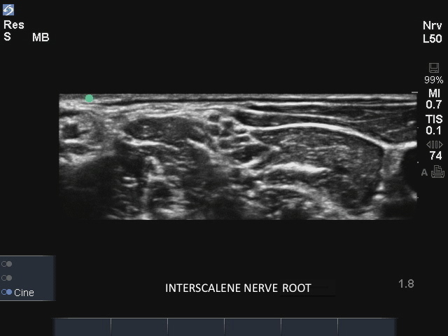
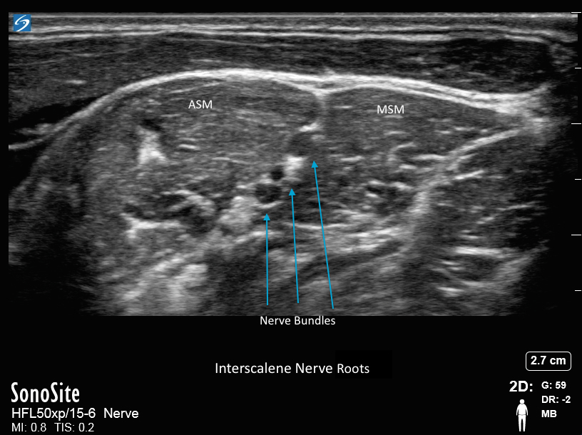
EDGE: Interscalene 2 EDGE: Interscalene 2 Read more about EDGE: Interscalene 2 /sites/default/files/201408_IMAGE_EDGE_INTERSCALENE_NERVE2.jpg Clinical Specialties Anesthesiology (Regional) Media Library Type Image Media Library Tag A Anesthesia Anesthesiology Anesthetic Anterior Scalene Muscle Block Brachial Plexus Bundles Edge Grapes Hypoechoic Image Injection Insertion Interscalene Groove Interscalene Nerve Lower Arm Medial Scalene Muscle Needle Nerve Bundles Plexus Procedure Sacrum Scanning Technique Shoulder Trunks Ultrasound Guidance
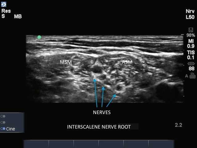
Edge: Interscalene Nerve Edge: Interscalene Nerve Read more about Edge: Interscalene Nerve /sites/default/files/201408_IMAGE_EDGE_INTERSCALENE_NERVE1.jpg Clinical Specialties Anesthesiology (Regional) Media Library Type Image Media Library Tag A Anesthesia Anesthesiology Anesthetic Anterior Scalene Muscle Block Brachial Plexus Bundles Edge Grapes Hypoechoic Image Injection Insertion Interscalene Groove Interscalene Nerve Lower Arm Medial Scalene Muscle Needle Nerve Bundles Plexus Procedure Sacrum Scanning Technique Shoulder Trunks Ultrasound Guidance Compatible Products Sonosite Edge
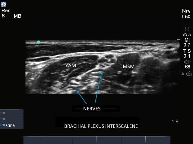
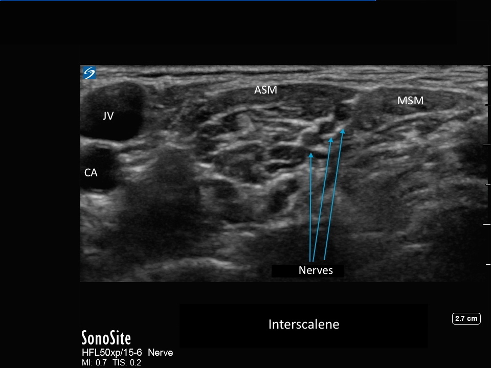
EDGE: Interscalene EDGE: Interscalene Read more about EDGE: Interscalene /sites/default/files/201408_IMAGE_EDGE_INTERSCALENE_NERVE.jpg Clinical Specialties Anesthesiology (Regional) Media Library Type Image Media Library Tag A Anesthesia Anesthesiology Anesthetic Anterior Scalene Muscle Block Brachial Plexus Bundles Edge Grapes Hypoechoic Image Injection Insertion Interscalene Groove Interscalene Nerve Lower Arm Medial Scalene Muscle Needle Nerve Bundles Plexus Procedure Sacrum Scanning Technique Shoulder Trunks Ultrasound Guidance
Supraclavicular Nerve Bundle Supraclavicular Nerve Bundle Read more about Supraclavicular Nerve Bundle /sites/default/files/201408_IMAGE_X-PORTE_SUPRACLAVICULAR_NERVE.jpg Clinical Specialties Anesthesiology (Regional) Media Library Type Image Media Library Tag Supraclavicular Nerve Block Scalene Subclavian Artery 1st Rib A Anesthesia Anesthetic Anesthesiology Nerve Bundles Brachial Plexus Rib Pleural Ultrasound Guidance Injection Trunks Divisions Plexus Sacrum Elbow Lower Arm Grapes Hypoechoic Bundles Insertion Needle Scanning Technique Image X-Porte Compatible Products Sonosite X-Porte
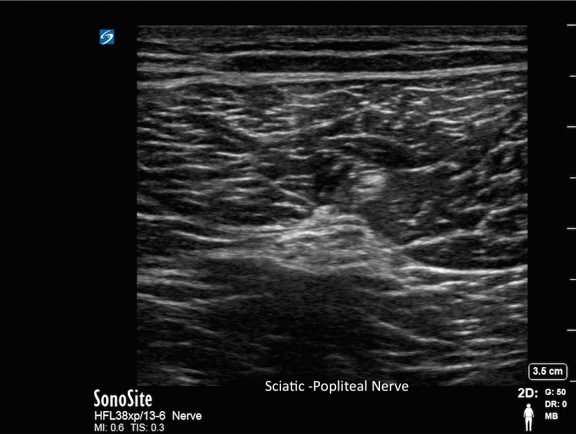
Sciatic-Popliteal Nerve Sciatic-Popliteal Nerve Read more about Sciatic-Popliteal Nerve /sites/default/files/201408_IMAGE_X-PORTE_SCIATIC_NERVE.jpg Clinical Specialties Anesthesiology (Regional) Media Library Type Image Media Library Tag A Anesthesia Anesthesiology Anesthetic Biceps Femoris Block Blockade Common Peroneal Nerve Hyperechoic Image Injection Insertion Knee Lower Extremity Needle Nerve Nerve Bundles Popliteal Popliteal Artery Popliteal Fossa Popliteal Vein Scanning Technique Sciatic Semimembranosus Muscual Semitendinosus Muscle Spread Surgery Tibia Nerve Ultrasound Guidance X-Porte
X-Porte: Interscalene Nerve 1 X-Porte: Interscalene Nerve 1 Read more about X-Porte: Interscalene Nerve 1 /sites/default/files/201408_IMAGE_X-PORTE_INTERSCALENE.jpg Clinical Specialties Anesthesiology (Regional) Media Library Type Image Media Library Tag A Anesthesia Anesthesiology Anesthetic Anterior Scalene Muscle Block Brachial Plexus Bundles Grapes Hypoechoic Image Injection Insertion Interscalene Groove Interscalene Nerve Lower Arm Medial Scalene Muscle Needle Nerve Bundles Plexus Procedure Sacrum Scanning Technique Shoulder Trunks Ultrasound Guidance X-Porte
X-Porte: Interscalene Nerve X-Porte: Interscalene Nerve Read more about X-Porte: Interscalene Nerve /sites/default/files/201408_IMAGE_X-PORTE_INTERSCALENE_NERVE.jpg Clinical Specialties Anesthesiology (Regional) Media Library Type Image Media Library Tag A Anesthesia Anesthesiology Anesthetic Anterior Scalene Muscle Block Brachial Plexus Bundles Grapes Hypoechoic Image Injection Insertion Interscalene Groove Interscalene Nerve Lower Arm Medial Scalene Muscle Needle Nerve Bundles Plexus Procedure Sacrum Scanning Technique Shoulder Trunks Ultrasound Guidance X-Porte
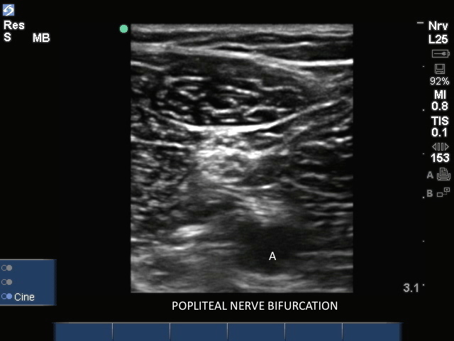
Popliteal Nerve Bifurcation1 Popliteal Nerve Bifurcation1 Read more about Popliteal Nerve Bifurcation1 /sites/default/files/201408_IMAGE_EDGE_POPLITEAL_NERVE_BIFURCATION1.jpg Clinical Specialties Anesthesiology (Regional) Media Library Type Image Media Library Tag A Anesthesia Anesthesiology Anesthetic Biceps Femoris Block Blockade Common Peroneal Nerve Edge Hyperechoic Image Injection Insertion Knee Lower Extremity Needle Nerve Nerve Bundles Popliteal Popliteal Artery Popliteal Fossa Popliteal Vein Scanning Technique Sciatic Sciatic Nerve Semimembranosus Muscual Semitendinosus Muscle Spread Surgery Tibia Nerve Ultrasound Guidance
Popliteal Nerve Bifurcation Popliteal Nerve Bifurcation Read more about Popliteal Nerve Bifurcation /sites/default/files/201408_IMAGE_EDGE_POPLITEAL_NERVE_BIFURCTION_0.jpg Clinical Specialties Anesthesiology (Regional) Media Library Type Image Media Library Tag A Anesthesia Anesthesiology Anesthetic Biceps Femoris Block Blockade Common Peroneal Nerve Edge Hyperechoic Image Injection Insertion Knee Lower Extremity Needle Nerve Nerve Bundles Popliteal Popliteal Artery Popliteal Fossa Popliteal Vein Scanning Technique Sciatic Sciatic Nerve Semimembranosus Muscual Semitendinosus Muscle Spread Surgery Tibia Nerve Ultrasound Guidance
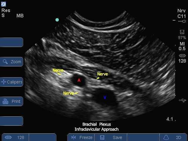
S Series: Brachial Plexus Infraclavicular S Series: Brachial Plexus Infraclavicular Read more about S Series: Brachial Plexus Infraclavicular /sites/default/files/201410_Image_S-System_Brachial_Plexus_Infraclavicular_Level_.jpg S Series: Brachial Plexus Infraclavicular . Clinical Specialties Anesthesiology (Regional) Media Library Type Image Media Library Tag A Anesthesia Anesthesiology Anesthetic Block Brachial Plexus Bundles Divisions Elbow Grapes Hypoechoic Infraclavicular Injection Insertion Lateral Posterior Lower Arm Medial Needle Nerve Nerve Bundles Pleural Plexus Sacrum Scalene Trunks Ultrasound Guidance