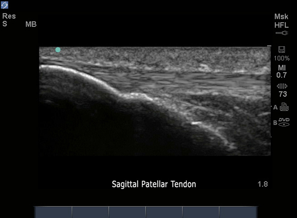
3D How To: Infra-Patellar Tendon Exam

3D How To: Infra-Patellar Tendon Exam

/sites/default/files/Infrapatellar_Discaimer_thumbnail_edu00550.jpg
3D animation demonstrating an ultrasound exam of the Infra-Patellar Tendon.
Clinical Specialties
Media Library Type
Subtitles
<p begin="00:00:07.188" end="00:00:08.845" style="s2">- [Voiceover] A linear array transducer</p>
<p begin="00:00:08.845" end="00:00:11.064" style="s2">with a musculoskeletal exam type</p>
<p begin="00:00:11.064" end="00:00:13.476" style="s2">is used to perform an<br />ultrasound examination</p>
<p begin="00:00:13.476" end="00:00:15.738" style="s2">of the infrapatellar tendon.</p>
<p begin="00:00:15.738" end="00:00:17.577" style="s2">The patient is in a supine position</p>
<p begin="00:00:17.577" end="00:00:20.176" style="s2">with the knee flexed 90 degrees.</p>
<p begin="00:00:20.176" end="00:00:22.815" style="s2">The transducer is placed longitudinally</p>
<p begin="00:00:22.815" end="00:00:25.613" style="s2">just inferior to the<br />patella in the mid-line</p>
<p begin="00:00:25.613" end="00:00:26.858" style="s2">with the orientation marker</p>
<p begin="00:00:26.858" end="00:00:29.102" style="s2">directed to the patient's head.</p>
<p begin="00:00:29.102" end="00:00:31.030" style="s2">The bright hyperechoic patella</p>
<p begin="00:00:31.030" end="00:00:33.007" style="s2">is seen on the left of the image.</p>
<p begin="00:00:33.007" end="00:00:34.572" style="s2">The infrapatellar tendon</p>
<p begin="00:00:34.572" end="00:00:38.046" style="s2">is seen as a compact<br />fibrillar pattern structure</p>
<p begin="00:00:38.046" end="00:00:40.679" style="s2">which extends from the<br />patellar insertion distally</p>
<p begin="00:00:40.679" end="00:00:43.517" style="s2">to the anterior tibial tuberosity.</p>
<p begin="00:00:43.517" end="00:00:45.316" style="s2">The long axis examination</p>
<p begin="00:00:45.316" end="00:00:47.519" style="s2">is repeated medially and laterally</p>
<p begin="00:00:47.519" end="00:00:49.785" style="s2">to fully evaluate the tendon.</p>
<p begin="00:00:49.785" end="00:00:53.546" style="s2">The transducer is then rotated<br />90 degrees counter clockwise</p>
<p begin="00:00:53.546" end="00:00:55.964" style="s2">to obtain a short axis view.</p>
<p begin="00:00:55.964" end="00:00:59.087" style="s2">The infrapatellar tendon is<br />seen as a speckled pattern,</p>
<p begin="00:00:59.087" end="00:01:01.662" style="s2">elliptical structure distal to the patella</p>
<p begin="00:01:01.662" end="00:01:03.829" style="s2">and anterior to the tibia.</p>
<p begin="00:01:04.822" end="00:01:07.231" style="s2">The transducer is slowly moved distally</p>
<p begin="00:01:07.231" end="00:01:08.992" style="s2">as the tendon gradually thins</p>
<p begin="00:01:08.992" end="00:01:11.992" style="s2">to insert on the tibial tuberosity.</p>
Brightcove ID
5752866924001
https://youtube.com/watch?v=uyFfdXwYzeg

How To: Patellar Tendon Exam

How To: Patellar Tendon Exam

/sites/default/files/Coach_sampson_patellar_tendon_exam_thumb.jpg
Dr. Steven Sampson demonstrates how to perform a patellar tendon exam.
Media Library Type
Subtitles
<p begin="00:00:09.961" end="00:00:12.478" style="s2">- Next we're going to<br />view the patella tendon</p>
<p begin="00:00:12.478" end="00:00:14.561" style="s2">in its longitudinal view.</p>
<p begin="00:00:15.568" end="00:00:19.401" style="s2">I'm going to place the<br />transducer just beneath</p>
<p begin="00:00:20.518" end="00:00:23.988" style="s2">the patient's patella, maintaining contact</p>
<p begin="00:00:23.988" end="00:00:25.821" style="s2">on the patient's knee.</p>
<p begin="00:00:26.918" end="00:00:29.447" style="s2">Here we see the fibers<br />of the patellar tendon,</p>
<p begin="00:00:29.447" end="00:00:31.794" style="s2">attaching on the patella.</p>
<p begin="00:00:31.794" end="00:00:34.143" style="s2">Next we're going to follow<br />these fibers distally</p>
<p begin="00:00:34.143" end="00:00:37.153" style="s2">as they attach on the tibial tubercle.</p>
<p begin="00:00:37.153" end="00:00:38.621" style="s2">And here we see the distal attachment</p>
<p begin="00:00:38.621" end="00:00:40.538" style="s2">on the tibial tubercle.</p>
<p begin="00:00:42.457" end="00:00:46.162" style="s2">Generally, I'll scan the<br />patellar tendon three times,</p>
<p begin="00:00:46.162" end="00:00:49.530" style="s2">once in the center, and<br />then once to each side,</p>
<p begin="00:00:49.530" end="00:00:51.869" style="s2">to view the entire tendon.</p>
<p begin="00:00:51.869" end="00:00:55.286" style="s2">In this case medially and next laterally.</p>
<p begin="00:01:05.601" end="00:01:07.440" style="s2">Next we're going to<br />view the patellar tendon</p>
<p begin="00:01:07.440" end="00:01:09.812" style="s2">in cross-sectional view.</p>
<p begin="00:01:09.812" end="00:01:11.617" style="s2">I'm going to place the transducer</p>
<p begin="00:01:11.617" end="00:01:14.824" style="s2">beneath the patient's patella,</p>
<p begin="00:01:14.824" end="00:01:17.741" style="s2">just overlying the patellar tendon.</p>
<p begin="00:01:19.514" end="00:01:22.200" style="s2">In here we're going to<br />see a circular pattern</p>
<p begin="00:01:22.200" end="00:01:24.182" style="s2">of the patellar tendon in cross-section,</p>
<p begin="00:01:24.182" end="00:01:26.881" style="s2">looking for any abnormalities.</p>
<p begin="00:01:26.881" end="00:01:29.906" style="s2">I'm going to scan distally<br />down to the tibial tubercle,</p>
<p begin="00:01:29.906" end="00:01:32.374" style="s2">slowly looking for irregularities.</p>
<p begin="00:01:32.374" end="00:01:34.045" style="s2">We'll begin to see the<br />tendon slowly disappearing</p>
<p begin="00:01:34.045" end="00:01:36.545" style="s2">and into the tibial tubercle.</p>
Brightcove ID
5752879796001
https://youtube.com/watch?v=Ba2l2LxU4AQ