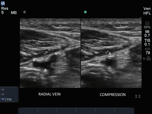
3D How To: Radial Artery Catheterization

3D How To: Radial Artery Catheterization

/sites/default/files/RadialArtery_edu00505_thumbnail.jpg
3D animation demonstrating an ultrasound guided radial artery catheterization transverse approach.
Applications
Clinical Specialties
Media Library Type
Subtitles
<p begin="00:00:07.217" end="00:00:08.450" style="s2">- [Voiceover] Linear array transducer</p>
<p begin="00:00:08.450" end="00:00:10.353" style="s2">with a vascular exam type,</p>
<p begin="00:00:10.353" end="00:00:13.051" style="s2">is used to perform an<br />ultrasound guided insertion</p>
<p begin="00:00:13.051" end="00:00:16.639" style="s2">of a radial artery catheter<br />via a transverse approach.</p>
<p begin="00:00:16.639" end="00:00:18.809" style="s2">The patient is in a supine position,</p>
<p begin="00:00:18.809" end="00:00:22.821" style="s2">with the arm abducted 45<br />degrees and externally rotated.</p>
<p begin="00:00:22.821" end="00:00:25.326" style="s2">The wrist is extended<br />by placing a small roll</p>
<p begin="00:00:25.326" end="00:00:26.974" style="s2">beneath the wrist crease.</p>
<p begin="00:00:26.974" end="00:00:29.218" style="s2">The transducer is placed transversely</p>
<p begin="00:00:29.218" end="00:00:31.187" style="s2">just proximal to the wrist crease,</p>
<p begin="00:00:31.187" end="00:00:34.397" style="s2">with the orientation marker<br />directed to the patient's right.</p>
<p begin="00:00:34.397" end="00:00:36.681" style="s2">The radial artery is superficial,</p>
<p begin="00:00:36.681" end="00:00:40.593" style="s2">and is seen as a dark anechoic,<br />round, pulsatile structure</p>
<p begin="00:00:40.593" end="00:00:42.541" style="s2">on the lateral aspect of the wrist,</p>
<p begin="00:00:42.541" end="00:00:46.056" style="s2">seen in the midst of<br />static, echodense material.</p>
<p begin="00:00:46.056" end="00:00:48.006" style="s2">There are often small venous vessels,</p>
<p begin="00:00:48.006" end="00:00:49.852" style="s2">which accompany the radial artery,</p>
<p begin="00:00:49.852" end="00:00:53.165" style="s2">which can be compressed with<br />gentle transducer pressure.</p>
<p begin="00:00:53.165" end="00:00:56.565" style="s2">The radius can be seen as<br />an anechoic, dark structure,</p>
<p begin="00:00:56.565" end="00:00:59.991" style="s2">approximately one centimeter<br />below the radial artery.</p>
<p begin="00:00:59.991" end="00:01:03.399" style="s2">Adjust the transducer so it<br />is centered over the artery.</p>
<p begin="00:01:03.399" end="00:01:06.422" style="s2">Follow the needle entry by<br />slowing sliding the transducer</p>
<p begin="00:01:06.422" end="00:01:08.929" style="s2">in the direction of needle advancement.</p>
<p begin="00:01:08.929" end="00:01:11.892" style="s2">The needle will appear<br />as a small, bright dot.</p>
<p begin="00:01:11.892" end="00:01:13.338" style="s2">When the needle tip appears,</p>
<p begin="00:01:13.338" end="00:01:16.666" style="s2">the transducer should be advanced<br />a short distance distally</p>
<p begin="00:01:16.666" end="00:01:19.079" style="s2">to follow the tip of<br />the needle trajectory,</p>
<p begin="00:01:19.079" end="00:01:21.525" style="s2">and stay in advance of the needle entry.</p>
<p begin="00:01:21.525" end="00:01:22.923" style="s2">The needle is slowly advanced,</p>
<p begin="00:01:22.923" end="00:01:25.176" style="s2">under direct ultrasound visualization,</p>
<p begin="00:01:25.176" end="00:01:28.168" style="s2">until the tip is seen to<br />puncture the radial artery.</p>
<p begin="00:01:28.168" end="00:01:30.016" style="s2">The transducer should be moved slightly</p>
<p begin="00:01:30.016" end="00:01:32.702" style="s2">proximally and distally<br />to confirm the needle tip</p>
<p begin="00:01:32.702" end="00:01:35.869" style="s2">lies in the mid-portion of the artery.</p>
Brightcove ID
5508123545001
https://youtube.com/watch?v=uHfeyAYiWOc