Case: FAST Exam - Suprapubic Views

Case: FAST Exam - Suprapubic Views

/sites/default/files/Cases_SB_FAST_SP_Thumb.jpg
This video details how using long-axis and short-axis suprapubic views during a trauma FAST ultrasound examination enables clinicians to identify fluid in a trauma patient's pelvic cavity.
Media Library Type
Subtitles
<p begin="00:00:18.083" end="00:00:20.666" style="s2">- Hello, my name is Phil<br />Perera and I'm the Emergency</p>
<p begin="00:00:20.666" end="00:00:22.183" style="s2">Ultrasound Coordinator at the New York</p>
<p begin="00:00:22.183" end="00:00:24.395" style="s2">Presbyterian Hospital in New York City.</p>
<p begin="00:00:24.395" end="00:00:27.632" style="s2">And welcome to Soundbytes Cases.</p>
<p begin="00:00:27.632" end="00:00:29.103" style="s2">In this Soundbytes module we'll continue</p>
<p begin="00:00:29.103" end="00:00:32.003" style="s2">our journey looking at<br />the Trauma Fast Exam.</p>
<p begin="00:00:32.003" end="00:00:34.120" style="s2">I hope you've had a<br />chance to join me prior</p>
<p begin="00:00:34.120" end="00:00:36.526" style="s2">for modules looking at<br />the right upper quadrant</p>
<p begin="00:00:36.526" end="00:00:39.414" style="s2">and left upper quadrant views<br />of the Trauma Fast Exam.</p>
<p begin="00:00:39.414" end="00:00:40.997" style="s2">In this module we will<br />specifically look at</p>
<p begin="00:00:40.997" end="00:00:44.087" style="s2">the Suprapubic view, also<br />known as the bladder or pelvic</p>
<p begin="00:00:44.087" end="00:00:46.495" style="s2">view of the Trauma Fast Exam.</p>
<p begin="00:00:46.495" end="00:00:48.203" style="s2">There is a lot of information we can gain</p>
<p begin="00:00:48.203" end="00:00:50.036" style="s2">from looking at the Suprapubic view,</p>
<p begin="00:00:50.036" end="00:00:52.369" style="s2">as we can potentially detect<br />a smaller amount of fluid</p>
<p begin="00:00:52.369" end="00:00:54.359" style="s2">than is required to make a positive right</p>
<p begin="00:00:54.359" end="00:00:56.845" style="s2">upper quadrant or left<br />upper quadrant view.</p>
<p begin="00:00:56.845" end="00:00:59.158" style="s2">Literature suggests that only about</p>
<p begin="00:00:59.158" end="00:01:02.141" style="s2">100 to 200 ccs of fluid can be detected</p>
<p begin="00:01:02.141" end="00:01:04.495" style="s2">accurately on the Suprapubic view.</p>
<p begin="00:01:04.495" end="00:01:06.073" style="s2">Here's a slide reviewing how to perform</p>
<p begin="00:01:06.073" end="00:01:08.586" style="s2">the Suprapubic view of<br />the Trauma Fast Exam.</p>
<p begin="00:01:08.586" end="00:01:10.468" style="s2">In contrast to the upper quadrant views,</p>
<p begin="00:01:10.468" end="00:01:12.676" style="s2">where we looked only in the<br />long axis configuration,</p>
<p begin="00:01:12.676" end="00:01:15.884" style="s2">the Suprapubic view is<br />made up of two planes.</p>
<p begin="00:01:15.884" end="00:01:18.987" style="s2">We want to look in both long<br />and short axis configurations.</p>
<p begin="00:01:18.987" end="00:01:21.086" style="s2">We'll begin at long<br />axis with the marker dot</p>
<p begin="00:01:21.086" end="00:01:22.829" style="s2">oriented toward the patient's head</p>
<p begin="00:01:22.829" end="00:01:24.523" style="s2">and complete our exam by moving the probe</p>
<p begin="00:01:24.523" end="00:01:26.092" style="s2">into short axis with the marker dot</p>
<p begin="00:01:26.092" end="00:01:27.933" style="s2">toward the patients right side.</p>
<p begin="00:01:27.933" end="00:01:30.004" style="s2">This way we can fully<br />scan through the pelvis</p>
<p begin="00:01:30.004" end="00:01:32.589" style="s2">and not miss any small<br />amounts of fluid there.</p>
<p begin="00:01:32.589" end="00:01:34.515" style="s2">It's optimal to perform the examination</p>
<p begin="00:01:34.515" end="00:01:35.876" style="s2">with a full bladder as an optimal</p>
<p begin="00:01:35.876" end="00:01:37.305" style="s2">acoustic window, so perform the</p>
<p begin="00:01:37.305" end="00:01:38.631" style="s2">exam prior to having the patient</p>
<p begin="00:01:38.631" end="00:01:41.619" style="s2">void or placing a foley catheter.</p>
<p begin="00:01:41.619" end="00:01:43.362" style="s2">Here's an illustration showing the anatomy</p>
<p begin="00:01:43.362" end="00:01:44.538" style="s2">that we'll need to know to perform</p>
<p begin="00:01:44.538" end="00:01:47.169" style="s2">the Suprapubic view of<br />the Trauma Fast Exam.</p>
<p begin="00:01:47.169" end="00:01:49.590" style="s2">We see the pattern of<br />fluid flow in a female,</p>
<p begin="00:01:49.590" end="00:01:52.355" style="s2">to the left here, and<br />a male, to the right.</p>
<p begin="00:01:52.355" end="00:01:54.565" style="s2">Let's look closer at the female pelvis,</p>
<p begin="00:01:54.565" end="00:01:56.497" style="s2">to the left, and what we<br />see is that fluid will</p>
<p begin="00:01:56.497" end="00:01:58.616" style="s2">preferentially develop in the pelvic</p>
<p begin="00:01:58.616" end="00:02:01.782" style="s2">Cul de Sac, located behind the uterus.</p>
<p begin="00:02:01.782" end="00:02:03.971" style="s2">Now, small amounts of fluid will only be</p>
<p begin="00:02:03.971" end="00:02:05.464" style="s2">located in the pelvic Cul de Sac,</p>
<p begin="00:02:05.464" end="00:02:07.247" style="s2">but as the amount of fluid enlarges</p>
<p begin="00:02:07.247" end="00:02:09.160" style="s2">it will come out and increase so</p>
<p begin="00:02:09.160" end="00:02:10.416" style="s2">that it will layer out on top of</p>
<p begin="00:02:10.416" end="00:02:12.848" style="s2">the uterus and on top of<br />the dome of the bladder.</p>
<p begin="00:02:12.848" end="00:02:15.141" style="s2">But small amounts of fluid<br />will only be found in</p>
<p begin="00:02:15.141" end="00:02:17.849" style="s2">that pelvic Cul de Sac<br />posterior to the uterus.</p>
<p begin="00:02:17.849" end="00:02:19.469" style="s2">Now let's take a look at the male</p>
<p begin="00:02:19.469" end="00:02:20.860" style="s2">pelvis to the right and we see</p>
<p begin="00:02:20.860" end="00:02:22.586" style="s2">small amounts of fluid<br />that will only be found</p>
<p begin="00:02:22.586" end="00:02:25.542" style="s2">in the Retrovesical<br />Space behind the bladder.</p>
<p begin="00:02:25.542" end="00:02:27.510" style="s2">As the amount of fluid enlarges it</p>
<p begin="00:02:27.510" end="00:02:29.212" style="s2">will come anterior to settle out</p>
<p begin="00:02:29.212" end="00:02:31.079" style="s2">over the top of the dome of the bladder.</p>
<p begin="00:02:31.079" end="00:02:33.070" style="s2">But as we emphasized in the female,</p>
<p begin="00:02:33.070" end="00:02:34.863" style="s2">small amounts of fluid will only be</p>
<p begin="00:02:34.863" end="00:02:36.807" style="s2">found in one place and in the male</p>
<p begin="00:02:36.807" end="00:02:38.486" style="s2">it will be in that Retrovesical Space</p>
<p begin="00:02:38.486" end="00:02:40.564" style="s2">immediately posterior to the bladder.</p>
<p begin="00:02:40.564" end="00:02:42.072" style="s2">Let's begin by looking at some normal</p>
<p begin="00:02:42.072" end="00:02:44.073" style="s2">video from the Suprapubic View.</p>
<p begin="00:02:44.073" end="00:02:46.775" style="s2">In this case, a long<br />axis view in a female,</p>
<p begin="00:02:46.775" end="00:02:49.374" style="s2">superior to the left,<br />inferior to the right.</p>
<p begin="00:02:49.374" end="00:02:50.767" style="s2">The first structure we identify is</p>
<p begin="00:02:50.767" end="00:02:53.356" style="s2">the bladder, the dark area, anteriorly</p>
<p begin="00:02:53.356" end="00:02:56.334" style="s2">and posterior to the<br />bladder we see the uterus.</p>
<p begin="00:02:56.334" end="00:02:58.100" style="s2">Now if we look into the potential space,</p>
<p begin="00:02:58.100" end="00:02:59.853" style="s2">the Pelvic Cul de Sac, posterior to the</p>
<p begin="00:02:59.853" end="00:03:02.531" style="s2">uterus for any dark fluid<br />collections, we see an</p>
<p begin="00:03:02.531" end="00:03:06.306" style="s2">absence of any fluid on<br />this normal video clip.</p>
<p begin="00:03:06.306" end="00:03:08.553" style="s2">Now let's inspect a<br />video clip from a male.</p>
<p begin="00:03:08.553" end="00:03:10.417" style="s2">In this case a short axis view.</p>
<p begin="00:03:10.417" end="00:03:12.959" style="s2">We see a large bladder there, anteriorly</p>
<p begin="00:03:12.959" end="00:03:14.474" style="s2">and behind the bladder we see two</p>
<p begin="00:03:14.474" end="00:03:16.173" style="s2">tubular structures making up the</p>
<p begin="00:03:16.173" end="00:03:19.355" style="s2">Seminal Vesicles, a<br />normal finding in a male.</p>
<p begin="00:03:19.355" end="00:03:20.896" style="s2">Now if we're looking for free fluid</p>
<p begin="00:03:20.896" end="00:03:22.389" style="s2">behind the bladder, we'd be looking</p>
<p begin="00:03:22.389" end="00:03:24.409" style="s2">for dark or anechoic fluid collection</p>
<p begin="00:03:24.409" end="00:03:26.564" style="s2">layering out behind the bladder.</p>
<p begin="00:03:26.564" end="00:03:29.942" style="s2">Notice this is a normal examination.</p>
<p begin="00:03:29.942" end="00:03:31.422" style="s2">Here's a positive examination</p>
<p begin="00:03:31.422" end="00:03:32.839" style="s2">in a female trauma patient.</p>
<p begin="00:03:32.839" end="00:03:34.414" style="s2">We're looking in the long axis view,</p>
<p begin="00:03:34.414" end="00:03:36.742" style="s2">superior to the left,<br />inferior to the right.</p>
<p begin="00:03:36.742" end="00:03:38.784" style="s2">The first structure we identify is</p>
<p begin="00:03:38.784" end="00:03:40.786" style="s2">the bladder, as seen inferior here.</p>
<p begin="00:03:40.786" end="00:03:42.849" style="s2">And notice the uterus, the solid organ,</p>
<p begin="00:03:42.849" end="00:03:45.268" style="s2">as seen superior to the bladder.</p>
<p begin="00:03:45.268" end="00:03:47.586" style="s2">We note the parts of<br />the uterus, the fundus</p>
<p begin="00:03:47.586" end="00:03:50.489" style="s2">anteriorly, and the<br />cervix more posteriorly.</p>
<p begin="00:03:50.489" end="00:03:52.557" style="s2">Now let's look into the Pelvic Cul de Sac</p>
<p begin="00:03:52.557" end="00:03:54.885" style="s2">immediately posterior to the uterus,</p>
<p begin="00:03:54.885" end="00:03:56.409" style="s2">and what we see here is the presence of a</p>
<p begin="00:03:56.409" end="00:03:59.243" style="s2">dark or anechoic fluid<br />collection just posterior</p>
<p begin="00:03:59.243" end="00:04:01.906" style="s2">to the cervix within<br />the pelvic Cul de Sac.</p>
<p begin="00:04:01.906" end="00:04:03.650" style="s2">So in the female trauma patient,</p>
<p begin="00:04:03.650" end="00:04:05.895" style="s2">this does denote a positive examination</p>
<p begin="00:04:05.895" end="00:04:07.406" style="s2">and can be a sign of ongoing bleeding</p>
<p begin="00:04:07.406" end="00:04:09.766" style="s2">within the abdominal pelvic cavity.</p>
<p begin="00:04:09.766" end="00:04:11.421" style="s2">So let's contrast this clip in which</p>
<p begin="00:04:11.421" end="00:04:12.604" style="s2">we see a small amount of fresh</p>
<p begin="00:04:12.604" end="00:04:14.786" style="s2">fluid within the pelvic Cul de Sac.</p>
<p begin="00:04:14.786" end="00:04:16.392" style="s2">With this one, in which we have a female</p>
<p begin="00:04:16.392" end="00:04:18.100" style="s2">trauma patient with a large amount</p>
<p begin="00:04:18.100" end="00:04:20.697" style="s2">of bleeding within the pelvic cavity.</p>
<p begin="00:04:20.697" end="00:04:23.006" style="s2">We see here, again, a<br />long axis scan superior</p>
<p begin="00:04:23.006" end="00:04:25.545" style="s2">to the left, inferior to<br />the right, the bladder</p>
<p begin="00:04:25.545" end="00:04:28.133" style="s2">we see as the dark structure inferiorly</p>
<p begin="00:04:28.133" end="00:04:30.566" style="s2">and the uterus superior to the bladder.</p>
<p begin="00:04:30.566" end="00:04:33.017" style="s2">Notice the fresh fluid as seen posterior</p>
<p begin="00:04:33.017" end="00:04:34.715" style="s2">to the uterus within the Cul de Sac,</p>
<p begin="00:04:34.715" end="00:04:36.744" style="s2">but note that the amount of fluid</p>
<p begin="00:04:36.744" end="00:04:38.768" style="s2">comes anterior to the uterus,</p>
<p begin="00:04:38.768" end="00:04:41.351" style="s2">as seen here between the<br />uterus and the bladder.</p>
<p begin="00:04:41.351" end="00:04:44.140" style="s2">So this denotes a large<br />amount of blood within</p>
<p begin="00:04:44.140" end="00:04:47.084" style="s2">the pelvic cavity in this<br />female trauma patient.</p>
<p begin="00:04:47.084" end="00:04:49.940" style="s2">If we now orient the probe<br />to the patient's right side,</p>
<p begin="00:04:49.940" end="00:04:52.849" style="s2">we obtain a short axis<br />view of the same patient.</p>
<p begin="00:04:52.849" end="00:04:54.604" style="s2">And what we see here is the uterus in the</p>
<p begin="00:04:54.604" end="00:04:56.937" style="s2">middle of the image and<br />notice the large amount</p>
<p begin="00:04:56.937" end="00:04:59.442" style="s2">of fresh fluid as seen both<br />to the top, or anterior,</p>
<p begin="00:04:59.442" end="00:05:01.807" style="s2">to the uterus and posterior to the uterus.</p>
<p begin="00:05:01.807" end="00:05:03.401" style="s2">Notice in this case we can see the</p>
<p begin="00:05:03.401" end="00:05:05.629" style="s2">broad ligaments of the<br />uterus well outlined</p>
<p begin="00:05:05.629" end="00:05:08.119" style="s2">by all the fresh fluid within the pelvis.</p>
<p begin="00:05:08.119" end="00:05:10.733" style="s2">So a large amount of<br />fresh fluid, or blood,</p>
<p begin="00:05:10.733" end="00:05:14.050" style="s2">in this case, within this<br />female trauma patient.</p>
<p begin="00:05:14.050" end="00:05:15.597" style="s2">Here's a positive examination</p>
<p begin="00:05:15.597" end="00:05:18.316" style="s2">Suprapubic View, short axis in a male.</p>
<p begin="00:05:18.316" end="00:05:20.673" style="s2">Probe is oriented towards<br />the patient's right and</p>
<p begin="00:05:20.673" end="00:05:23.066" style="s2">anteriorly we see a large,<br />fluid filled bladder.</p>
<p begin="00:05:23.066" end="00:05:25.238" style="s2">Posterior to the bladder,<br />in the retrovesical</p>
<p begin="00:05:25.238" end="00:05:27.863" style="s2">space we appreciate the<br />presence of free fluid,</p>
<p begin="00:05:27.863" end="00:05:29.509" style="s2">as shown by that dark or anechoic</p>
<p begin="00:05:29.509" end="00:05:30.992" style="s2">fluid collection there.</p>
<p begin="00:05:30.992" end="00:05:32.868" style="s2">Now this gives a finding known as</p>
<p begin="00:05:32.868" end="00:05:34.767" style="s2">the double wall sign, and we see</p>
<p begin="00:05:34.767" end="00:05:36.319" style="s2">the wall of the bladder, outlined by</p>
<p begin="00:05:36.319" end="00:05:38.435" style="s2">the urine inside the bladder, and the</p>
<p begin="00:05:38.435" end="00:05:40.589" style="s2">blood, in this case, outside the bladder</p>
<p begin="00:05:40.589" end="00:05:43.180" style="s2">in the area of the retrovesical space.</p>
<p begin="00:05:43.180" end="00:05:44.850" style="s2">To further confirm that the last patient</p>
<p begin="00:05:44.850" end="00:05:46.105" style="s2">had a positive exam and that we're</p>
<p begin="00:05:46.105" end="00:05:48.688" style="s2">not mistaking areas of<br />fluid as seminal vesicles,</p>
<p begin="00:05:48.688" end="00:05:51.348" style="s2">we'll re-scan the patient<br />in the long axis plane,</p>
<p begin="00:05:51.348" end="00:05:53.555" style="s2">superior to the left,<br />inferior to the right.</p>
<p begin="00:05:53.555" end="00:05:55.425" style="s2">We see the large circular bladder,</p>
<p begin="00:05:55.425" end="00:05:57.655" style="s2">as seen anteriorly and superior</p>
<p begin="00:05:57.655" end="00:05:59.269" style="s2">and posterior to the bladder in the</p>
<p begin="00:05:59.269" end="00:06:01.039" style="s2">retrovesical space, we can see</p>
<p begin="00:06:01.039" end="00:06:02.783" style="s2">free fluid layering out there.</p>
<p begin="00:06:02.783" end="00:06:04.257" style="s2">This confirms that indeed the patient</p>
<p begin="00:06:04.257" end="00:06:06.125" style="s2">has a positive exam, with blood</p>
<p begin="00:06:06.125" end="00:06:07.804" style="s2">layering out behind the bladder.</p>
<p begin="00:06:07.804" end="00:06:09.731" style="s2">And, again, we see the double wall sign,</p>
<p begin="00:06:09.731" end="00:06:11.733" style="s2">urine outlining the inner wall of the</p>
<p begin="00:06:11.733" end="00:06:13.318" style="s2">bladder, and blood, in this case,</p>
<p begin="00:06:13.318" end="00:06:15.776" style="s2">outlining the outer wall of the bladder.</p>
<p begin="00:06:15.776" end="00:06:17.531" style="s2">Here we're scanning a<br />male trauma patient with</p>
<p begin="00:06:17.531" end="00:06:19.874" style="s2">a long axis configuration<br />and we see a large</p>
<p begin="00:06:19.874" end="00:06:22.323" style="s2">amount of free fluid within the pelvis.</p>
<p begin="00:06:22.323" end="00:06:23.837" style="s2">We note the bladder inferiorly,</p>
<p begin="00:06:23.837" end="00:06:25.709" style="s2">and note all the free fluid layering</p>
<p begin="00:06:25.709" end="00:06:27.434" style="s2">out both posterior to the bladder</p>
<p begin="00:06:27.434" end="00:06:29.429" style="s2">in the retrovesical space and coming</p>
<p begin="00:06:29.429" end="00:06:31.048" style="s2">anteriorly onto the dome of the</p>
<p begin="00:06:31.048" end="00:06:33.234" style="s2">bladder as seen to the left here.</p>
<p begin="00:06:33.234" end="00:06:37.485" style="s2">So a large amount of free fluid<br />in this male trauma patient.</p>
<p begin="00:06:37.485" end="00:06:38.615" style="s2">In conclusion, I'm glad I could</p>
<p begin="00:06:38.615" end="00:06:39.898" style="s2">share with you the Soundbytes Module</p>
<p begin="00:06:39.898" end="00:06:43.131" style="s2">covering the Suprapubic View<br />of the Trauma Fast Exam.</p>
<p begin="00:06:43.131" end="00:06:44.365" style="s2">This view is a very important</p>
<p begin="00:06:44.365" end="00:06:45.997" style="s2">one to add onto the exam of your</p>
<p begin="00:06:45.997" end="00:06:47.497" style="s2">trauma patient as we can potentially</p>
<p begin="00:06:47.497" end="00:06:49.670" style="s2">detect a smaller amount of fluid here,</p>
<p begin="00:06:49.670" end="00:06:51.524" style="s2">within the pelvis, than it takes to</p>
<p begin="00:06:51.524" end="00:06:53.064" style="s2">make a positive right upper quadrant</p>
<p begin="00:06:53.064" end="00:06:54.985" style="s2">or left upper quadrant view.</p>
<p begin="00:06:54.985" end="00:06:56.869" style="s2">Remember that this is a two-step exam,</p>
<p begin="00:06:56.869" end="00:06:58.041" style="s2">we'll be looking in both short</p>
<p begin="00:06:58.041" end="00:06:59.924" style="s2">and long axis configurations to</p>
<p begin="00:06:59.924" end="00:07:01.442" style="s2">verify fluid, and also remember</p>
<p begin="00:07:01.442" end="00:07:03.712" style="s2">the differences between<br />the female, where we're</p>
<p begin="00:07:03.712" end="00:07:05.578" style="s2">looking into the pelvic<br />Cul de Sac for fluid,</p>
<p begin="00:07:05.578" end="00:07:06.898" style="s2">and the male, where we're looking into</p>
<p begin="00:07:06.898" end="00:07:09.175" style="s2">the retrovesical space for fluid.</p>
<p begin="00:07:09.175" end="00:07:10.741" style="s2">So I hope to see you back in the</p>
<p begin="00:07:10.741" end="00:07:13.324" style="s2">future as Soundbytes continues.</p>
Brightcove ID
5508114789001
https://youtube.com/watch?v=Pa3z9zWNfB8

How to: Female Pelvis: Transvaginal View

How to: Female Pelvis: Transvaginal View

/sites/default/files/ST_Female_Pelvis_Transvaginal_Thumb.jpg
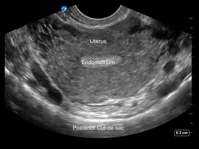
Obtaining a transvaginal view of the female pelvis
Clinical Specialties
Media Library Type
Subtitles
<p begin="00:00:14.018" end="00:00:16.081" style="s2">- If you do a transabdominal examination</p>
<p begin="00:00:16.081" end="00:00:18.045" style="s2">of the pelvis for early pregnancy</p>
<p begin="00:00:18.045" end="00:00:19.706" style="s2">and you cannot find a definitive</p>
<p begin="00:00:19.706" end="00:00:21.944" style="s2">gestational sack inside of the uterus,</p>
<p begin="00:00:21.944" end="00:00:23.733" style="s2">the next step you need to do is</p>
<p begin="00:00:23.733" end="00:00:27.982" style="s2">an intracavitary examination of the uterus</p>
<p begin="00:00:27.982" end="00:00:30.294" style="s2">and cervix and the adnexal area</p>
<p begin="00:00:30.294" end="00:00:31.923" style="s2">to determine where the pregnancy is,</p>
<p begin="00:00:31.923" end="00:00:34.086" style="s2">whether it's intrauterine or extrauterine.</p>
<p begin="00:00:34.086" end="00:00:35.668" style="s2">So what I'm gonna do right now</p>
<p begin="00:00:35.668" end="00:00:37.365" style="s2">is walk through the steps on how</p>
<p begin="00:00:37.365" end="00:00:38.996" style="s2">you would do that examination</p>
<p begin="00:00:38.996" end="00:00:41.674" style="s2">and we're gonna use this<br />Phantom by Blue Phantom,</p>
<p begin="00:00:41.674" end="00:00:46.628" style="s2">that will actually demonstrate<br />an ectopic pregnancy.</p>
<p begin="00:00:46.628" end="00:00:49.180" style="s2">So the first stage of the prep</p>
<p begin="00:00:49.180" end="00:00:51.637" style="s2">for the intracavitary transducer</p>
<p begin="00:00:51.637" end="00:00:55.137" style="s2">is that we need to cover it with a sheath.</p>
<p begin="00:00:56.388" end="00:00:58.073" style="s2">When you do this, we need to remove</p>
<p begin="00:00:58.073" end="00:01:01.424" style="s2">any air gaps that are inside the sheath,</p>
<p begin="00:01:01.424" end="00:01:03.468" style="s2">next to the transducer face.</p>
<p begin="00:01:03.468" end="00:01:05.523" style="s2">So what we will do is put a little</p>
<p begin="00:01:05.523" end="00:01:07.856" style="s2">bit of gel inside the cover,</p>
<p begin="00:01:11.529" end="00:01:14.696" style="s2">position that over the<br />tip of the transducer</p>
<p begin="00:01:14.696" end="00:01:18.497" style="s2">and then just pull down the<br />covering over the transducer</p>
<p begin="00:01:18.497" end="00:01:20.370" style="s2">and pull this down right over the handle,</p>
<p begin="00:01:20.370" end="00:01:23.401" style="s2">so you've got good coverage.</p>
<p begin="00:01:23.401" end="00:01:25.724" style="s2">And then just make sure that<br />you examine the tip here</p>
<p begin="00:01:25.724" end="00:01:27.025" style="s2">and make sure you eliminate any</p>
<p begin="00:01:27.025" end="00:01:31.848" style="s2">air bubbles that are<br />covering the transducer face.</p>
<p begin="00:01:31.848" end="00:01:34.168" style="s2">So once you've got the<br />air bubbles eliminated,</p>
<p begin="00:01:34.168" end="00:01:36.871" style="s2">then you can move on to the next step.</p>
<p begin="00:01:36.871" end="00:01:37.704" style="s2">We're going to put a little bit</p>
<p begin="00:01:37.704" end="00:01:41.454" style="s2">of gel on the outside<br />of the latex cover now.</p>
<p begin="00:01:44.847" end="00:01:46.257" style="s2">And again, your orientation for</p>
<p begin="00:01:46.257" end="00:01:48.341" style="s2">this examination is very important.</p>
<p begin="00:01:48.341" end="00:01:51.441" style="s2">So for the long ax or sagittal view,</p>
<p begin="00:01:51.441" end="00:01:54.325" style="s2">we want the orientation marker up</p>
<p begin="00:01:54.325" end="00:01:55.967" style="s2">and then when we go to the coronal view,</p>
<p begin="00:01:55.967" end="00:02:00.569" style="s2">we're gonna turn that so it<br />faces the patient's right.</p>
<p begin="00:02:00.569" end="00:02:04.081" style="s2">After we've inserted the<br />transducer in a long axis view,</p>
<p begin="00:02:04.081" end="00:02:05.111" style="s2">the first thing that we're gonna</p>
<p begin="00:02:05.111" end="00:02:08.323" style="s2">see is a long ax of the uterus.</p>
<p begin="00:02:08.323" end="00:02:11.648" style="s2">In this Phantom<br />representation of the uterus,</p>
<p begin="00:02:11.648" end="00:02:14.512" style="s2">we see an endometrial stripe,</p>
<p begin="00:02:14.512" end="00:02:17.010" style="s2">which is represented by the white line.</p>
<p begin="00:02:17.010" end="00:02:19.884" style="s2">So if I was doing this<br />as a real pelvic exam,</p>
<p begin="00:02:19.884" end="00:02:22.635" style="s2">I would scan to the right of the patient,</p>
<p begin="00:02:22.635" end="00:02:25.844" style="s2">all the way through to the right adnexa</p>
<p begin="00:02:25.844" end="00:02:30.081" style="s2">and then back over, all the<br />way back through the uterus,</p>
<p begin="00:02:30.081" end="00:02:31.768" style="s2">over to the left adnexa.</p>
<p begin="00:02:31.768" end="00:02:32.940" style="s2">And the reason I'm doing this</p>
<p begin="00:02:32.940" end="00:02:37.204" style="s2">is because I don't see the<br />pregnancy inside the uterus,</p>
<p begin="00:02:37.204" end="00:02:39.476" style="s2">so I'm suspecting that it's extrauterine</p>
<p begin="00:02:39.476" end="00:02:41.811" style="s2">or an ectopic pregnancy.</p>
<p begin="00:02:41.811" end="00:02:44.414" style="s2">I do have some hints here<br />though, using this Phantom.</p>
<p begin="00:02:44.414" end="00:02:46.721" style="s2">In the posterior cul de sac region,</p>
<p begin="00:02:46.721" end="00:02:49.013" style="s2">I'm seeing a black anechoic area,</p>
<p begin="00:02:49.013" end="00:02:52.513" style="s2">which would represent free fluid or blood.</p>
<p begin="00:02:54.243" end="00:02:58.377" style="s2">If I scan all the way over to<br />the left adnexa, in this case,</p>
<p begin="00:02:58.377" end="00:03:00.228" style="s2">the first structure I come across,</p>
<p begin="00:03:00.228" end="00:03:02.611" style="s2">this echogenic area represents the ovary.</p>
<p begin="00:03:02.611" end="00:03:06.444" style="s2">If I keep scanning to<br />the left, in the pelvis,</p>
<p begin="00:03:09.227" end="00:03:11.560" style="s2">I encounter this other area</p>
<p begin="00:03:14.662" end="00:03:18.908" style="s2">and this represents, in this<br />case, our ectopic pregnancy.</p>
<p begin="00:03:18.908" end="00:03:21.964" style="s2">When I'm medial, just<br />right beside the uterus,</p>
<p begin="00:03:21.964" end="00:03:25.780" style="s2">I see the ovary and I<br />scan out a little bit more</p>
<p begin="00:03:25.780" end="00:03:29.280" style="s2">and this represents the ectopic pregnancy.</p>
<p begin="00:03:31.412" end="00:03:32.613" style="s2">And this is what you should see,</p>
<p begin="00:03:32.613" end="00:03:34.864" style="s2">a round, circular structure like this,</p>
<p begin="00:03:34.864" end="00:03:37.520" style="s2">with a bright, echogenic brim.</p>
<p begin="00:03:37.520" end="00:03:39.301" style="s2">And it is possible to sometimes</p>
<p begin="00:03:39.301" end="00:03:42.336" style="s2">to see a fetal heartbeat inside</p>
<p begin="00:03:42.336" end="00:03:44.836" style="s2">the ectopic pregnancy as well.</p>
<p begin="00:03:46.410" end="00:03:49.351" style="s2">Now, I'm gonna change the<br />orientation of the transducer,</p>
<p begin="00:03:49.351" end="00:03:50.637" style="s2">so I'm in a coronal view.</p>
<p begin="00:03:50.637" end="00:03:55.568" style="s2">To do that, I'm gonna turn the<br />transducer counterclockwise.</p>
<p begin="00:03:55.568" end="00:03:57.688" style="s2">The uterus will appear circular,</p>
<p begin="00:03:57.688" end="00:04:00.765" style="s2">because I'm cutting a<br />cross sectional view of it</p>
<p begin="00:04:00.765" end="00:04:04.128" style="s2">and I tilt the handle<br />of the transducer up,</p>
<p begin="00:04:04.128" end="00:04:08.295" style="s2">to move inferiorly, down<br />to the region of the cervix</p>
<p begin="00:04:09.749" end="00:04:11.291" style="s2">and then bring the transducer handle</p>
<p begin="00:04:11.291" end="00:04:13.838" style="s2">down to scan superiorly,</p>
<p begin="00:04:13.838" end="00:04:17.275" style="s2">into the area of the fundus of the uterus.</p>
<p begin="00:04:17.275" end="00:04:18.594" style="s2">Again, in this case, I can see that</p>
<p begin="00:04:18.594" end="00:04:23.112" style="s2">there's no pregnancy<br />inside the uterus itself.</p>
<p begin="00:04:23.112" end="00:04:26.207" style="s2">If I scan over to the left of the Phantom,</p>
<p begin="00:04:26.207" end="00:04:30.059" style="s2">again, we see the left ovary<br />and then right beside it,</p>
<p begin="00:04:30.059" end="00:04:34.314" style="s2">we see our representation<br />of the ectopic pregnancy.</p>
<p begin="00:04:34.314" end="00:04:36.564" style="s2">This bright, circular area.</p>
<p begin="00:04:42.023" end="00:04:43.748" style="s2">Moving back towards the midline,</p>
<p begin="00:04:43.748" end="00:04:47.995" style="s2">the anechoic area just<br />posterior to the uterus</p>
<p begin="00:04:47.995" end="00:04:52.162" style="s2">is represented as free fluid<br />or blood, in this case.</p>
<p begin="00:04:53.025" end="00:04:54.214" style="s2">So those are the views that you</p>
<p begin="00:04:54.214" end="00:04:55.624" style="s2">would need to do a thorough assessment</p>
<p begin="00:04:55.624" end="00:04:58.457" style="s2">of the pelvis for early pregnancy.</p>
Brightcove ID
5750481386001
https://youtube.com/watch?v=0CqicUl0hw8

How To: Female Pelvis: Transabdominal View

How To: Female Pelvis: Transabdominal View

/sites/default/files/09_Female_Pelvis_Transabdominal_Scanning_Technique.jpg
Learn the basics of female transabdominal pelvic scanning.
Clinical Specialties
Media Library Type
Subtitles
<p begin="00:00:07.318" end="00:00:09.711" style="s2">- [Voiceover] A curved, or<br />phased array transducer,</p>
<p begin="00:00:09.711" end="00:00:11.431" style="s2">with a pelvis exam type,</p>
<p begin="00:00:11.431" end="00:00:14.568" style="s2">is used to perform the<br />pelvis ultrasound exam.</p>
<p begin="00:00:14.568" end="00:00:16.987" style="s2">A full bladder is used<br />as an acoustic window</p>
<p begin="00:00:16.987" end="00:00:18.989" style="s2">to view the pelvic organs.</p>
<p begin="00:00:18.989" end="00:00:22.579" style="s2">The pelvis is evaluated in two plains.</p>
<p begin="00:00:22.579" end="00:00:25.534" style="s2">Place the transducer<br />in a long axis position</p>
<p begin="00:00:25.534" end="00:00:28.254" style="s2">with the orientation marker<br />to the patient's head,</p>
<p begin="00:00:28.254" end="00:00:31.254" style="s2">at the level of the symphysis pubis.</p>
<p begin="00:00:32.384" end="00:00:36.384" style="s2">Angle the transducer<br />inferiorly into the pelvis.</p>
<p begin="00:00:38.036" end="00:00:40.705" style="s2">The bladder appears in the<br />near-field of the image,</p>
<p begin="00:00:40.705" end="00:00:43.616" style="s2">as a hypoechoic triangular structure.</p>
<p begin="00:00:43.616" end="00:00:45.800" style="s2">The uterus is gray in appearance</p>
<p begin="00:00:45.800" end="00:00:48.233" style="s2">and located either directly posterior</p>
<p begin="00:00:48.233" end="00:00:50.087" style="s2">or superior to the bladder.</p>
<p begin="00:00:50.087" end="00:00:52.139" style="s2">The endometrial stripe will appear as</p>
<p begin="00:00:52.139" end="00:00:55.945" style="s2">a bright echogenic line from<br />the fundus to the cervix.</p>
<p begin="00:00:55.945" end="00:00:58.877" style="s2">The uterus does not always<br />lay directly in the midline</p>
<p begin="00:00:58.877" end="00:01:02.209" style="s2">so it may be necessary to<br />slightly rotate the transducer</p>
<p begin="00:01:02.209" end="00:01:04.732" style="s2">to view the entire length of the uterus.</p>
<p begin="00:01:04.732" end="00:01:07.015" style="s2">Sweep the transducer from side to side</p>
<p begin="00:01:07.015" end="00:01:09.219" style="s2">to see the entire uterus.</p>
<p begin="00:01:09.219" end="00:01:12.192" style="s2">The ovaries may be seen<br />by sweeping the transducer</p>
<p begin="00:01:12.192" end="00:01:14.615" style="s2">to the lateral aspects of the pelvis.</p>
<p begin="00:01:14.615" end="00:01:18.536" style="s2">They are almond-shaped and<br />slightly hypoechoic structures.</p>
<p begin="00:01:18.536" end="00:01:20.605" style="s2">Follicles may appear as multiple</p>
<p begin="00:01:20.605" end="00:01:24.688" style="s2">hypoechoic, cystic structures<br />within the ovaries.</p>
<p begin="00:01:27.166" end="00:01:29.314" style="s2">Some follicles may be quite prominent,</p>
<p begin="00:01:29.314" end="00:01:31.981" style="s2">depending upon the luteal stage.</p>
<p begin="00:01:34.855" end="00:01:37.375" style="s2">To obtain a transverse view of the uterus,</p>
<p begin="00:01:37.375" end="00:01:39.869" style="s2">rotate the transducer 90 degrees,</p>
<p begin="00:01:39.869" end="00:01:42.788" style="s2">so the orientation marker<br />is to the patient's right.</p>
<p begin="00:01:42.788" end="00:01:46.581" style="s2">The bladder appears more<br />rectangular in shape in this view.</p>
<p begin="00:01:46.581" end="00:01:48.775" style="s2">Sweep the transducer superiorly</p>
<p begin="00:01:48.775" end="00:01:51.181" style="s2">from the level of the cervix to the fundus</p>
<p begin="00:01:51.181" end="00:01:53.284" style="s2">to see the entire uterus.</p>
<p begin="00:01:53.284" end="00:01:56.324" style="s2">The ovaries will be seen on<br />either side of the uterus</p>
<p begin="00:01:56.324" end="00:01:57.941" style="s2">and can vary in location,</p>
<p begin="00:01:57.941" end="00:02:01.024" style="s2">from a superior to inferior position.</p>
Brightcove ID
5750473717001
https://youtube.com/watch?v=ebpcUlQVmLE

How To: FAST Exam: Female Pelvis

How To: FAST Exam: Female Pelvis

/sites/default/files/08_FAST_Exam_Female_Pelvic_View_Scanning_Technique.jpg
Learn to examine the female pelvis for free fluid.
Media Library Type
Subtitles
<p begin="00:00:15.338" end="00:00:17.481" style="s2">- We're gonna do the FAST exam,</p>
<p begin="00:00:17.481" end="00:00:19.857" style="s2">the pelvic view in a female now.</p>
<p begin="00:00:19.857" end="00:00:22.104" style="s2">Again we have the phase array transducer,</p>
<p begin="00:00:22.104" end="00:00:24.440" style="s2">here's our probe marker right here,</p>
<p begin="00:00:24.440" end="00:00:26.319" style="s2">I'm gonna start off with a sagittal view.</p>
<p begin="00:00:26.319" end="00:00:29.870" style="s2">And ideally, the<br />(slurred) bladder is full.</p>
<p begin="00:00:29.870" end="00:00:32.183" style="s2">So you go just above the symphysis pubis,</p>
<p begin="00:00:32.183" end="00:00:34.754" style="s2">so we're scanning down here sagittaly,</p>
<p begin="00:00:34.754" end="00:00:36.570" style="s2">and some of the anatomic landmarks</p>
<p begin="00:00:36.570" end="00:00:37.842" style="s2">that we're looking at,</p>
<p begin="00:00:37.842" end="00:00:39.802" style="s2">at the very top of the<br />screen is the bladder,</p>
<p begin="00:00:39.802" end="00:00:41.482" style="s2">which is an anechoic structure,</p>
<p begin="00:00:41.482" end="00:00:43.363" style="s2">fluid filled, obviously with urine,</p>
<p begin="00:00:43.363" end="00:00:45.292" style="s2">it's got nice walls around it.</p>
<p begin="00:00:45.292" end="00:00:47.500" style="s2">Just below that is the uterus,</p>
<p begin="00:00:47.500" end="00:00:49.548" style="s2">which is a pear-shaped organ.</p>
<p begin="00:00:49.548" end="00:00:50.908" style="s2">You can see it well visualized</p>
<p begin="00:00:50.908" end="00:00:52.772" style="s2">because the bladder is full.</p>
<p begin="00:00:52.772" end="00:00:53.605" style="s2">And just below that</p>
<p begin="00:00:53.605" end="00:00:55.772" style="s2">is a potential space, the Cul de Sac,</p>
<p begin="00:00:55.772" end="00:00:58.125" style="s2">where we can look for, very carefully,</p>
<p begin="00:00:58.125" end="00:01:01.186" style="s2">small amounts of free fluid.</p>
<p begin="00:01:01.186" end="00:01:02.495" style="s2">When we scan this patient,</p>
<p begin="00:01:02.495" end="00:01:04.192" style="s2">in this view, in the sagittal view,</p>
<p begin="00:01:04.192" end="00:01:06.871" style="s2">from the patient's left<br />to the patient's right.</p>
<p begin="00:01:06.871" end="00:01:09.234" style="s2">And we're slowly slowly scanning to see</p>
<p begin="00:01:09.234" end="00:01:11.808" style="s2">if there's any evidence of free fluid.</p>
<p begin="00:01:11.808" end="00:01:13.816" style="s2">Once we finish the sagittal view,</p>
<p begin="00:01:13.816" end="00:01:16.087" style="s2">we want to go ahead and<br />do a transverse view.</p>
<p begin="00:01:16.087" end="00:01:17.141" style="s2">What we're gonna do now</p>
<p begin="00:01:17.141" end="00:01:20.830" style="s2">is point the transducer marker<br />toward the patient's right.</p>
<p begin="00:01:20.830" end="00:01:23.677" style="s2">So we're gonna rotate that transducer,</p>
<p begin="00:01:23.677" end="00:01:24.941" style="s2">toward the patient's right.</p>
<p begin="00:01:24.941" end="00:01:27.734" style="s2">Again, we see the bladder<br />in the center of the screen.</p>
<p begin="00:01:27.734" end="00:01:30.381" style="s2">We're now gonna scan from inferior,</p>
<p begin="00:01:30.381" end="00:01:34.269" style="s2">just below the symphysis,<br />to more superior,</p>
<p begin="00:01:34.269" end="00:01:38.726" style="s2">and in between we're also<br />gonna identify the uterus.</p>
<p begin="00:01:38.726" end="00:01:41.965" style="s2">So we're going right to the<br />very top of the bladder,</p>
<p begin="00:01:41.965" end="00:01:42.880" style="s2">to about mid-bladder,</p>
<p begin="00:01:42.880" end="00:01:46.769" style="s2">we can see the uterus here in the center.</p>
<p begin="00:01:46.769" end="00:01:49.936" style="s2">A very small amount of free fluid here,</p>
<p begin="00:01:49.936" end="00:01:52.576" style="s2">which can be physiological as well.</p>
<p begin="00:01:52.576" end="00:01:55.993" style="s2">So we're scanning all the way inferiorly,</p>
<p begin="00:01:57.937" end="00:02:00.672" style="s2">to more superiorly, we see the uterus,</p>
<p begin="00:02:00.672" end="00:02:03.792" style="s2">we actually see part of the ovary there,</p>
<p begin="00:02:03.792" end="00:02:07.959" style="s2">and then we're gonna go all<br />the way more superiorly.</p>
Brightcove ID
5508120185001
https://youtube.com/watch?v=HxQE2gYH3Sk

3D How To: Female Pelvis Exam

3D How To: Female Pelvis Exam

/sites/default/files/youtube_ebpcUlQVmLE.jpg
3D animation demonstrating a Female Pelvis ultrasound exam.
Publication Date
Media Library Type
Subtitles
<p begin="00:00:07.318" end="00:00:09.711" style="s2">- [Voiceover] A curved, or<br />phased array transducer,</p>
<p begin="00:00:09.711" end="00:00:11.431" style="s2">with a pelvis exam type,</p>
<p begin="00:00:11.431" end="00:00:14.568" style="s2">is used to perform the<br />pelvis ultrasound exam.</p>
<p begin="00:00:14.568" end="00:00:16.987" style="s2">A full bladder is used<br />as an acoustic window</p>
<p begin="00:00:16.987" end="00:00:18.989" style="s2">to view the pelvic organs.</p>
<p begin="00:00:18.989" end="00:00:22.579" style="s2">The pelvis is evaluated in two plains.</p>
<p begin="00:00:22.579" end="00:00:25.534" style="s2">Place the transducer<br />in a long axis position</p>
<p begin="00:00:25.534" end="00:00:28.254" style="s2">with the orientation marker<br />to the patient's head,</p>
<p begin="00:00:28.254" end="00:00:31.254" style="s2">at the level of the symphysis pubis.</p>
<p begin="00:00:32.384" end="00:00:36.384" style="s2">Angle the transducer<br />inferiorly into the pelvis.</p>
<p begin="00:00:38.036" end="00:00:40.705" style="s2">The bladder appears in the<br />near-field of the image,</p>
<p begin="00:00:40.705" end="00:00:43.616" style="s2">as a hypoechoic triangular structure.</p>
<p begin="00:00:43.616" end="00:00:45.800" style="s2">The uterus is gray in appearance</p>
<p begin="00:00:45.800" end="00:00:48.233" style="s2">and located either directly posterior</p>
<p begin="00:00:48.233" end="00:00:50.087" style="s2">or superior to the bladder.</p>
<p begin="00:00:50.087" end="00:00:52.139" style="s2">The endometrial stripe will appear as</p>
<p begin="00:00:52.139" end="00:00:55.945" style="s2">a bright echogenic line from<br />the fundus to the cervix.</p>
<p begin="00:00:55.945" end="00:00:58.877" style="s2">The uterus does not always<br />lay directly in the midline</p>
<p begin="00:00:58.877" end="00:01:02.209" style="s2">so it may be necessary to<br />slightly rotate the transducer</p>
<p begin="00:01:02.209" end="00:01:04.732" style="s2">to view the entire length of the uterus.</p>
<p begin="00:01:04.732" end="00:01:07.015" style="s2">Sweep the transducer from side to side</p>
<p begin="00:01:07.015" end="00:01:09.219" style="s2">to see the entire uterus.</p>
<p begin="00:01:09.219" end="00:01:12.192" style="s2">The ovaries may be seen<br />by sweeping the transducer</p>
<p begin="00:01:12.192" end="00:01:14.615" style="s2">to the lateral aspects of the pelvis.</p>
<p begin="00:01:14.615" end="00:01:18.536" style="s2">They are almond-shaped and<br />slightly hypoechoic structures.</p>
<p begin="00:01:18.536" end="00:01:20.605" style="s2">Follicles may appear as multiple</p>
<p begin="00:01:20.605" end="00:01:24.688" style="s2">hypoechoic, cystic structures<br />within the ovaries.</p>
<p begin="00:01:27.166" end="00:01:29.314" style="s2">Some follicles may be quite prominent,</p>
<p begin="00:01:29.314" end="00:01:31.981" style="s2">depending upon the luteal stage.</p>
<p begin="00:01:34.855" end="00:01:37.375" style="s2">To obtain a transverse view of the uterus,</p>
<p begin="00:01:37.375" end="00:01:39.869" style="s2">rotate the transducer 90 degrees,</p>
<p begin="00:01:39.869" end="00:01:42.788" style="s2">so the orientation marker<br />is to the patient's right.</p>
<p begin="00:01:42.788" end="00:01:46.581" style="s2">The bladder appears more<br />rectangular in shape in this view.</p>
<p begin="00:01:46.581" end="00:01:48.775" style="s2">Sweep the transducer superiorly</p>
<p begin="00:01:48.775" end="00:01:51.181" style="s2">from the level of the cervix to the fundus</p>
<p begin="00:01:51.181" end="00:01:53.284" style="s2">to see the entire uterus.</p>
<p begin="00:01:53.284" end="00:01:56.324" style="s2">The ovaries will be seen on<br />either side of the uterus</p>
<p begin="00:01:56.324" end="00:01:57.941" style="s2">and can vary in location,</p>
<p begin="00:01:57.941" end="00:02:01.024" style="s2">from a superior to inferior position.</p>
Brightcove ID
5750473717001
https://youtube.com/watch?v=ebpcUlQVmLE

3D How To: eFAST Pelvis

3D How To: eFAST Pelvis

/sites/default/files/EFast_Pelvis_EDU00457_Thumbnail.jpg

3D animation demonstrating the pelvis view while performing an eFAST exam.

Media Library Type
Subtitles
<p begin="00:00:07.326" end="00:00:08.966" style="s2">- [Voiceover] A phased array transducer</p>
<p begin="00:00:08.966" end="00:00:10.768" style="s2">with an abdomen exam type</p>
<p begin="00:00:10.768" end="00:00:14.463" style="s2">is used to perform the<br />pelvis view of the fast exam.</p>
<p begin="00:00:14.463" end="00:00:17.238" style="s2">Place the transducer in<br />a transverse position</p>
<p begin="00:00:17.238" end="00:00:19.326" style="s2">with the orientation marker to the right</p>
<p begin="00:00:19.326" end="00:00:22.046" style="s2">at the level of the symphysis pubis.</p>
<p begin="00:00:22.046" end="00:00:25.136" style="s2">The pelvis is evaluated in two planes.</p>
<p begin="00:00:25.136" end="00:00:26.975" style="s2">It is easier to perform this exam</p>
<p begin="00:00:26.975" end="00:00:28.840" style="s2">when the bladder is filled.</p>
<p begin="00:00:28.840" end="00:00:30.951" style="s2">The bladder is used as an acoustic window</p>
<p begin="00:00:30.951" end="00:00:32.302" style="s2">to view the cul de sac</p>
<p begin="00:00:32.302" end="00:00:35.478" style="s2">or retrovesicular space for free fluid.</p>
<p begin="00:00:35.478" end="00:00:37.113" style="s2">To visualize the bladder,</p>
<p begin="00:00:37.113" end="00:00:40.830" style="s2">angle the transducer<br />inferiorly into the pelvis.</p>
<p begin="00:00:40.830" end="00:00:42.879" style="s2">If it is difficult to<br />visualize the bladder,</p>
<p begin="00:00:42.879" end="00:00:46.456" style="s2">slide to the left or right<br />of the symphysis pubis</p>
<p begin="00:00:46.456" end="00:00:48.646" style="s2">to bring the bladder into view.</p>
<p begin="00:00:48.646" end="00:00:51.313" style="s2">To evaluate the pelvis for free fluid,</p>
<p begin="00:00:51.313" end="00:00:55.480" style="s2">sweep the transducer from an<br />inferior to superior position.</p>
<p begin="00:00:56.314" end="00:00:59.499" style="s2">Fluid will appear hyperechoic or anechoic</p>
<p begin="00:00:59.499" end="00:01:01.994" style="s2">and accumulate posterior to the bladder,</p>
<p begin="00:01:01.994" end="00:01:05.569" style="s2">posterior to the uterus,<br />and between loops of bowel.</p>
<p begin="00:01:05.569" end="00:01:07.508" style="s2">To obtain a long access view,</p>
<p begin="00:01:07.508" end="00:01:09.952" style="s2">rotate the transducer 90 degrees</p>
<p begin="00:01:09.952" end="00:01:11.440" style="s2">with the orientation marker</p>
<p begin="00:01:11.440" end="00:01:14.273" style="s2">pointed toward the patient's head.</p>
<p begin="00:01:11.440" end="00:01:22.000" style="s2">Sweep the transducer across the pelvis from left to right</p>
<p begin="00:01:22.000" end="00:01:26.000" style="s2">to evaluate the pelvis for free fluid.</p>

Brightcove ID
5508134284001
https://youtube.com/watch?v=pFtpx-yZfe0
Body

3D animation demonstrating the pelvis view while performing an eFAST exam.

Case: Intrauterine Pregnancy - Part 2

Case: Intrauterine Pregnancy - Part 2

/sites/default/files/perera_intrauterine_2.jpg
This video discusses how to use ultrasound to determine the gestational age of a normal pregnancy, determine a fetal heart rate, and identify markers for an abnormal pregnancy and fetal demise.
Clinical Specialties
Media Library Type
Subtitles
<p begin="00:00:14.315" end="00:00:15.904" style="s2">- [Voiceover] Hello,<br />my name is Phil Perera</p>
<p begin="00:00:15.904" end="00:00:17.729" style="s2">and I am the emergency<br />ultrasound coordinator</p>
<p begin="00:00:17.729" end="00:00:20.635" style="s2">at the New York Presbyterian<br />Hospital in New York City.</p>
<p begin="00:00:20.635" end="00:00:22.968" style="s2">Welcome to Soundbytes Cases.</p>
<p begin="00:00:24.474" end="00:00:26.970" style="s2">In this module entitled<br />Emergency OB/GYN Ultrasound:</p>
<p begin="00:00:26.970" end="00:00:29.090" style="s2">Part 2 of Intrauterine Pregnancy,</p>
<p begin="00:00:29.090" end="00:00:30.881" style="s2">we're going to focus on<br />the further assessment</p>
<p begin="00:00:30.881" end="00:00:32.548" style="s2">of normal pregnancy.</p>
<p begin="00:00:33.740" end="00:00:35.121" style="s2">We'll look at two further things that</p>
<p begin="00:00:35.121" end="00:00:37.308" style="s2">are important to assess<br />in your pregnancies.</p>
<p begin="00:00:37.308" end="00:00:39.978" style="s2">First of all, learning how<br />to date the gestational age</p>
<p begin="00:00:39.978" end="00:00:42.169" style="s2">of the pregnancy, as well as learning</p>
<p begin="00:00:42.169" end="00:00:44.932" style="s2">how to determine the fetal heart rate.</p>
<p begin="00:00:44.932" end="00:00:47.966" style="s2">Then we'll wrap up the module<br />by examining further findings</p>
<p begin="00:00:47.966" end="00:00:50.804" style="s2">in abnormal pregnancies and<br />learning how to differentiate</p>
<p begin="00:00:50.804" end="00:00:55.012" style="s2">these findings from a normal<br />intrauterine pregnancy.</p>
<p begin="00:00:55.012" end="00:00:56.776" style="s2">The first concept that we'll focus on</p>
<p begin="00:00:56.776" end="00:00:59.182" style="s2">is dating fetal gestational age.</p>
<p begin="00:00:59.182" end="00:01:01.741" style="s2">In the first trimester, we're<br />going to use an assessment</p>
<p begin="00:01:01.741" end="00:01:03.491" style="s2">of the crown rump length.</p>
<p begin="00:01:03.491" end="00:01:05.427" style="s2">Interestingly, dating<br />in the first trimester</p>
<p begin="00:01:05.427" end="00:01:07.491" style="s2">is actually probably the<br />most accurate during all</p>
<p begin="00:01:07.491" end="00:01:09.503" style="s2">phases of pregnancy, as<br />there's a difference in</p>
<p begin="00:01:09.503" end="00:01:12.446" style="s2">the growth curve as the fetus develops.</p>
<p begin="00:01:12.446" end="00:01:14.004" style="s2">In the second trimester, we'll measure</p>
<p begin="00:01:14.004" end="00:01:16.495" style="s2">the skull biparietal diameter.</p>
<p begin="00:01:16.495" end="00:01:18.839" style="s2">And the third trimester,<br />the dating is composed</p>
<p begin="00:01:18.839" end="00:01:22.124" style="s2">of the biophysical profile,<br />focusing on the femur length,</p>
<p begin="00:01:22.124" end="00:01:25.624" style="s2">as well as other biophysical measurements.</p>
<p begin="00:01:26.709" end="00:01:28.975" style="s2">This is an image of a<br />first trimester pregnancy,</p>
<p begin="00:01:28.975" end="00:01:30.796" style="s2">and we're going to evaluate<br />the gestational age by</p>
<p begin="00:01:30.796" end="00:01:32.990" style="s2">measuring the crown rump length.</p>
<p begin="00:01:32.990" end="00:01:34.557" style="s2">Here we see the fetal<br />pole stretched across</p>
<p begin="00:01:34.557" end="00:01:37.173" style="s2">the gestational sac and<br />we see the crown located</p>
<p begin="00:01:37.173" end="00:01:40.680" style="s2">over towards the right and<br />the rump towards the left.</p>
<p begin="00:01:40.680" end="00:01:43.567" style="s2">If we put the calipers<br />down from the crown across</p>
<p begin="00:01:43.567" end="00:01:47.102" style="s2">to the rump we get a<br />measurement of 1.46cm.</p>
<p begin="00:01:47.102" end="00:01:49.584" style="s2">By selecting Crown Rump<br />Length in the software package</p>
<p begin="00:01:49.584" end="00:01:51.565" style="s2">on the ultrasound machine,<br />we'll get an assessment</p>
<p begin="00:01:51.565" end="00:01:54.084" style="s2">of gestational age which<br />we can see here towards</p>
<p begin="00:01:54.084" end="00:01:57.915" style="s2">the bottom left, seven weeks and six days.</p>
<p begin="00:01:57.915" end="00:01:59.967" style="s2">As first trimester dating is<br />considered one of the most</p>
<p begin="00:01:59.967" end="00:02:02.531" style="s2">accurate during the entire<br />pregnancy, it's nice to print</p>
<p begin="00:02:02.531" end="00:02:04.920" style="s2">this image out and give to<br />your patient to take for</p>
<p begin="00:02:04.920" end="00:02:09.087" style="s2">their followup visit with their OB/GYN.</p>
<p begin="00:02:09.946" end="00:02:12.130" style="s2">In the second trimester,<br />dating of gestational age</p>
<p begin="00:02:12.130" end="00:02:14.439" style="s2">focuses on skull circumference<br />or measurement of</p>
<p begin="00:02:14.439" end="00:02:16.688" style="s2">the biparietal diameter.</p>
<p begin="00:02:16.688" end="00:02:18.285" style="s2">We want to measure the<br />skull at about the level</p>
<p begin="00:02:18.285" end="00:02:22.579" style="s2">of the thalamus in an axial<br />orientation with the face down.</p>
<p begin="00:02:22.579" end="00:02:25.066" style="s2">As we can see here,<br />replacing the calipers from</p>
<p begin="00:02:25.066" end="00:02:28.005" style="s2">the outer skull table<br />proximally to the inner skull</p>
<p begin="00:02:28.005" end="00:02:32.771" style="s2">table distally, and we have<br />a measurement of 3.26cm</p>
<p begin="00:02:32.771" end="00:02:36.854" style="s2">correlating to a 16 week<br />one day gestational age.</p>
<p begin="00:02:38.641" end="00:02:41.207" style="s2">In addition to measuring the<br />gestational age of the fetus,</p>
<p begin="00:02:41.207" end="00:02:43.443" style="s2">another very important concept<br />is to get a measurement</p>
<p begin="00:02:43.443" end="00:02:45.285" style="s2">of the fetal heart rate.</p>
<p begin="00:02:45.285" end="00:02:48.431" style="s2">Normal fetal heart rates<br />will range from 120-160</p>
<p begin="00:02:48.431" end="00:02:50.747" style="s2">beats per minute, but lower<br />rates down to 90 beats</p>
<p begin="00:02:50.747" end="00:02:53.022" style="s2">per minute can be seen<br />in early pregnancy in</p>
<p begin="00:02:53.022" end="00:02:55.804" style="s2">the early parts of the first trimester.</p>
<p begin="00:02:55.804" end="00:02:58.897" style="s2">M-Mode is the best method for<br />determining fetal heart rate.</p>
<p begin="00:02:58.897" end="00:03:01.710" style="s2">Power Doppler and Contrast<br />gives more ultrasonic energy</p>
<p begin="00:03:01.710" end="00:03:04.874" style="s2">to the developing heart,<br />thus M-Mode is the preferred</p>
<p begin="00:03:04.874" end="00:03:09.487" style="s2">way of measuring the fetal<br />heart rate at this time.</p>
<p begin="00:03:09.487" end="00:03:11.120" style="s2">Here, we're going to<br />use M-Mode to determine</p>
<p begin="00:03:11.120" end="00:03:12.421" style="s2">the fetal heart rate.</p>
<p begin="00:03:12.421" end="00:03:14.750" style="s2">Notice we have the fetus<br />zoomed up towards the top</p>
<p begin="00:03:14.750" end="00:03:17.127" style="s2">of the image and replacing<br />the M-Mode caliper directly</p>
<p begin="00:03:17.127" end="00:03:18.841" style="s2">over the fetal heart.</p>
<p begin="00:03:18.841" end="00:03:21.036" style="s2">Towards the bottom we see<br />the M-Mode Motion strip</p>
<p begin="00:03:21.036" end="00:03:23.675" style="s2">and notice the little<br />waves showing the motion</p>
<p begin="00:03:23.675" end="00:03:25.603" style="s2">of the fetal heart.</p>
<p begin="00:03:25.603" end="00:03:27.781" style="s2">In this particular ultrasound<br />machine, we need to</p>
<p begin="00:03:27.781" end="00:03:29.965" style="s2">measure between each<br />peak, and we see here that</p>
<p begin="00:03:29.965" end="00:03:32.800" style="s2">we get a heart rate<br />determination towards the bottom,</p>
<p begin="00:03:32.800" end="00:03:34.550" style="s2">158 beats per minute.</p>
<p begin="00:03:35.920" end="00:03:38.223" style="s2">This is something we can print<br />out and place on the chart</p>
<p begin="00:03:38.223" end="00:03:41.298" style="s2">to show that at the time<br />we saw the baby there was</p>
<p begin="00:03:41.298" end="00:03:43.048" style="s2">an actual heart beat.</p>
<p begin="00:03:44.545" end="00:03:46.320" style="s2">While fortunately most<br />pregnancies have a successful</p>
<p begin="00:03:46.320" end="00:03:49.155" style="s2">outcome, unfortunately<br />there are going to be some</p>
<p begin="00:03:49.155" end="00:03:51.226" style="s2">abnormal pregnancies that<br />we'll see in the emergency</p>
<p begin="00:03:51.226" end="00:03:54.077" style="s2">department, consistent with fetal demise.</p>
<p begin="00:03:54.077" end="00:03:55.398" style="s2">Some of the measurements<br />we'll use to determine</p>
<p begin="00:03:55.398" end="00:03:58.151" style="s2">abnormal pregnancy with<br />fetal demise is a very large</p>
<p begin="00:03:58.151" end="00:04:00.698" style="s2">gestational sac greater<br />than 10 millimeters if</p>
<p begin="00:04:00.698" end="00:04:02.918" style="s2">no yolk sac is seen.</p>
<p begin="00:04:02.918" end="00:04:05.644" style="s2">Once the gestational sac is<br />greater than 18 millimeters,</p>
<p begin="00:04:05.644" end="00:04:08.172" style="s2">we should see a fetal pole<br />or else this is an abnormal</p>
<p begin="00:04:08.172" end="00:04:09.689" style="s2">pregnancy.</p>
<p begin="00:04:09.689" end="00:04:11.939" style="s2">And many times the<br />gestational sac in an abnormal</p>
<p begin="00:04:11.939" end="00:04:14.697" style="s2">pregnancy will have an<br />irregular shape with a scallop</p>
<p begin="00:04:14.697" end="00:04:16.530" style="s2">type appearance to it.</p>
<p begin="00:04:17.517" end="00:04:19.920" style="s2">Here's video from an abnormal pregnancy.</p>
<p begin="00:04:19.920" end="00:04:22.274" style="s2">The first thing we notice is a very large</p>
<p begin="00:04:22.274" end="00:04:24.461" style="s2">gestational sac without<br />a yolk sac or discernable</p>
<p begin="00:04:24.461" end="00:04:26.817" style="s2">fetal pole with heart beat.</p>
<p begin="00:04:26.817" end="00:04:28.698" style="s2">We also see the presence<br />of subchorionic hemorrhage</p>
<p begin="00:04:28.698" end="00:04:31.468" style="s2">to the superior aspect<br />of the gestational sac.</p>
<p begin="00:04:31.468" end="00:04:34.724" style="s2">That's that area of dark or anechoic fluid</p>
<p begin="00:04:34.724" end="00:04:37.053" style="s2">surrounding the gestational sac.</p>
<p begin="00:04:37.053" end="00:04:39.552" style="s2">This is seen commonly<br />with abnormal pregnancies</p>
<p begin="00:04:39.552" end="00:04:42.570" style="s2">or spontaneous miscarriage.</p>
<p begin="00:04:42.570" end="00:04:44.506" style="s2">Here, we'll put the calipers<br />down to measure the diameter</p>
<p begin="00:04:44.506" end="00:04:46.060" style="s2">of the gestational sac.</p>
<p begin="00:04:46.060" end="00:04:48.759" style="s2">Note that it's very large<br />at 2.8 centimeters by</p>
<p begin="00:04:48.759" end="00:04:52.659" style="s2">1.6 centimeters, much larger<br />than the one centimeter</p>
<p begin="00:04:52.659" end="00:04:56.087" style="s2">mark that we said defined<br />an abnormal pregnancy</p>
<p begin="00:04:56.087" end="00:04:58.559" style="s2">if there was no yolk sac<br />or 18 millimeters if no</p>
<p begin="00:04:58.559" end="00:05:01.056" style="s2">fetal pole was seen.</p>
<p begin="00:05:01.056" end="00:05:03.262" style="s2">Other indicators of an<br />abnormal pregnancy with fetal</p>
<p begin="00:05:03.262" end="00:05:05.888" style="s2">demise is a gestation<br />greater than seven weeks,</p>
<p begin="00:05:05.888" end="00:05:09.095" style="s2">which is abnormal if no<br />fetal heart beat is seen.</p>
<p begin="00:05:09.095" end="00:05:11.503" style="s2">And if the fetal pole is<br />greater than five millimeters</p>
<p begin="00:05:11.503" end="00:05:14.094" style="s2">in dimension this is abnormal if no fetal</p>
<p begin="00:05:14.094" end="00:05:15.594" style="s2">heartbeat is seen.</p>
<p begin="00:05:16.567" end="00:05:18.222" style="s2">This was an unfortunate<br />case in which we see</p>
<p begin="00:05:18.222" end="00:05:20.477" style="s2">a large a fetal pole,<br />greater than five millimeters</p>
<p begin="00:05:20.477" end="00:05:22.364" style="s2">without a heart beat.</p>
<p begin="00:05:22.364" end="00:05:25.662" style="s2">This is indicative of<br />embryonic demise and we also</p>
<p begin="00:05:25.662" end="00:05:29.369" style="s2">see a large circular amnion<br />within the gestational sac.</p>
<p begin="00:05:29.369" end="00:05:31.668" style="s2">While I do think it's<br />important we're able to pick up</p>
<p begin="00:05:31.668" end="00:05:33.904" style="s2">the findings of the abnormal<br />pregnancy, I'm always</p>
<p begin="00:05:33.904" end="00:05:37.046" style="s2">going to get a confirmatory<br />ultrasound and/or OB/GYN</p>
<p begin="00:05:37.046" end="00:05:39.475" style="s2">consultation before giving<br />the patient the news that</p>
<p begin="00:05:39.475" end="00:05:41.893" style="s2">there is a fetal demise.</p>
<p begin="00:05:41.893" end="00:05:43.638" style="s2">I'd like to conclude this<br />module with another form</p>
<p begin="00:05:43.638" end="00:05:46.486" style="s2">of abnormal pregnancy,<br />which is a molar pregnancy,</p>
<p begin="00:05:46.486" end="00:05:49.992" style="s2">which is a form of Gestational<br />Trophoblastic Disease.</p>
<p begin="00:05:49.992" end="00:05:52.301" style="s2">Gestational Trophoblastic<br />Disease ranges from</p>
<p begin="00:05:52.301" end="00:05:55.493" style="s2">a spectrum from a Benign<br />Hydatidiform Mole to</p>
<p begin="00:05:55.493" end="00:05:59.926" style="s2">Invasive Choriocarcinoma, a<br />form of metastatic disease.</p>
<p begin="00:05:59.926" end="00:06:02.003" style="s2">The majority of these<br />are derived from paternal</p>
<p begin="00:06:02.003" end="00:06:05.598" style="s2">chromosomes; there is no maternal<br />chromosomes in the embryo.</p>
<p begin="00:06:05.598" end="00:06:07.432" style="s2">The ultrasound appearance<br />will be a cyst-like bunch</p>
<p begin="00:06:07.432" end="00:06:09.594" style="s2">of grapes with a<br />snowstorm-type appearance,</p>
<p begin="00:06:09.594" end="00:06:14.110" style="s2">and classically the serum<br />Beta-HCG will be very elevated.</p>
<p begin="00:06:14.110" end="00:06:16.156" style="s2">Here's video from a<br />patient who presented with</p>
<p begin="00:06:16.156" end="00:06:17.394" style="s2">a Molar Pregnancy.</p>
<p begin="00:06:17.394" end="00:06:20.232" style="s2">Her presenting symptoms were<br />uncontrolled hypertension</p>
<p begin="00:06:20.232" end="00:06:24.211" style="s2">during the pregnancy, as well<br />as vaginal bleeding, and pain.</p>
<p begin="00:06:24.211" end="00:06:26.855" style="s2">What we see here is the<br />presence of a molar pregnancy</p>
<p begin="00:06:26.855" end="00:06:29.024" style="s2">within the fundal region of the uterus.</p>
<p begin="00:06:29.024" end="00:06:31.707" style="s2">Notice it has a cyst-like<br />type of appearance.</p>
<p begin="00:06:31.707" end="00:06:33.665" style="s2">Very different from the normal appearance</p>
<p begin="00:06:33.665" end="00:06:35.918" style="s2">of a intrauterine pregnancy.</p>
<p begin="00:06:35.918" end="00:06:37.918" style="s2">As we scan back and forth,<br />it almost looks like</p>
<p begin="00:06:37.918" end="00:06:41.205" style="s2">a bunch of grapes within<br />the fundus of the uterus.</p>
<p begin="00:06:41.205" end="00:06:44.286" style="s2">So a diagnosis of a molar<br />pregnancy and my next move</p>
<p begin="00:06:44.286" end="00:06:47.671" style="s2">was to get an OB/GYN consultation stat.</p>
<p begin="00:06:47.671" end="00:06:50.437" style="s2">So thanks for tuning in to<br />Part 2 of Emergency OB/GYN</p>
<p begin="00:06:50.437" end="00:06:53.921" style="s2">Ultrasound, focusing on<br />intrauterine pregnancy.</p>
<p begin="00:06:53.921" end="00:06:55.725" style="s2">Hopefully you now have a<br />better understanding on</p>
<p begin="00:06:55.725" end="00:06:58.082" style="s2">how to further assess a normal<br />pregnancy by determining</p>
<p begin="00:06:58.082" end="00:07:01.344" style="s2">gestational age and fetal heart rate.</p>
<p begin="00:07:01.344" end="00:07:02.992" style="s2">I hope also I've been<br />able to give you some of</p>
<p begin="00:07:02.992" end="00:07:04.858" style="s2">the ultrasound findings<br />that you may see in an</p>
<p begin="00:07:04.858" end="00:07:06.994" style="s2">abnormal pregnancy to<br />know when you need to get</p>
<p begin="00:07:06.994" end="00:07:10.218" style="s2">an OB/GYN consultation in the ED.</p>
<p begin="00:07:10.218" end="00:07:13.722" style="s2">I hope to see you back as we<br />move on to Ectopic Pregnancy,</p>
<p begin="00:07:13.722" end="00:07:15.849" style="s2">and two modules in which<br />we'll discuss the various</p>
<p begin="00:07:15.849" end="00:07:18.311" style="s2">findings of ectopic<br />pregnancies that we may see</p>
<p begin="00:07:18.311" end="00:07:20.525" style="s2">in the emergency department.</p>
<p begin="00:07:20.525" end="00:07:24.025" style="s2">I'll see you back as Soundbytes continues.</p>
Brightcove ID
5750480594001
https://youtube.com/watch?v=4clxpcVLOS0

Case: Intrauterine Pregnancy - Part 1

Case: Intrauterine Pregnancy - Part 1

/sites/default/files/perera_intrauterine_part1.jpg
This video discusses the use of transvaginal and transabdominal ultrasound for detecting intrauterine pregnancies.
Clinical Specialties
Media Library Type
Subtitles
<p begin="00:00:16.058" end="00:00:17.654" style="s2">- Hello, my name is Phil Perera,</p>
<p begin="00:00:17.654" end="00:00:19.576" style="s2">and I'm the emergency<br />ultrasound coordinator</p>
<p begin="00:00:19.576" end="00:00:22.550" style="s2">at the New York Presbyterian<br />Hospital in New York City.</p>
<p begin="00:00:22.550" end="00:00:26.001" style="s2">And welcome to SoundBytes Cases.</p>
<p begin="00:00:26.001" end="00:00:29.766" style="s2">In this module entitled Emergency<br />OB/GYN Ultrasound: Part I,</p>
<p begin="00:00:29.766" end="00:00:31.349" style="s2">we're going to focus entirely on the</p>
<p begin="00:00:31.349" end="00:00:34.318" style="s2">ultrasound findings of<br />intrauterine pregnancy.</p>
<p begin="00:00:34.318" end="00:00:37.134" style="s2">Now patients with early<br />pregnancy and vaginal bleeding</p>
<p begin="00:00:37.134" end="00:00:38.680" style="s2">with or without abdominal pain</p>
<p begin="00:00:38.680" end="00:00:41.270" style="s2">are frequently seen in<br />the emergency department.</p>
<p begin="00:00:41.270" end="00:00:43.925" style="s2">Luckily for us, emergency<br />OB/GYN ultrasound</p>
<p begin="00:00:43.925" end="00:00:46.267" style="s2">has evolved to be one of the most helpful</p>
<p begin="00:00:46.267" end="00:00:47.793" style="s2">applications of sonography</p>
<p begin="00:00:47.793" end="00:00:50.131" style="s2">in a busy emergency medicine practice.</p>
<p begin="00:00:50.131" end="00:00:51.482" style="s2">So this module will be focused</p>
<p begin="00:00:51.482" end="00:00:54.803" style="s2">primarily on the detection<br />of intrauterine pregnancy</p>
<p begin="00:00:54.803" end="00:00:57.083" style="s2">and we'll examine the<br />ultrasound findings that define</p>
<p begin="00:00:57.083" end="00:01:01.250" style="s2">a normal pregnancy for an<br />emergency physician sonographer.</p>
<p begin="00:01:02.122" end="00:01:03.969" style="s2">Before launching into<br />the sonographic findings</p>
<p begin="00:01:03.969" end="00:01:05.793" style="s2">of a normal intrauterine pregnancy,</p>
<p begin="00:01:05.793" end="00:01:07.282" style="s2">let's take a moment to quickly review</p>
<p begin="00:01:07.282" end="00:01:10.369" style="s2">the OB/GYN anatomy important<br />for this application.</p>
<p begin="00:01:10.369" end="00:01:13.561" style="s2">We see the uterus to the<br />left and adnexa to the right.</p>
<p begin="00:01:13.561" end="00:01:14.939" style="s2">Notice the areas of the uterus.</p>
<p begin="00:01:14.939" end="00:01:16.281" style="s2">We see the lower cervix,</p>
<p begin="00:01:16.281" end="00:01:17.761" style="s2">the intermediate body,</p>
<p begin="00:01:17.761" end="00:01:20.609" style="s2">and the fundal region towards<br />the top of the uterus.</p>
<p begin="00:01:20.609" end="00:01:22.713" style="s2">Now, the fundal region is where we define</p>
<p begin="00:01:22.713" end="00:01:25.353" style="s2">an intrauterine pregnancy to be located.</p>
<p begin="00:01:25.353" end="00:01:27.097" style="s2">We see the area where the fallopian tube</p>
<p begin="00:01:27.097" end="00:01:28.809" style="s2">enters into the uterus,</p>
<p begin="00:01:28.809" end="00:01:31.809" style="s2">which is the interstitial<br />region in a normal uterus</p>
<p begin="00:01:31.809" end="00:01:34.295" style="s2">and the cornual region<br />in a bicornuate uterus.</p>
<p begin="00:01:34.295" end="00:01:37.806" style="s2">And this is where some variants<br />of ectopics can implant.</p>
<p begin="00:01:37.806" end="00:01:39.663" style="s2">Notice the areas of the<br />fallopian tube to the right,</p>
<p begin="00:01:39.663" end="00:01:41.422" style="s2">which we'll concentrate more on</p>
<p begin="00:01:41.422" end="00:01:43.790" style="s2">with regard to ectopic pregnancy.</p>
<p begin="00:01:43.790" end="00:01:46.230" style="s2">And we see the broad<br />ligament there encasing</p>
<p begin="00:01:46.230" end="00:01:50.397" style="s2">the fallopian tube and the<br />ovary as seen to the right.</p>
<p begin="00:01:51.431" end="00:01:52.598" style="s2">When taking care of a patient</p>
<p begin="00:01:52.598" end="00:01:54.591" style="s2">who has vaginal bleeding in pregnancy,</p>
<p begin="00:01:54.591" end="00:01:57.355" style="s2">there's four main<br />classifications of diagnoses.</p>
<p begin="00:01:57.355" end="00:01:59.295" style="s2">The first is a Threatened Abortion,</p>
<p begin="00:01:59.295" end="00:02:00.759" style="s2">which is defined as the presence</p>
<p begin="00:02:00.759" end="00:02:03.414" style="s2">of an intrauterine<br />pregnancy with bleeding.</p>
<p begin="00:02:03.414" end="00:02:04.809" style="s2">The second main classification</p>
<p begin="00:02:04.809" end="00:02:07.657" style="s2">encompasses several different terms.</p>
<p begin="00:02:07.657" end="00:02:09.144" style="s2">The terms that are commonly used are,</p>
<p begin="00:02:09.144" end="00:02:10.230" style="s2">Incomplete Abortion,</p>
<p begin="00:02:10.230" end="00:02:11.607" style="s2">Missed Abortion,</p>
<p begin="00:02:11.607" end="00:02:12.830" style="s2">Blighted Ovum,</p>
<p begin="00:02:12.830" end="00:02:14.344" style="s2">and Fetal Demise.</p>
<p begin="00:02:14.344" end="00:02:15.998" style="s2">Basically, all of these mean the presence</p>
<p begin="00:02:15.998" end="00:02:17.502" style="s2">of fetal membranes or parts,</p>
<p begin="00:02:17.502" end="00:02:21.550" style="s2">without expected fetal<br />growth or cardiac activity.</p>
<p begin="00:02:21.550" end="00:02:24.030" style="s2">The third main classification<br />is a Completed Abortion,</p>
<p begin="00:02:24.030" end="00:02:25.993" style="s2">in which there is no further presence</p>
<p begin="00:02:25.993" end="00:02:27.488" style="s2">of fetal membranes or parts,</p>
<p begin="00:02:27.488" end="00:02:31.739" style="s2">and on examination, usually<br />the cervical os will be closed.</p>
<p begin="00:02:31.739" end="00:02:34.559" style="s2">The fourth main classification<br />is the most dangerous,</p>
<p begin="00:02:34.559" end="00:02:36.309" style="s2">is Ectopic Pregnancy.</p>
<p begin="00:02:38.407" end="00:02:40.657" style="s2">Here's a table showing the<br />structures in pregnancy</p>
<p begin="00:02:40.657" end="00:02:42.875" style="s2">and about the time that<br />they're seen on transvaginal</p>
<p begin="00:02:42.875" end="00:02:45.215" style="s2">versus transabdominal sonography.</p>
<p begin="00:02:45.215" end="00:02:47.975" style="s2">As we look in the Embryonic<br />Structure column to the left,</p>
<p begin="00:02:47.975" end="00:02:49.088" style="s2">we see the first structure that appears</p>
<p begin="00:02:49.088" end="00:02:52.350" style="s2">is a gestational sac, seen<br />on transvaginal sonography</p>
<p begin="00:02:52.350" end="00:02:54.331" style="s2">at about 4.5 to 5 weeks,</p>
<p begin="00:02:54.331" end="00:02:57.560" style="s2">and about a week later on<br />transabdominal sonography.</p>
<p begin="00:02:57.560" end="00:03:00.696" style="s2">The yolk sac is seen<br />at about 5 to 5.5 weeks</p>
<p begin="00:03:00.696" end="00:03:02.432" style="s2">on transvaginal sonography</p>
<p begin="00:03:02.432" end="00:03:05.224" style="s2">and a week later on<br />transabdominal sonography.</p>
<p begin="00:03:05.224" end="00:03:06.432" style="s2">I have this circled in red,</p>
<p begin="00:03:06.432" end="00:03:08.328" style="s2">as this is really the way we diagnose</p>
<p begin="00:03:08.328" end="00:03:10.417" style="s2">an intrauterine pregnancy.</p>
<p begin="00:03:10.417" end="00:03:13.896" style="s2">Then note the fetal pole is<br />seen at about 5.5 to 6 weeks</p>
<p begin="00:03:13.896" end="00:03:15.368" style="s2">on transvaginal sonography</p>
<p begin="00:03:15.368" end="00:03:18.928" style="s2">and a week later on<br />transabdominal sonography.</p>
<p begin="00:03:18.928" end="00:03:21.216" style="s2">The last main finding,<br />which is a fetal heart beat,</p>
<p begin="00:03:21.216" end="00:03:24.308" style="s2">is seen at about six weeks<br />on transvaginal sonography</p>
<p begin="00:03:24.308" end="00:03:28.088" style="s2">and about at seven weeks on<br />transabdominal sonography.</p>
<p begin="00:03:28.088" end="00:03:30.584" style="s2">Another important concept<br />for OB/GYN sonography,</p>
<p begin="00:03:30.584" end="00:03:32.929" style="s2">is the correlation of the serum beta HCG</p>
<p begin="00:03:32.929" end="00:03:35.288" style="s2">to the findings of a normal pregnancy.</p>
<p begin="00:03:35.288" end="00:03:37.552" style="s2">As we see here for<br />transvaginal sonography,</p>
<p begin="00:03:37.552" end="00:03:40.267" style="s2">the discriminatory zone<br />at which we will see</p>
<p begin="00:03:40.267" end="00:03:42.232" style="s2">findings of an intrauterine pregnancy</p>
<p begin="00:03:42.232" end="00:03:44.649" style="s2">are about 1,500 to 2,000 mIU.</p>
<p begin="00:03:46.498" end="00:03:48.013" style="s2">For transabdominal sonography,</p>
<p begin="00:03:48.013" end="00:03:51.596" style="s2">the discriminatory zone<br />is about 6,500 mIU.</p>
<p begin="00:03:52.595" end="00:03:54.984" style="s2">Now, this rule does not<br />apply to ectopic pregnancies,</p>
<p begin="00:03:54.984" end="00:03:57.364" style="s2">which secrete beta HCG at atypical levels</p>
<p begin="00:03:57.364" end="00:04:00.424" style="s2">and are commonly seen with<br />betas all over the map.</p>
<p begin="00:04:00.424" end="00:04:03.018" style="s2">They can be seen with<br />betas lower than 1,000</p>
<p begin="00:04:03.018" end="00:04:04.851" style="s2">and as high as 30,000.</p>
<p begin="00:04:06.608" end="00:04:07.824" style="s2">The first finding that will occur</p>
<p begin="00:04:07.824" end="00:04:09.144" style="s2">during an intrauterine pregnancy</p>
<p begin="00:04:09.144" end="00:04:11.344" style="s2">is going to be a gestational sac.</p>
<p begin="00:04:11.344" end="00:04:13.608" style="s2">As we see here in the<br />ultrasound picture to the right,</p>
<p begin="00:04:13.608" end="00:04:16.120" style="s2">it's a small, round circle that's dark</p>
<p begin="00:04:16.120" end="00:04:19.561" style="s2">or hypoechoic in relation<br />to the rest of the uterus.</p>
<p begin="00:04:19.561" end="00:04:21.617" style="s2">We actually see a<br />gestational sac below that</p>
<p begin="00:04:21.617" end="00:04:22.922" style="s2">that came out of a patient.</p>
<p begin="00:04:22.922" end="00:04:26.089" style="s2">Notice that it has a translucent,<br />membrane-type appearance.</p>
<p begin="00:04:26.089" end="00:04:28.112" style="s2">Unfortunately, gestational sac</p>
<p begin="00:04:28.112" end="00:04:30.761" style="s2">is not diagnostic of an<br />intrauterine pregnancy,</p>
<p begin="00:04:30.761" end="00:04:33.186" style="s2">as a pseudogestational<br />sac of ectopic pregnancy</p>
<p begin="00:04:33.186" end="00:04:35.736" style="s2">can be seen from hormonal stimulation.</p>
<p begin="00:04:35.736" end="00:04:37.971" style="s2">As a general rule of emergency ultrasound,</p>
<p begin="00:04:37.971" end="00:04:40.104" style="s2">is that visualization of a gestational sac</p>
<p begin="00:04:40.104" end="00:04:44.464" style="s2">is not adequate to call<br />an intrauterine pregnancy.</p>
<p begin="00:04:44.464" end="00:04:46.624" style="s2">Here's two video clips<br />showing the gestational sac.</p>
<p begin="00:04:46.624" end="00:04:47.912" style="s2">Long Axis to the left,</p>
<p begin="00:04:47.912" end="00:04:49.984" style="s2">and Short Axis to the right.</p>
<p begin="00:04:49.984" end="00:04:53.456" style="s2">We see here a very small<br />diameter gestational sac</p>
<p begin="00:04:53.456" end="00:04:55.312" style="s2">in both of these orientations.</p>
<p begin="00:04:55.312" end="00:04:57.496" style="s2">Unfortunately, this can be seen with a</p>
<p begin="00:04:57.496" end="00:05:00.307" style="s2">pseudogestational sac<br />of ectopic pregnancy.</p>
<p begin="00:05:00.307" end="00:05:02.216" style="s2">So a small gestational sac, like this,</p>
<p begin="00:05:02.216" end="00:05:05.058" style="s2">is in no way diagnostic of<br />an intrauterine pregnancy</p>
<p begin="00:05:05.058" end="00:05:07.392" style="s2">for the emergency physician sonographer.</p>
<p begin="00:05:07.392" end="00:05:09.296" style="s2">Remember that the gestational sac is seen</p>
<p begin="00:05:09.296" end="00:05:12.659" style="s2">at about 4.5 to 5 weeks on<br />transvaginal sonography,</p>
<p begin="00:05:12.659" end="00:05:17.267" style="s2">and about a week later on<br />transabdominal sonography.</p>
<p begin="00:05:17.267" end="00:05:18.680" style="s2">Here are the findings that we define</p>
<p begin="00:05:18.680" end="00:05:20.914" style="s2">as indicative of an intrauterine pregnancy</p>
<p begin="00:05:20.914" end="00:05:22.898" style="s2">for an emergency physician sonographer,</p>
<p begin="00:05:22.898" end="00:05:25.313" style="s2">and that is the presence<br />of a gestational sac</p>
<p begin="00:05:25.313" end="00:05:27.448" style="s2">with a yolk sac inside.</p>
<p begin="00:05:27.448" end="00:05:29.000" style="s2">As we see in the picture to the right,</p>
<p begin="00:05:29.000" end="00:05:31.225" style="s2">the yolk sac has a<br />circular-type appearance</p>
<p begin="00:05:31.225" end="00:05:34.296" style="s2">that we call the Positive Cheerio Sign.</p>
<p begin="00:05:34.296" end="00:05:37.272" style="s2">Let's just remember,<br />gestational sac plus yolk sac</p>
<p begin="00:05:37.272" end="00:05:39.905" style="s2">is indicative of intrauterine pregnancy.</p>
<p begin="00:05:39.905" end="00:05:41.765" style="s2">However, bonus points are given</p>
<p begin="00:05:41.765" end="00:05:43.897" style="s2">if you see a fetal pole with a heart beat</p>
<p begin="00:05:43.897" end="00:05:47.192" style="s2">for confirmation of<br />intrauterine pregnancy.</p>
<p begin="00:05:47.192" end="00:05:48.672" style="s2">Here's a video clip showing a definitive</p>
<p begin="00:05:48.672" end="00:05:50.168" style="s2">intrauterine pregnancy.</p>
<p begin="00:05:50.168" end="00:05:53.088" style="s2">What we see here is a<br />larger gestational sac</p>
<p begin="00:05:53.088" end="00:05:55.160" style="s2">and as we look inside the gestational sac,</p>
<p begin="00:05:55.160" end="00:05:59.569" style="s2">we see the positive yolk<br />sac or Cheerio Sign.</p>
<p begin="00:05:59.569" end="00:06:01.560" style="s2">Notice the circular yolk sac is seen</p>
<p begin="00:06:01.560" end="00:06:04.528" style="s2">towards the inferior aspect<br />of this gestational sac.</p>
<p begin="00:06:04.528" end="00:06:07.504" style="s2">This would be diagnostic of<br />an intrauterine pregnancy,</p>
<p begin="00:06:07.504" end="00:06:09.744" style="s2">effectively ruling out<br />an ectopic pregnancy</p>
<p begin="00:06:09.744" end="00:06:11.592" style="s2">in the vast majority of patients.</p>
<p begin="00:06:11.592" end="00:06:13.122" style="s2">Remember that the yolk sac is seen</p>
<p begin="00:06:13.122" end="00:06:16.569" style="s2">at about 5 to 5.5 weeks on<br />transvaginal sonography,</p>
<p begin="00:06:16.569" end="00:06:20.770" style="s2">and about a week later on<br />transabdominal sonography.</p>
<p begin="00:06:20.770" end="00:06:23.328" style="s2">Here we see a pregnancy that<br />is a bit further advanced.</p>
<p begin="00:06:23.328" end="00:06:25.888" style="s2">Note we have a larger gestational sac,</p>
<p begin="00:06:25.888" end="00:06:27.841" style="s2">that darker or hypoechoic area,</p>
<p begin="00:06:27.841" end="00:06:29.664" style="s2">within the fundal region of the uterus,</p>
<p begin="00:06:29.664" end="00:06:31.826" style="s2">and as we look inside the gestational sac,</p>
<p begin="00:06:31.826" end="00:06:34.650" style="s2">we see the positive<br />yolk sac or the Cheerio,</p>
<p begin="00:06:34.650" end="00:06:37.010" style="s2">and looking just to the<br />left of the yolk sac,</p>
<p begin="00:06:37.010" end="00:06:40.000" style="s2">we see a tiny little fetal pole there.</p>
<p begin="00:06:40.000" end="00:06:42.752" style="s2">Interestingly enough, as we<br />zoomed up on that fetal pole,</p>
<p begin="00:06:42.752" end="00:06:45.141" style="s2">we could make out the<br />flicker of a heart beat.</p>
<p begin="00:06:45.141" end="00:06:47.482" style="s2">So, a definitive intrauterine pregnancy.</p>
<p begin="00:06:47.482" end="00:06:49.221" style="s2">Recall that the fetal pole is seen</p>
<p begin="00:06:49.221" end="00:06:52.742" style="s2">at about 5.5 to 6 weeks on<br />transvaginal sonography,</p>
<p begin="00:06:52.742" end="00:06:56.818" style="s2">and about a week later on<br />transabdominal sonography.</p>
<p begin="00:06:56.818" end="00:06:58.838" style="s2">Here's a transvaginal short axis view</p>
<p begin="00:06:58.838" end="00:07:01.287" style="s2">of a seven week intrautertine pregnancy.</p>
<p begin="00:07:01.287" end="00:07:03.645" style="s2">We see the gestational sac here.</p>
<p begin="00:07:03.645" end="00:07:04.985" style="s2">Notice that the gestational sac</p>
<p begin="00:07:04.985" end="00:07:06.790" style="s2">is located in the center of the uterus</p>
<p begin="00:07:06.790" end="00:07:08.318" style="s2">as seen here in short axis,</p>
<p begin="00:07:08.318" end="00:07:10.246" style="s2">and there's a good amount<br />of myometrial mantle</p>
<p begin="00:07:10.246" end="00:07:11.958" style="s2">surrounding the gestational sac,</p>
<p begin="00:07:11.958" end="00:07:14.623" style="s2">signifying a fundal location.</p>
<p begin="00:07:14.623" end="00:07:17.070" style="s2">We see the positive<br />Cheerio sign, or yolk sac,</p>
<p begin="00:07:17.070" end="00:07:19.830" style="s2">to the upper right aspect<br />of the gestational sac,</p>
<p begin="00:07:19.830" end="00:07:22.639" style="s2">and right below, we see the<br />fetal pole stretched out.</p>
<p begin="00:07:22.639" end="00:07:24.654" style="s2">Notice the positive cardiac activity</p>
<p begin="00:07:24.654" end="00:07:28.142" style="s2">as we scan back and forth<br />through the fetal pole.</p>
<p begin="00:07:28.142" end="00:07:30.575" style="s2">Here's another intrauterine<br />pregnancy at about seven weeks,</p>
<p begin="00:07:30.575" end="00:07:33.191" style="s2">again in the transvaginal short axis view.</p>
<p begin="00:07:33.191" end="00:07:34.422" style="s2">We note the good amount of uterus</p>
<p begin="00:07:34.422" end="00:07:36.349" style="s2">surrounding the gestational sac,</p>
<p begin="00:07:36.349" end="00:07:38.822" style="s2">signifying the fundal location.</p>
<p begin="00:07:38.822" end="00:07:41.750" style="s2">We see here the yolk sac or Cheerio sign,</p>
<p begin="00:07:41.750" end="00:07:44.694" style="s2">and the fetal pole is stretched<br />out below the yolk sac.</p>
<p begin="00:07:44.694" end="00:07:46.413" style="s2">Notice the positive cardiac activity</p>
<p begin="00:07:46.413" end="00:07:47.981" style="s2">within the fetal pole.</p>
<p begin="00:07:47.981" end="00:07:49.933" style="s2">Now we see another very<br />important finding here</p>
<p begin="00:07:49.933" end="00:07:51.008" style="s2">on this ultrasound,</p>
<p begin="00:07:51.008" end="00:07:52.825" style="s2">which is the amniotic membrane,</p>
<p begin="00:07:52.825" end="00:07:55.046" style="s2">billowing out from around the fetal pole.</p>
<p begin="00:07:55.046" end="00:07:56.549" style="s2">Eventually the amniotic membrane</p>
<p begin="00:07:56.549" end="00:08:00.098" style="s2">will plaster down on the<br />margins of the gestational sac</p>
<p begin="00:08:00.098" end="00:08:02.059" style="s2">to form the amniotic cavity,</p>
<p begin="00:08:02.059" end="00:08:04.877" style="s2">in which further growth<br />of the fetus will occur.</p>
<p begin="00:08:04.877" end="00:08:06.298" style="s2">Here's an interesting video clip</p>
<p begin="00:08:06.298" end="00:08:08.050" style="s2">showing a twin pregnancy.</p>
<p begin="00:08:08.050" end="00:08:10.793" style="s2">What we see here are two gestational sacs</p>
<p begin="00:08:10.793" end="00:08:13.090" style="s2">signifying dichorionic twins,</p>
<p begin="00:08:13.090" end="00:08:14.769" style="s2">and within each of the gestational sacs</p>
<p begin="00:08:14.769" end="00:08:16.273" style="s2">we can see little fetal poles</p>
<p begin="00:08:16.273" end="00:08:18.433" style="s2">with a flicker of heart beats.</p>
<p begin="00:08:18.433" end="00:08:19.979" style="s2">Recall that fetal heart activity</p>
<p begin="00:08:19.979" end="00:08:23.218" style="s2">is seen at about six weeks<br />on transvaginal sonography</p>
<p begin="00:08:23.218" end="00:08:28.099" style="s2">and about seven weeks on<br />transabdominal sonography.</p>
<p begin="00:08:28.099" end="00:08:30.284" style="s2">Here's an early second<br />trimester pregnancy.</p>
<p begin="00:08:30.284" end="00:08:33.089" style="s2">What we see here is the<br />next Oscar De La Hoya.</p>
<p begin="00:08:33.089" end="00:08:36.381" style="s2">Note the mean right hook on the baby here.</p>
<p begin="00:08:36.381" end="00:08:37.690" style="s2">The important finding here is that</p>
<p begin="00:08:37.690" end="00:08:39.170" style="s2">this is an intrauterine pregnancy</p>
<p begin="00:08:39.170" end="00:08:41.324" style="s2">as we can define a good mantle of uterus</p>
<p begin="00:08:41.324" end="00:08:43.139" style="s2">surrounding the pregnancy.</p>
<p begin="00:08:43.139" end="00:08:45.652" style="s2">That's very important as<br />there are some ectopics</p>
<p begin="00:08:45.652" end="00:08:47.088" style="s2">that can grow to an advanced stage,</p>
<p begin="00:08:47.088" end="00:08:49.580" style="s2">but they're discerned by a lack of uterus</p>
<p begin="00:08:49.580" end="00:08:51.330" style="s2">around the pregnancy.</p>
<p begin="00:08:53.118" end="00:08:54.741" style="s2">Here's another second trimester baby</p>
<p begin="00:08:54.741" end="00:08:56.844" style="s2">and as I work in Northern Manhattan,</p>
<p begin="00:08:56.844" end="00:09:00.275" style="s2">I refer to this baby as the Merengue baby.</p>
<p begin="00:09:00.275" end="00:09:04.655" style="s2">Note the baby moving around<br />fluidly within the amniotic sac.</p>
<p begin="00:09:04.655" end="00:09:08.843" style="s2">A sure sign that this kid will<br />grow up to be a slick dancer.</p>
<p begin="00:09:08.843" end="00:09:10.456" style="s2">In conclusion, I'm glad<br />I could share with you</p>
<p begin="00:09:10.456" end="00:09:11.605" style="s2">this SoundBytes module</p>
<p begin="00:09:11.605" end="00:09:14.460" style="s2">going over Emergency<br />OB/GYN Ultrasound: Part I</p>
<p begin="00:09:14.460" end="00:09:16.357" style="s2">of intrauterine pregnancy.</p>
<p begin="00:09:16.357" end="00:09:18.515" style="s2">Emergency OB/GYN ultrasound is definitely</p>
<p begin="00:09:18.515" end="00:09:20.708" style="s2">one of the most helpful<br />sonographic applications</p>
<p begin="00:09:20.708" end="00:09:22.942" style="s2">in a busy emergency medicine practice</p>
<p begin="00:09:22.942" end="00:09:24.483" style="s2">and hopefully by going through the module</p>
<p begin="00:09:24.483" end="00:09:25.524" style="s2">you now have an understanding</p>
<p begin="00:09:25.524" end="00:09:29.612" style="s2">of the ultrasound findings<br />diagnostic of a normal pregnancy.</p>
<p begin="00:09:29.612" end="00:09:31.758" style="s2">I hope to see you back as we return</p>
<p begin="00:09:31.758" end="00:09:35.012" style="s2">in OB/GYN Ultrasound Pregnancy Part 2,</p>
<p begin="00:09:35.012" end="00:09:37.531" style="s2">focusing on further<br />assessment of normal pregnancy</p>
<p begin="00:09:37.531" end="00:09:39.515" style="s2">as well as looking further into</p>
<p begin="00:09:39.515" end="00:09:43.598" style="s2">the ultrasound findings<br />of an abnormal pregnancy.</p>
Brightcove ID
5508114751001
https://youtube.com/watch?v=gv4q8ZB25JM