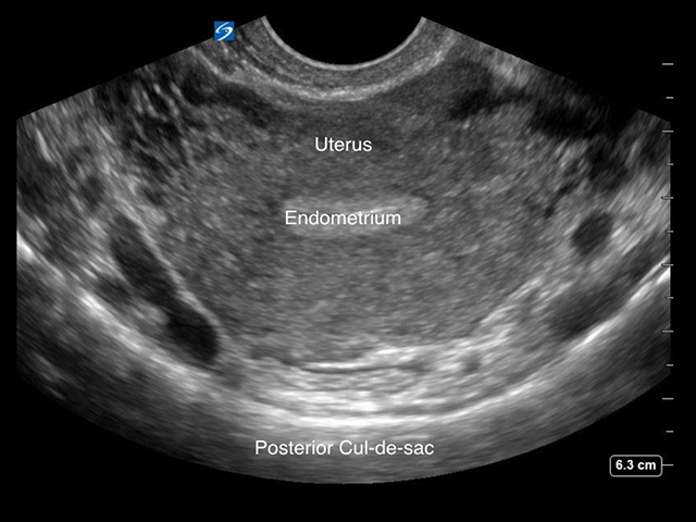
This video details how using long-axis and short-axis suprapubic views during a trauma FAST ultrasound examination enables clinicians to identify fluid in a trauma patient's pelvic cavity.
Subtitles
<p begin="00:00:18.083" end="00:00:20.666" style="s2">- Hello, my name is Phil<br />Perera and I'm the Emergency</p>
<p begin="00:00:20.666" end="00:00:22.183" style="s2">Ultrasound Coordinator at the New York</p>
<p begin="00:00:22.183" end="00:00:24.395" style="s2">Presbyterian Hospital in New York City.</p>
<p begin="00:00:24.395" end="00:00:27.632" style="s2">And welcome to Soundbytes Cases.</p>
<p begin="00:00:27.632" end="00:00:29.103" style="s2">In this Soundbytes module we'll continue</p>
<p begin="00:00:29.103" end="00:00:32.003" style="s2">our journey looking at<br />the Trauma Fast Exam.</p>
<p begin="00:00:32.003" end="00:00:34.120" style="s2">I hope you've had a<br />chance to join me prior</p>
<p begin="00:00:34.120" end="00:00:36.526" style="s2">for modules looking at<br />the right upper quadrant</p>
<p begin="00:00:36.526" end="00:00:39.414" style="s2">and left upper quadrant views<br />of the Trauma Fast Exam.</p>
<p begin="00:00:39.414" end="00:00:40.997" style="s2">In this module we will<br />specifically look at</p>
<p begin="00:00:40.997" end="00:00:44.087" style="s2">the Suprapubic view, also<br />known as the bladder or pelvic</p>
<p begin="00:00:44.087" end="00:00:46.495" style="s2">view of the Trauma Fast Exam.</p>
<p begin="00:00:46.495" end="00:00:48.203" style="s2">There is a lot of information we can gain</p>
<p begin="00:00:48.203" end="00:00:50.036" style="s2">from looking at the Suprapubic view,</p>
<p begin="00:00:50.036" end="00:00:52.369" style="s2">as we can potentially detect<br />a smaller amount of fluid</p>
<p begin="00:00:52.369" end="00:00:54.359" style="s2">than is required to make a positive right</p>
<p begin="00:00:54.359" end="00:00:56.845" style="s2">upper quadrant or left<br />upper quadrant view.</p>
<p begin="00:00:56.845" end="00:00:59.158" style="s2">Literature suggests that only about</p>
<p begin="00:00:59.158" end="00:01:02.141" style="s2">100 to 200 ccs of fluid can be detected</p>
<p begin="00:01:02.141" end="00:01:04.495" style="s2">accurately on the Suprapubic view.</p>
<p begin="00:01:04.495" end="00:01:06.073" style="s2">Here's a slide reviewing how to perform</p>
<p begin="00:01:06.073" end="00:01:08.586" style="s2">the Suprapubic view of<br />the Trauma Fast Exam.</p>
<p begin="00:01:08.586" end="00:01:10.468" style="s2">In contrast to the upper quadrant views,</p>
<p begin="00:01:10.468" end="00:01:12.676" style="s2">where we looked only in the<br />long axis configuration,</p>
<p begin="00:01:12.676" end="00:01:15.884" style="s2">the Suprapubic view is<br />made up of two planes.</p>
<p begin="00:01:15.884" end="00:01:18.987" style="s2">We want to look in both long<br />and short axis configurations.</p>
<p begin="00:01:18.987" end="00:01:21.086" style="s2">We'll begin at long<br />axis with the marker dot</p>
<p begin="00:01:21.086" end="00:01:22.829" style="s2">oriented toward the patient's head</p>
<p begin="00:01:22.829" end="00:01:24.523" style="s2">and complete our exam by moving the probe</p>
<p begin="00:01:24.523" end="00:01:26.092" style="s2">into short axis with the marker dot</p>
<p begin="00:01:26.092" end="00:01:27.933" style="s2">toward the patients right side.</p>
<p begin="00:01:27.933" end="00:01:30.004" style="s2">This way we can fully<br />scan through the pelvis</p>
<p begin="00:01:30.004" end="00:01:32.589" style="s2">and not miss any small<br />amounts of fluid there.</p>
<p begin="00:01:32.589" end="00:01:34.515" style="s2">It's optimal to perform the examination</p>
<p begin="00:01:34.515" end="00:01:35.876" style="s2">with a full bladder as an optimal</p>
<p begin="00:01:35.876" end="00:01:37.305" style="s2">acoustic window, so perform the</p>
<p begin="00:01:37.305" end="00:01:38.631" style="s2">exam prior to having the patient</p>
<p begin="00:01:38.631" end="00:01:41.619" style="s2">void or placing a foley catheter.</p>
<p begin="00:01:41.619" end="00:01:43.362" style="s2">Here's an illustration showing the anatomy</p>
<p begin="00:01:43.362" end="00:01:44.538" style="s2">that we'll need to know to perform</p>
<p begin="00:01:44.538" end="00:01:47.169" style="s2">the Suprapubic view of<br />the Trauma Fast Exam.</p>
<p begin="00:01:47.169" end="00:01:49.590" style="s2">We see the pattern of<br />fluid flow in a female,</p>
<p begin="00:01:49.590" end="00:01:52.355" style="s2">to the left here, and<br />a male, to the right.</p>
<p begin="00:01:52.355" end="00:01:54.565" style="s2">Let's look closer at the female pelvis,</p>
<p begin="00:01:54.565" end="00:01:56.497" style="s2">to the left, and what we<br />see is that fluid will</p>
<p begin="00:01:56.497" end="00:01:58.616" style="s2">preferentially develop in the pelvic</p>
<p begin="00:01:58.616" end="00:02:01.782" style="s2">Cul de Sac, located behind the uterus.</p>
<p begin="00:02:01.782" end="00:02:03.971" style="s2">Now, small amounts of fluid will only be</p>
<p begin="00:02:03.971" end="00:02:05.464" style="s2">located in the pelvic Cul de Sac,</p>
<p begin="00:02:05.464" end="00:02:07.247" style="s2">but as the amount of fluid enlarges</p>
<p begin="00:02:07.247" end="00:02:09.160" style="s2">it will come out and increase so</p>
<p begin="00:02:09.160" end="00:02:10.416" style="s2">that it will layer out on top of</p>
<p begin="00:02:10.416" end="00:02:12.848" style="s2">the uterus and on top of<br />the dome of the bladder.</p>
<p begin="00:02:12.848" end="00:02:15.141" style="s2">But small amounts of fluid<br />will only be found in</p>
<p begin="00:02:15.141" end="00:02:17.849" style="s2">that pelvic Cul de Sac<br />posterior to the uterus.</p>
<p begin="00:02:17.849" end="00:02:19.469" style="s2">Now let's take a look at the male</p>
<p begin="00:02:19.469" end="00:02:20.860" style="s2">pelvis to the right and we see</p>
<p begin="00:02:20.860" end="00:02:22.586" style="s2">small amounts of fluid<br />that will only be found</p>
<p begin="00:02:22.586" end="00:02:25.542" style="s2">in the Retrovesical<br />Space behind the bladder.</p>
<p begin="00:02:25.542" end="00:02:27.510" style="s2">As the amount of fluid enlarges it</p>
<p begin="00:02:27.510" end="00:02:29.212" style="s2">will come anterior to settle out</p>
<p begin="00:02:29.212" end="00:02:31.079" style="s2">over the top of the dome of the bladder.</p>
<p begin="00:02:31.079" end="00:02:33.070" style="s2">But as we emphasized in the female,</p>
<p begin="00:02:33.070" end="00:02:34.863" style="s2">small amounts of fluid will only be</p>
<p begin="00:02:34.863" end="00:02:36.807" style="s2">found in one place and in the male</p>
<p begin="00:02:36.807" end="00:02:38.486" style="s2">it will be in that Retrovesical Space</p>
<p begin="00:02:38.486" end="00:02:40.564" style="s2">immediately posterior to the bladder.</p>
<p begin="00:02:40.564" end="00:02:42.072" style="s2">Let's begin by looking at some normal</p>
<p begin="00:02:42.072" end="00:02:44.073" style="s2">video from the Suprapubic View.</p>
<p begin="00:02:44.073" end="00:02:46.775" style="s2">In this case, a long<br />axis view in a female,</p>
<p begin="00:02:46.775" end="00:02:49.374" style="s2">superior to the left,<br />inferior to the right.</p>
<p begin="00:02:49.374" end="00:02:50.767" style="s2">The first structure we identify is</p>
<p begin="00:02:50.767" end="00:02:53.356" style="s2">the bladder, the dark area, anteriorly</p>
<p begin="00:02:53.356" end="00:02:56.334" style="s2">and posterior to the<br />bladder we see the uterus.</p>
<p begin="00:02:56.334" end="00:02:58.100" style="s2">Now if we look into the potential space,</p>
<p begin="00:02:58.100" end="00:02:59.853" style="s2">the Pelvic Cul de Sac, posterior to the</p>
<p begin="00:02:59.853" end="00:03:02.531" style="s2">uterus for any dark fluid<br />collections, we see an</p>
<p begin="00:03:02.531" end="00:03:06.306" style="s2">absence of any fluid on<br />this normal video clip.</p>
<p begin="00:03:06.306" end="00:03:08.553" style="s2">Now let's inspect a<br />video clip from a male.</p>
<p begin="00:03:08.553" end="00:03:10.417" style="s2">In this case a short axis view.</p>
<p begin="00:03:10.417" end="00:03:12.959" style="s2">We see a large bladder there, anteriorly</p>
<p begin="00:03:12.959" end="00:03:14.474" style="s2">and behind the bladder we see two</p>
<p begin="00:03:14.474" end="00:03:16.173" style="s2">tubular structures making up the</p>
<p begin="00:03:16.173" end="00:03:19.355" style="s2">Seminal Vesicles, a<br />normal finding in a male.</p>
<p begin="00:03:19.355" end="00:03:20.896" style="s2">Now if we're looking for free fluid</p>
<p begin="00:03:20.896" end="00:03:22.389" style="s2">behind the bladder, we'd be looking</p>
<p begin="00:03:22.389" end="00:03:24.409" style="s2">for dark or anechoic fluid collection</p>
<p begin="00:03:24.409" end="00:03:26.564" style="s2">layering out behind the bladder.</p>
<p begin="00:03:26.564" end="00:03:29.942" style="s2">Notice this is a normal examination.</p>
<p begin="00:03:29.942" end="00:03:31.422" style="s2">Here's a positive examination</p>
<p begin="00:03:31.422" end="00:03:32.839" style="s2">in a female trauma patient.</p>
<p begin="00:03:32.839" end="00:03:34.414" style="s2">We're looking in the long axis view,</p>
<p begin="00:03:34.414" end="00:03:36.742" style="s2">superior to the left,<br />inferior to the right.</p>
<p begin="00:03:36.742" end="00:03:38.784" style="s2">The first structure we identify is</p>
<p begin="00:03:38.784" end="00:03:40.786" style="s2">the bladder, as seen inferior here.</p>
<p begin="00:03:40.786" end="00:03:42.849" style="s2">And notice the uterus, the solid organ,</p>
<p begin="00:03:42.849" end="00:03:45.268" style="s2">as seen superior to the bladder.</p>
<p begin="00:03:45.268" end="00:03:47.586" style="s2">We note the parts of<br />the uterus, the fundus</p>
<p begin="00:03:47.586" end="00:03:50.489" style="s2">anteriorly, and the<br />cervix more posteriorly.</p>
<p begin="00:03:50.489" end="00:03:52.557" style="s2">Now let's look into the Pelvic Cul de Sac</p>
<p begin="00:03:52.557" end="00:03:54.885" style="s2">immediately posterior to the uterus,</p>
<p begin="00:03:54.885" end="00:03:56.409" style="s2">and what we see here is the presence of a</p>
<p begin="00:03:56.409" end="00:03:59.243" style="s2">dark or anechoic fluid<br />collection just posterior</p>
<p begin="00:03:59.243" end="00:04:01.906" style="s2">to the cervix within<br />the pelvic Cul de Sac.</p>
<p begin="00:04:01.906" end="00:04:03.650" style="s2">So in the female trauma patient,</p>
<p begin="00:04:03.650" end="00:04:05.895" style="s2">this does denote a positive examination</p>
<p begin="00:04:05.895" end="00:04:07.406" style="s2">and can be a sign of ongoing bleeding</p>
<p begin="00:04:07.406" end="00:04:09.766" style="s2">within the abdominal pelvic cavity.</p>
<p begin="00:04:09.766" end="00:04:11.421" style="s2">So let's contrast this clip in which</p>
<p begin="00:04:11.421" end="00:04:12.604" style="s2">we see a small amount of fresh</p>
<p begin="00:04:12.604" end="00:04:14.786" style="s2">fluid within the pelvic Cul de Sac.</p>
<p begin="00:04:14.786" end="00:04:16.392" style="s2">With this one, in which we have a female</p>
<p begin="00:04:16.392" end="00:04:18.100" style="s2">trauma patient with a large amount</p>
<p begin="00:04:18.100" end="00:04:20.697" style="s2">of bleeding within the pelvic cavity.</p>
<p begin="00:04:20.697" end="00:04:23.006" style="s2">We see here, again, a<br />long axis scan superior</p>
<p begin="00:04:23.006" end="00:04:25.545" style="s2">to the left, inferior to<br />the right, the bladder</p>
<p begin="00:04:25.545" end="00:04:28.133" style="s2">we see as the dark structure inferiorly</p>
<p begin="00:04:28.133" end="00:04:30.566" style="s2">and the uterus superior to the bladder.</p>
<p begin="00:04:30.566" end="00:04:33.017" style="s2">Notice the fresh fluid as seen posterior</p>
<p begin="00:04:33.017" end="00:04:34.715" style="s2">to the uterus within the Cul de Sac,</p>
<p begin="00:04:34.715" end="00:04:36.744" style="s2">but note that the amount of fluid</p>
<p begin="00:04:36.744" end="00:04:38.768" style="s2">comes anterior to the uterus,</p>
<p begin="00:04:38.768" end="00:04:41.351" style="s2">as seen here between the<br />uterus and the bladder.</p>
<p begin="00:04:41.351" end="00:04:44.140" style="s2">So this denotes a large<br />amount of blood within</p>
<p begin="00:04:44.140" end="00:04:47.084" style="s2">the pelvic cavity in this<br />female trauma patient.</p>
<p begin="00:04:47.084" end="00:04:49.940" style="s2">If we now orient the probe<br />to the patient's right side,</p>
<p begin="00:04:49.940" end="00:04:52.849" style="s2">we obtain a short axis<br />view of the same patient.</p>
<p begin="00:04:52.849" end="00:04:54.604" style="s2">And what we see here is the uterus in the</p>
<p begin="00:04:54.604" end="00:04:56.937" style="s2">middle of the image and<br />notice the large amount</p>
<p begin="00:04:56.937" end="00:04:59.442" style="s2">of fresh fluid as seen both<br />to the top, or anterior,</p>
<p begin="00:04:59.442" end="00:05:01.807" style="s2">to the uterus and posterior to the uterus.</p>
<p begin="00:05:01.807" end="00:05:03.401" style="s2">Notice in this case we can see the</p>
<p begin="00:05:03.401" end="00:05:05.629" style="s2">broad ligaments of the<br />uterus well outlined</p>
<p begin="00:05:05.629" end="00:05:08.119" style="s2">by all the fresh fluid within the pelvis.</p>
<p begin="00:05:08.119" end="00:05:10.733" style="s2">So a large amount of<br />fresh fluid, or blood,</p>
<p begin="00:05:10.733" end="00:05:14.050" style="s2">in this case, within this<br />female trauma patient.</p>
<p begin="00:05:14.050" end="00:05:15.597" style="s2">Here's a positive examination</p>
<p begin="00:05:15.597" end="00:05:18.316" style="s2">Suprapubic View, short axis in a male.</p>
<p begin="00:05:18.316" end="00:05:20.673" style="s2">Probe is oriented towards<br />the patient's right and</p>
<p begin="00:05:20.673" end="00:05:23.066" style="s2">anteriorly we see a large,<br />fluid filled bladder.</p>
<p begin="00:05:23.066" end="00:05:25.238" style="s2">Posterior to the bladder,<br />in the retrovesical</p>
<p begin="00:05:25.238" end="00:05:27.863" style="s2">space we appreciate the<br />presence of free fluid,</p>
<p begin="00:05:27.863" end="00:05:29.509" style="s2">as shown by that dark or anechoic</p>
<p begin="00:05:29.509" end="00:05:30.992" style="s2">fluid collection there.</p>
<p begin="00:05:30.992" end="00:05:32.868" style="s2">Now this gives a finding known as</p>
<p begin="00:05:32.868" end="00:05:34.767" style="s2">the double wall sign, and we see</p>
<p begin="00:05:34.767" end="00:05:36.319" style="s2">the wall of the bladder, outlined by</p>
<p begin="00:05:36.319" end="00:05:38.435" style="s2">the urine inside the bladder, and the</p>
<p begin="00:05:38.435" end="00:05:40.589" style="s2">blood, in this case, outside the bladder</p>
<p begin="00:05:40.589" end="00:05:43.180" style="s2">in the area of the retrovesical space.</p>
<p begin="00:05:43.180" end="00:05:44.850" style="s2">To further confirm that the last patient</p>
<p begin="00:05:44.850" end="00:05:46.105" style="s2">had a positive exam and that we're</p>
<p begin="00:05:46.105" end="00:05:48.688" style="s2">not mistaking areas of<br />fluid as seminal vesicles,</p>
<p begin="00:05:48.688" end="00:05:51.348" style="s2">we'll re-scan the patient<br />in the long axis plane,</p>
<p begin="00:05:51.348" end="00:05:53.555" style="s2">superior to the left,<br />inferior to the right.</p>
<p begin="00:05:53.555" end="00:05:55.425" style="s2">We see the large circular bladder,</p>
<p begin="00:05:55.425" end="00:05:57.655" style="s2">as seen anteriorly and superior</p>
<p begin="00:05:57.655" end="00:05:59.269" style="s2">and posterior to the bladder in the</p>
<p begin="00:05:59.269" end="00:06:01.039" style="s2">retrovesical space, we can see</p>
<p begin="00:06:01.039" end="00:06:02.783" style="s2">free fluid layering out there.</p>
<p begin="00:06:02.783" end="00:06:04.257" style="s2">This confirms that indeed the patient</p>
<p begin="00:06:04.257" end="00:06:06.125" style="s2">has a positive exam, with blood</p>
<p begin="00:06:06.125" end="00:06:07.804" style="s2">layering out behind the bladder.</p>
<p begin="00:06:07.804" end="00:06:09.731" style="s2">And, again, we see the double wall sign,</p>
<p begin="00:06:09.731" end="00:06:11.733" style="s2">urine outlining the inner wall of the</p>
<p begin="00:06:11.733" end="00:06:13.318" style="s2">bladder, and blood, in this case,</p>
<p begin="00:06:13.318" end="00:06:15.776" style="s2">outlining the outer wall of the bladder.</p>
<p begin="00:06:15.776" end="00:06:17.531" style="s2">Here we're scanning a<br />male trauma patient with</p>
<p begin="00:06:17.531" end="00:06:19.874" style="s2">a long axis configuration<br />and we see a large</p>
<p begin="00:06:19.874" end="00:06:22.323" style="s2">amount of free fluid within the pelvis.</p>
<p begin="00:06:22.323" end="00:06:23.837" style="s2">We note the bladder inferiorly,</p>
<p begin="00:06:23.837" end="00:06:25.709" style="s2">and note all the free fluid layering</p>
<p begin="00:06:25.709" end="00:06:27.434" style="s2">out both posterior to the bladder</p>
<p begin="00:06:27.434" end="00:06:29.429" style="s2">in the retrovesical space and coming</p>
<p begin="00:06:29.429" end="00:06:31.048" style="s2">anteriorly onto the dome of the</p>
<p begin="00:06:31.048" end="00:06:33.234" style="s2">bladder as seen to the left here.</p>
<p begin="00:06:33.234" end="00:06:37.485" style="s2">So a large amount of free fluid<br />in this male trauma patient.</p>
<p begin="00:06:37.485" end="00:06:38.615" style="s2">In conclusion, I'm glad I could</p>
<p begin="00:06:38.615" end="00:06:39.898" style="s2">share with you the Soundbytes Module</p>
<p begin="00:06:39.898" end="00:06:43.131" style="s2">covering the Suprapubic View<br />of the Trauma Fast Exam.</p>
<p begin="00:06:43.131" end="00:06:44.365" style="s2">This view is a very important</p>
<p begin="00:06:44.365" end="00:06:45.997" style="s2">one to add onto the exam of your</p>
<p begin="00:06:45.997" end="00:06:47.497" style="s2">trauma patient as we can potentially</p>
<p begin="00:06:47.497" end="00:06:49.670" style="s2">detect a smaller amount of fluid here,</p>
<p begin="00:06:49.670" end="00:06:51.524" style="s2">within the pelvis, than it takes to</p>
<p begin="00:06:51.524" end="00:06:53.064" style="s2">make a positive right upper quadrant</p>
<p begin="00:06:53.064" end="00:06:54.985" style="s2">or left upper quadrant view.</p>
<p begin="00:06:54.985" end="00:06:56.869" style="s2">Remember that this is a two-step exam,</p>
<p begin="00:06:56.869" end="00:06:58.041" style="s2">we'll be looking in both short</p>
<p begin="00:06:58.041" end="00:06:59.924" style="s2">and long axis configurations to</p>
<p begin="00:06:59.924" end="00:07:01.442" style="s2">verify fluid, and also remember</p>
<p begin="00:07:01.442" end="00:07:03.712" style="s2">the differences between<br />the female, where we're</p>
<p begin="00:07:03.712" end="00:07:05.578" style="s2">looking into the pelvic<br />Cul de Sac for fluid,</p>
<p begin="00:07:05.578" end="00:07:06.898" style="s2">and the male, where we're looking into</p>
<p begin="00:07:06.898" end="00:07:09.175" style="s2">the retrovesical space for fluid.</p>
<p begin="00:07:09.175" end="00:07:10.741" style="s2">So I hope to see you back in the</p>
<p begin="00:07:10.741" end="00:07:13.324" style="s2">future as Soundbytes continues.</p>