
Paediatric and internal medicine resident Marie Arnaout, DO, and Assistant Professor of Internal Medicine Prashant Patel, DO, recently returned from Madagascar on a mission for Western Michigan University School of Medicine. They were accompanied by internal physician Richard Roach, MD (a long-serving senior faculty member who goes to Madagascar annually) and other paediatric and internal specialists. The mission worked with local SALFA (Lutheran) clinicians at clinics and hospitals in three communities. The team borrowed a Sonosite M-Turbo from the Global Health Loaner Pool. Drs. Arnaout and Patel sent us this joint report from their mission.
Challenging Conditions
Every year, our school sends a team to work with the SALFA doctors in Madagascar for one month. We work in their clinics and hospital to help them and teach them as well as learn from them. There are many challenges for patients and doctors there. They have very limited resources for diagnostics and therapeutics, and the resources they do have are very old or don't always work well.
Patients travel for many miles just to be seen by physicians who serve a large catchment area. The only place with a CT scanner, MRI, and echocardiogram is in Madagascar’s capital of Antananarivo, so most of the other facilities rely on X-ray or ultrasound for primary imaging modalities. Patients usually wait until disease progression is severe to seek medical care due to cost and distance, which unfortunately results in patients presenting in extremis.
The entire team setting is collaborative, with a strong focus on bedside patient care, team rounding and teaching, and supplementing reasoning toward medical management. The core and unique aspect of our trip is to work with physicians who were trained in and continue working in Madagascar, thus having longer-lasting impact within the country.
We flew from Detroit to Paris to Madagascar. Once we arrived in country, everyone had their temperature checked to prevent spread of Ebola. Our sites during the trip were Antananarivo, the former French resort city of Antsirabe, and the northwest coastal city of Mahajanga. Getting from Antananarivo to Mahajanga was challenging, a cross-country bus trip over a single mountainside highway, taking about 15 hours to get there.
Ultrasound Helps Identify Wide Range of Pathologies
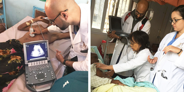
At Mahajanga, we used a Sonosite M-Turbo often at bedside and in clinic to direct our decision making after looking at the lungs, the heart, and the abdomen. It helped the residents to learn how to visualise and diagnose different pathologies, including dilated cardiomyopathy, liver cirrhosis, cholecystitis, polycystic kidneys, hydronephrosis, heart failure exacerbations, pulmonary edoema, and fluid status based on views of the inferior vena cava. Patients loved being able to have some answers at bedside, instead of having to wait for diagnostics or travelling. We also used it to teach the local doctors how to look at the heart and read it from different locations on the chest. They were excited to be able to see their own hearts as well.
Cardiac, Renal, and Pulmonary Applications
Heart failure is very prevalent in Madagascar. We had two cases that really stand out to me. Two men presented with shortness of breath. Looking at their echo using the phased-array probe, we could tell their heart function was about 15-20%. We were able to recommend some medications that would help relieve symptoms. We had another case of a young girl with bilateral polycystic kidney disease that we diagnosed at bedside. Her prognosis was very poor, but the immediacy of diagnostic results initiated the discussion. Unfortunately, the hard reality was that dialysis was not an option for this patient due to lack of resources.
On the other hand, we had a patient with worsening dyspepsia on exertion over a period of multiple weeks who presented to the outpatient clinic. Using the phased-array probe, we were able to visualise a pleural effusions with what appeared to be loculations. The Malagasy physicians ultimately used this information to dive deeper into his history, in which he stated he had unexpectedly lost weight, had intermittent hemoptysis, and had a worsening cough. This diagnosis led to our timely discussion of a probable TB effusion.
Thank you so much for the ultrasound machine. It was a great tool to help patients and teach the Malagasy doctors.