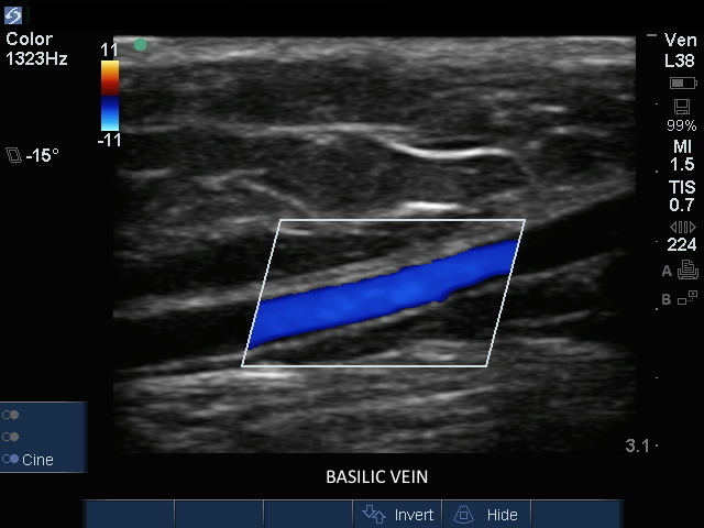
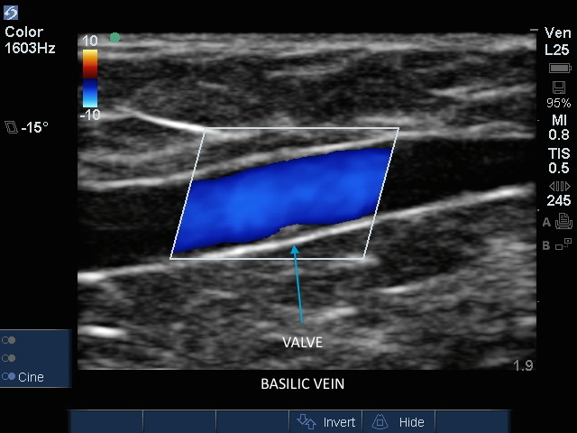
How to: Peripheral Arm Veins for Vascular Access

How to: Peripheral Arm Veins for Vascular Access

/sites/default/files/ST_Peripheral_Arm_Veins_for_Vascular_Access_Thumb.jpg
An overview of ultrasound landmarks and scanning techniques used for peripheral arm vein access.
Applications
Clinical Specialties
Media Library Type
Subtitles
<p begin="00:00:13.967" end="00:00:15.927" style="s2">- Ultrasound can be a great adjunct</p>
<p begin="00:00:15.927" end="00:00:17.673" style="s2">for peripheral vein access.</p>
<p begin="00:00:17.673" end="00:00:20.002" style="s2">Normally, our nurses can<br />get pretty good access</p>
<p begin="00:00:20.002" end="00:00:22.303" style="s2">to the peripheral vein<br />through standard palpations,</p>
<p begin="00:00:22.303" end="00:00:25.191" style="s2">but sometimes the patient<br />may have an edematous arm,</p>
<p begin="00:00:25.191" end="00:00:26.276" style="s2">their arm may be obese,</p>
<p begin="00:00:26.276" end="00:00:29.121" style="s2">you may not be able to feel<br />an antecubital vein easily.</p>
<p begin="00:00:29.121" end="00:00:32.451" style="s2">In addition, if the antecubital<br />veins cannot be accessed,</p>
<p begin="00:00:32.451" end="00:00:35.354" style="s2">we may want to access<br />deeper veins of the arm,</p>
<p begin="00:00:35.354" end="00:00:37.564" style="s2">those being the basilic<br />and brachial veins.</p>
<p begin="00:00:37.564" end="00:00:40.056" style="s2">Ultrasound provides great visualization</p>
<p begin="00:00:40.056" end="00:00:41.549" style="s2">of all of those veins.</p>
<p begin="00:00:41.549" end="00:00:43.741" style="s2">We're gonna go ahead and choose<br />the linear array transducer.</p>
<p begin="00:00:43.741" end="00:00:45.881" style="s2">This is going to allow<br />high-frequency imaging</p>
<p begin="00:00:45.881" end="00:00:48.073" style="s2">to give us really good image quality</p>
<p begin="00:00:48.073" end="00:00:49.893" style="s2">for superficial structures.</p>
<p begin="00:00:49.893" end="00:00:51.943" style="s2">Remember, these veins are superficial</p>
<p begin="00:00:51.943" end="00:00:55.245" style="s2">so we're gonna to want to get<br />as good image as possible.</p>
<p begin="00:00:55.245" end="00:00:57.085" style="s2">I'm gonna go ahead and<br />choose the exam type.</p>
<p begin="00:00:57.085" end="00:00:58.546" style="s2">Once I've chosen this transducer,</p>
<p begin="00:00:58.546" end="00:01:01.158" style="s2">I do have different exam<br />types I can choose from.</p>
<p begin="00:01:01.158" end="00:01:04.075" style="s2">I'm gonna use the Exam button</p>
<p begin="00:01:04.075" end="00:01:06.408" style="s2">to choose the Venous preset,</p>
<p begin="00:01:07.971" end="00:01:09.375" style="s2">hit Select.</p>
<p begin="00:01:09.375" end="00:01:11.857" style="s2">I've already got some gel<br />at the antecubital fossa.</p>
<p begin="00:01:11.857" end="00:01:13.255" style="s2">I already have a tourniquet up.</p>
<p begin="00:01:13.255" end="00:01:15.420" style="s2">So let me now take a look<br />at his antecubital fossa</p>
<p begin="00:01:15.420" end="00:01:18.017" style="s2">and see what veins we can identify.</p>
<p begin="00:01:18.017" end="00:01:19.703" style="s2">Here we have the transducer marker here,</p>
<p begin="00:01:19.703" end="00:01:21.371" style="s2">I'm gonna put it to the patient's right.</p>
<p begin="00:01:21.371" end="00:01:24.183" style="s2">Again, it doesn't really matter<br />when we do vascular access</p>
<p begin="00:01:24.183" end="00:01:25.550" style="s2">if it's to the right or left,</p>
<p begin="00:01:25.550" end="00:01:29.217" style="s2">as long as the target<br />vein is in the center.</p>
<p begin="00:01:30.742" end="00:01:32.190" style="s2">First thing I notice is</p>
<p begin="00:01:32.190" end="00:01:35.900" style="s2">that I can probably decrease<br />my Depth a little bit.</p>
<p begin="00:01:35.900" end="00:01:38.538" style="s2">So I'm going to go ahead and do that.</p>
<p begin="00:01:38.538" end="00:01:41.771" style="s2">I'm going to turn my Gain up a little bit.</p>
<p begin="00:01:41.771" end="00:01:45.438" style="s2">And I notice here, right in<br />the center of the screen,</p>
<p begin="00:01:45.438" end="00:01:49.183" style="s2">is an an anechoic structure,<br />that is easily collapsible.</p>
<p begin="00:01:49.183" end="00:01:53.520" style="s2">And as you can see, with<br />simple pressure with my hand,</p>
<p begin="00:01:53.520" end="00:01:56.592" style="s2">this vessel easily collapses.</p>
<p begin="00:01:56.592" end="00:02:00.528" style="s2">There's no pulsatile motion<br />and it easily collapses.</p>
<p begin="00:02:00.528" end="00:02:02.397" style="s2">It is also quite superficial.</p>
<p begin="00:02:02.397" end="00:02:03.490" style="s2">If you look at the marks here</p>
<p begin="00:02:03.490" end="00:02:05.529" style="s2">on the right hand of the screen,</p>
<p begin="00:02:05.529" end="00:02:06.854" style="s2">this is a half centimeter mark,</p>
<p begin="00:02:06.854" end="00:02:08.412" style="s2">this is a one centimeter mark.</p>
<p begin="00:02:08.412" end="00:02:12.579" style="s2">This vein is only about four<br />to five millimeters deep.</p>
<p begin="00:02:13.840" end="00:02:16.378" style="s2">So it'd be very easy to access.</p>
<p begin="00:02:16.378" end="00:02:18.513" style="s2">If I'm looking at the screen right now,</p>
<p begin="00:02:18.513" end="00:02:20.634" style="s2">and I put the vein directly in the center,</p>
<p begin="00:02:20.634" end="00:02:22.691" style="s2">that vein will be directly in the center</p>
<p begin="00:02:22.691" end="00:02:25.441" style="s2">of the transducer below the skin.</p>
<p begin="00:02:27.253" end="00:02:29.917" style="s2">If you can not find anything<br />in the antecubital veins,</p>
<p begin="00:02:29.917" end="00:02:32.415" style="s2">you may want to take a look at<br />the deeper veins of the arm.</p>
<p begin="00:02:32.415" end="00:02:34.320" style="s2">So now let's take a look at the brachial</p>
<p begin="00:02:34.320" end="00:02:36.748" style="s2">and the basilic veins.</p>
<p begin="00:02:36.748" end="00:02:40.748" style="s2">I'm gonna put a little<br />gel, a little more medial</p>
<p begin="00:02:42.784" end="00:02:46.879" style="s2">of the elbow, and a<br />little bit more proximal.</p>
<p begin="00:02:46.879" end="00:02:50.388" style="s2">I'm gonna scan transversely<br />across the arm.</p>
<p begin="00:02:50.388" end="00:02:51.971" style="s2">I see a large vein,</p>
<p begin="00:02:53.529" end="00:02:56.279" style="s2">here in the center of the screen,</p>
<p begin="00:02:58.694" end="00:03:00.086" style="s2">which is collapsible.</p>
<p begin="00:03:00.086" end="00:03:02.586" style="s2">In fact, I see multiple veins.</p>
<p begin="00:03:07.630" end="00:03:09.504" style="s2">You can put a pretty<br />large catheter into here.</p>
<p begin="00:03:09.504" end="00:03:12.792" style="s2">You can easily place a 14<br />or a 16 gauge catheter.</p>
<p begin="00:03:12.792" end="00:03:15.179" style="s2">I will recommend you<br />use a two inch catheter</p>
<p begin="00:03:15.179" end="00:03:17.008" style="s2">in all of these cannulations.</p>
<p begin="00:03:17.008" end="00:03:20.069" style="s2">If you use a standard angiocath,<br />you will get a flashback.</p>
<p begin="00:03:20.069" end="00:03:24.236" style="s2">But unfortunately, most likely<br />the catheter will dislodge.</p>
<p begin="00:03:25.734" end="00:03:27.629" style="s2">So this, this is a good vein here.</p>
<p begin="00:03:27.629" end="00:03:31.546" style="s2">If you look around a<br />little bit more laterally,</p>
<p begin="00:03:33.041" end="00:03:35.144" style="s2">we can see other veins,</p>
<p begin="00:03:35.144" end="00:03:38.079" style="s2">and we can see the artery as well.</p>
<p begin="00:03:38.079" end="00:03:39.859" style="s2">Here in the center of the screen,</p>
<p begin="00:03:39.859" end="00:03:43.954" style="s2">you see a pulsatile structure,<br />which represents the artery.</p>
<p begin="00:03:43.954" end="00:03:46.975" style="s2">If you're unsure, if<br />it's a very small vessel,</p>
<p begin="00:03:46.975" end="00:03:51.142" style="s2">is arterial venous, you<br />can put the color flow on.</p>
<p begin="00:03:52.021" end="00:03:55.104" style="s2">And we see classic color flow profile</p>
<p begin="00:03:56.440" end="00:03:58.523" style="s2">of an arterial structure.</p>
<p begin="00:03:59.685" end="00:04:03.321" style="s2">So we're now scanning just<br />above the antecubital fossa,</p>
<p begin="00:04:03.321" end="00:04:04.854" style="s2">a little bit medially.</p>
<p begin="00:04:04.854" end="00:04:06.704" style="s2">We have a pretty good image here,</p>
<p begin="00:04:06.704" end="00:04:10.272" style="s2">just above the antecubital<br />fossa, a little bit medially.</p>
<p begin="00:04:10.272" end="00:04:13.884" style="s2">And we see a few structures<br />here I want to point out.</p>
<p begin="00:04:13.884" end="00:04:17.008" style="s2">We can see an artery, here in the center.</p>
<p begin="00:04:17.008" end="00:04:19.530" style="s2">Again, when I put a<br />little compression on it,</p>
<p begin="00:04:19.530" end="00:04:21.780" style="s2">you can see pulsatile flow.</p>
<p begin="00:04:22.927" end="00:04:24.917" style="s2">Adjacent to that,</p>
<p begin="00:04:24.917" end="00:04:28.477" style="s2">you can see a vessel<br />which easily collapses.</p>
<p begin="00:04:28.477" end="00:04:31.143" style="s2">This represents a venous structure,</p>
<p begin="00:04:31.143" end="00:04:32.538" style="s2">which could be cannulated,</p>
<p begin="00:04:32.538" end="00:04:35.108" style="s2">but it doesn't look like<br />it's in a great location</p>
<p begin="00:04:35.108" end="00:04:38.475" style="s2">because it sits right<br />adjacent to the artery.</p>
<p begin="00:04:38.475" end="00:04:41.061" style="s2">In addition, on the other side of it,</p>
<p begin="00:04:41.061" end="00:04:44.680" style="s2">is a slightly hypoechoic structure.</p>
<p begin="00:04:44.680" end="00:04:46.458" style="s2">This represents a nerve.</p>
<p begin="00:04:46.458" end="00:04:50.148" style="s2">So if I was choosing a<br />location for venous access,</p>
<p begin="00:04:50.148" end="00:04:51.776" style="s2">I would not choose this vein,</p>
<p begin="00:04:51.776" end="00:04:55.943" style="s2">given the proximity both to<br />the artery and the nerve.</p>
Brightcove ID
5508134285001
https://youtube.com/watch?v=Pga4SAD-J9U

3D How To: Femoral Artery Catheter Insertion

3D How To: Femoral Artery Catheter Insertion

/sites/default/files/CentralVenous_FemoralArtery_edu00502_thumbnail.jpg
3D animation demonstrating an ultrasound guided insertion of a Femoral Artery Catheter.
Applications
Clinical Specialties
Media Library Type
Subtitles
<p begin="00:00:07.403" end="00:00:09.316" style="s2">- [Voiceover] A linear array transducer</p>
<p begin="00:00:09.316" end="00:00:10.984" style="s2">with a vascular exam type,</p>
<p begin="00:00:10.984" end="00:00:13.876" style="s2">is used to perform an<br />ultrasound guided insertion</p>
<p begin="00:00:13.876" end="00:00:17.790" style="s2">of a femoral artery catheter<br />via a transverse approach.</p>
<p begin="00:00:17.790" end="00:00:19.896" style="s2">The patient is in a supine position,</p>
<p begin="00:00:19.896" end="00:00:22.611" style="s2">with the operator at the right side.</p>
<p begin="00:00:22.611" end="00:00:24.986" style="s2">The transducer is placed transversely,</p>
<p begin="00:00:24.986" end="00:00:27.338" style="s2">just inferior to the inguinal crease,</p>
<p begin="00:00:27.338" end="00:00:29.254" style="s2">with the orientation marker direction</p>
<p begin="00:00:29.254" end="00:00:32.996" style="s2">to the patient's right<br />at a 9 o'clock position.</p>
<p begin="00:00:32.996" end="00:00:35.117" style="s2">The superficial femoral artery</p>
<p begin="00:00:35.117" end="00:00:38.847" style="s2">is seen as a dark, anechoic,<br />pulsatile, circular structure</p>
<p begin="00:00:38.847" end="00:00:41.307" style="s2">in the mid-portion of<br />the ultrasound image.</p>
<p begin="00:00:41.307" end="00:00:43.629" style="s2">Deeper, and slightly to<br />the right of the image,</p>
<p begin="00:00:43.629" end="00:00:45.526" style="s2">the femoral vein will appear</p>
<p begin="00:00:45.526" end="00:00:48.165" style="s2">as a dark, anechoic, elliptical shape</p>
<p begin="00:00:48.165" end="00:00:50.972" style="s2">that is compressible<br />with transducer pressure.</p>
<p begin="00:00:50.972" end="00:00:53.170" style="s2">Adjust the transducer so it is centered</p>
<p begin="00:00:53.170" end="00:00:56.471" style="s2">over the femoral artery,<br />lateral to the vein.</p>
<p begin="00:00:56.471" end="00:00:59.547" style="s2">Follow the needle entry by<br />slowing sliding the transducer</p>
<p begin="00:00:59.547" end="00:01:01.993" style="s2">in the direction of needle advancement.</p>
<p begin="00:01:01.993" end="00:01:05.135" style="s2">The needle will appear<br />as a small, bright dot.</p>
<p begin="00:01:05.135" end="00:01:06.672" style="s2">When the needle tip appears,</p>
<p begin="00:01:06.672" end="00:01:10.067" style="s2">the transducer should be advanced<br />a short distance distally,</p>
<p begin="00:01:10.067" end="00:01:12.259" style="s2">to follow the tip of the needle trajectory</p>
<p begin="00:01:12.259" end="00:01:15.350" style="s2">and stay in advance of the needle entry.</p>
<p begin="00:01:15.350" end="00:01:16.591" style="s2">The needle is slowly advanced</p>
<p begin="00:01:16.591" end="00:01:19.257" style="s2">under direct ultrasound visualization,</p>
<p begin="00:01:19.257" end="00:01:23.008" style="s2">until the tip is seen to<br />puncture the femoral artery.</p>
<p begin="00:01:23.008" end="00:01:24.972" style="s2">The transducer should be moved slightly</p>
<p begin="00:01:24.972" end="00:01:28.121" style="s2">proximally and distally to<br />confirm that the needle tip</p>
<p begin="00:01:28.121" end="00:01:31.288" style="s2">lies in the mid-portion of the artery.</p>
Brightcove ID
5508128530001
https://youtube.com/watch?v=n7knHl9pSas

3D How To: Peripherally Inserted Venous Catheter

3D How To: Peripherally Inserted Venous Catheter

/sites/default/files/PeripheralVenous_edu00475_thumbnail.jpg
3D animation demonstrating an ultrasound guided Peripheral Inserted Venous Catheter (upper extremity).
Applications
Clinical Specialties
Media Library Type
Subtitles
<p begin="00:00:07.465" end="00:00:09.053" style="s2">- [Voiceover] A liner<br />array transducer with</p>
<p begin="00:00:09.053" end="00:00:11.160" style="s2">a venous exam type is used to preform</p>
<p begin="00:00:11.160" end="00:00:13.097" style="s2">an ultrasound guided insertion</p>
<p begin="00:00:13.097" end="00:00:15.733" style="s2">of a peripherally<br />inserted central catheter</p>
<p begin="00:00:15.733" end="00:00:18.144" style="s2">via a transverse approach.</p>
<p begin="00:00:18.144" end="00:00:19.835" style="s2">The patient is in a supine position</p>
<p begin="00:00:19.835" end="00:00:23.522" style="s2">with the arm extended 90<br />degrees at the patient's side</p>
<p begin="00:00:23.522" end="00:00:24.977" style="s2">and externally rotated.</p>
<p begin="00:00:24.977" end="00:00:27.961" style="s2">The transducer is placed<br />transversely just proximal</p>
<p begin="00:00:27.961" end="00:00:30.980" style="s2">to the medial condyle<br />in the bicipital groove</p>
<p begin="00:00:30.980" end="00:00:34.207" style="s2">with the orientation marker<br />directed to the patient's right.</p>
<p begin="00:00:34.207" end="00:00:37.505" style="s2">The basilic vein is seen as<br />a dark anechoic circular,</p>
<p begin="00:00:37.505" end="00:00:40.124" style="s2">compressible structure<br />in the mid-portion of</p>
<p begin="00:00:40.124" end="00:00:43.985" style="s2">the ultrasound image, between<br />the biceps and triceps muscle.</p>
<p begin="00:00:43.985" end="00:00:46.285" style="s2">Deeper, and slightly to<br />the right of the screen,</p>
<p begin="00:00:46.285" end="00:00:49.383" style="s2">the bright hyper-echoic<br />humerus can be seen.</p>
<p begin="00:00:49.383" end="00:00:51.365" style="s2">Adjust the transducer so it is</p>
<p begin="00:00:51.365" end="00:00:53.544" style="s2">centered over the basilic vein.</p>
<p begin="00:00:53.544" end="00:00:55.856" style="s2">Follow the needle entry by slowly sliding</p>
<p begin="00:00:55.856" end="00:00:58.889" style="s2">the transducer in the direction<br />of needle advancement.</p>
<p begin="00:00:58.889" end="00:01:01.720" style="s2">The needle will appear<br />as a small bright dot.</p>
<p begin="00:01:01.720" end="00:01:03.472" style="s2">When the needle tip appears,</p>
<p begin="00:01:03.472" end="00:01:06.443" style="s2">the transducer should be<br />advanced a short distance</p>
<p begin="00:01:06.443" end="00:01:08.519" style="s2">to follow the tip of the needle trajectory</p>
<p begin="00:01:08.519" end="00:01:11.159" style="s2">and stay in advance of the needle entry.</p>
<p begin="00:01:11.159" end="00:01:12.892" style="s2">The needle is slowly advanced under</p>
<p begin="00:01:12.892" end="00:01:15.533" style="s2">direct ultrasound<br />visualization until the tip</p>
<p begin="00:01:15.533" end="00:01:19.306" style="s2">is seen to indent and then<br />puncture the basilic vein.</p>
<p begin="00:01:19.306" end="00:01:21.919" style="s2">The transducer should be<br />moved slightly proximally</p>
<p begin="00:01:21.919" end="00:01:24.040" style="s2">and distally to confirm<br />that the needle tip</p>
<p begin="00:01:24.040" end="00:01:26.679" style="s2">lies in the mid portion<br />of the basilic vein.</p>
<p begin="00:01:26.679" end="00:01:28.592" style="s2">This technique can also be used</p>
<p begin="00:01:28.592" end="00:01:31.925" style="s2">with the deep brachial or cephalic vein.</p>
Brightcove ID
5741617671001
https://youtube.com/watch?v=rWxN4JFBEJc

3D How To: Infraclavicular Nerve Block

3D How To: Infraclavicular Nerve Block

/sites/default/files/Infraclavicular_edu00493_thumbnail.jpg
3D animation demonstrating an ultrasound guided infraclavicular nerve block.
Clinical Specialties
Media Library Type
Subtitles
<p begin="00:00:07.354" end="00:00:09.128" style="s2">- [Voiceover] A linear array transducer</p>
<p begin="00:00:09.128" end="00:00:11.643" style="s2">with a nerve exam type is used to perform</p>
<p begin="00:00:11.643" end="00:00:15.694" style="s2">an ultrasound-guided,<br />infraclavicular regional nerve block.</p>
<p begin="00:00:15.694" end="00:00:19.134" style="s2">The target depth is approximately<br />three to four centmeters</p>
<p begin="00:00:19.134" end="00:00:21.341" style="s2">in an 80 kilogram adult.</p>
<p begin="00:00:21.341" end="00:00:23.542" style="s2">The patient is positioned supine</p>
<p begin="00:00:23.542" end="00:00:25.523" style="s2">with the arm abducted 90 degrees</p>
<p begin="00:00:25.523" end="00:00:27.616" style="s2">and the elbow bent 90 degrees</p>
<p begin="00:00:27.616" end="00:00:29.525" style="s2">to move the clavicle posterior,</p>
<p begin="00:00:29.525" end="00:00:32.434" style="s2">and permit a shallow<br />angle of needle insertion.</p>
<p begin="00:00:32.434" end="00:00:34.126" style="s2">The patient's head should be rotated</p>
<p begin="00:00:34.126" end="00:00:36.427" style="s2">toward the contralateral side.</p>
<p begin="00:00:36.427" end="00:00:40.380" style="s2">The transducer is placed on the<br />chest in a paramedian plane,</p>
<p begin="00:00:40.380" end="00:00:42.936" style="s2">inferior to the midpoint of the clavicle</p>
<p begin="00:00:42.936" end="00:00:46.372" style="s2">with the orientation marker<br />directed to the patient's head.</p>
<p begin="00:00:46.372" end="00:00:48.368" style="s2">The axillary artery is seen as</p>
<p begin="00:00:48.368" end="00:00:52.247" style="s2">a dark, round pulsatile<br />structure with a hyperechoic wall</p>
<p begin="00:00:52.247" end="00:00:56.144" style="s2">inferior to the pectoralis<br />major and minor muscles.</p>
<p begin="00:00:56.144" end="00:00:59.668" style="s2">The axillary vein lies<br />beside the axillary artery</p>
<p begin="00:00:59.668" end="00:01:01.751" style="s2">and collapses with compression.</p>
<p begin="00:01:01.751" end="00:01:04.801" style="s2">The nerve cords appear<br />as hyperechoic circles</p>
<p begin="00:01:04.801" end="00:01:06.193" style="s2">with a honeycomb appearance</p>
<p begin="00:01:06.193" end="00:01:10.370" style="s2">within the bright hyperechoic<br />fascia of the brachial plexus.</p>
<p begin="00:01:10.370" end="00:01:13.618" style="s2">The transducer is slowly<br />moved in a lateral direction,</p>
<p begin="00:01:13.618" end="00:01:16.454" style="s2">following the course<br />of the axillary artery.</p>
<p begin="00:01:16.454" end="00:01:18.722" style="s2">The nerves will split into a lateral,</p>
<p begin="00:01:18.722" end="00:01:21.793" style="s2">medial, and posterior cord.</p>
<p begin="00:01:21.793" end="00:01:24.069" style="s2">The optimum position of the transducer</p>
<p begin="00:01:24.069" end="00:01:26.136" style="s2">for the infraclavicular nerve block</p>
<p begin="00:01:26.136" end="00:01:28.306" style="s2">is as lateral as possible.</p>
<p begin="00:01:28.306" end="00:01:31.280" style="s2">The needle is positioned<br />two centimeters cephalad</p>
<p begin="00:01:31.280" end="00:01:35.432" style="s2">to the transducer and advanced<br />using an in-plane technique.</p>
<p begin="00:01:35.432" end="00:01:37.913" style="s2">The needle path is<br />directed over the clavicle</p>
<p begin="00:01:37.913" end="00:01:40.181" style="s2">and through the pectoral muscles.</p>
<p begin="00:01:40.181" end="00:01:42.143" style="s2">The initial endpoint for the needle is</p>
<p begin="00:01:42.143" end="00:01:45.627" style="s2">immediately posterior to<br />the midpoint of the artery.</p>
<p begin="00:01:45.627" end="00:01:48.770" style="s2">The spread of local<br />anesthetic should be observed.</p>
<p begin="00:01:48.770" end="00:01:52.364" style="s2">Supplemental injections can<br />be made anterior to the artery</p>
<p begin="00:01:52.364" end="00:01:53.954" style="s2">to deposit local anesthetic</p>
<p begin="00:01:53.954" end="00:01:57.954" style="s2">around the lateral and<br />medial cords if required.</p>
Brightcove ID
5745561363001
https://youtube.com/watch?v=8xRDVLKb5BY