

M-Turbo: IVC long Axis
M-Turbo: IVC long Axis
/sites/default/files/201410_Image_M-Turbo_P21_IVC_Long.jpg
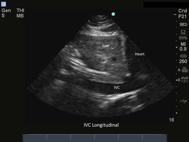
M-Turbo: IVC Long Axis.
Media Library Type
Compatible Products
Body
M-Turbo: IVC Long Axis.