Fetal Heart Fetal Heart Read more about Fetal Heart /sites/default/files/201408_IMAGE_EDGE_FETAL_HEART.jpg Clinical Specialties FP/GP Media Library Type Image Media Library Tag Fetal Heart Fetus Heart Heartrate Biometrics Gestational Age Viable Pregnancy Endovaginal M-Mode Measurement Image Edge Compatible Products Sonosite Edge
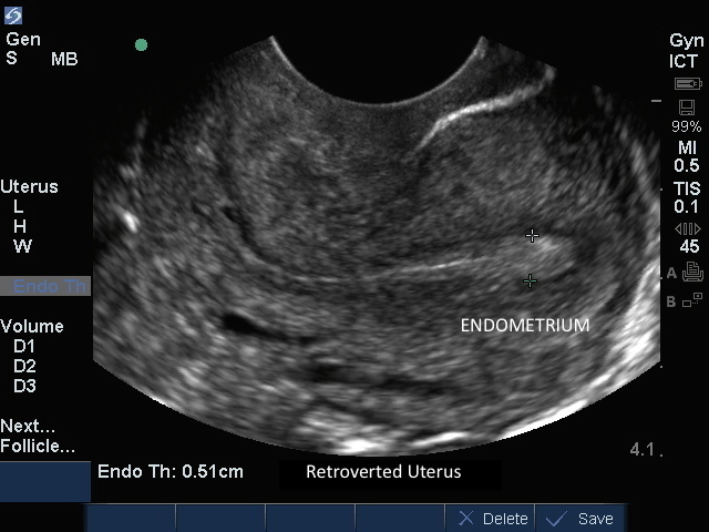
Endovag Uterus: Endometrium Measurement Endovag Uterus: Endometrium Measurement Read more about Endovag Uterus: Endometrium Measurement /sites/default/files/201408_IMAGE_EDGE_ENDOVAG_UTERUS_ENDO_MEASUREMENT.jpg Media Library Type Image Media Library Tag Bleeding Cervix Ectopic Edge Endometrium Fundus Gynocology Image Intrauterine Measurement Normal Transvaginal Uterus