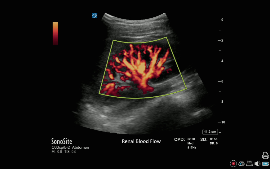
Right Kidney with Color Power Doppler Right Kidney with Color Power Doppler Read more about Right Kidney with Color Power Doppler /sites/default/files/201408_IMAGE_X-PORTE_RIGHT_KIDNEY_COLOR_POWER.jpg Applications Cardiology Media Library Type Image Media Library Tag Blood Flow Calcification Color Doppler Cortex Hydronephrosis Hypertension Image Kidney Kidney Stone Liver Medium Mild Morrison's Pouch Parachema Power Doppler Renal Pelvis Renals Severe Shadow Spleen Hepatorenal Spleenorenal Ureter Urology X-Porte
3D How To: Right Kidney Ultrasound 3D How To: Right Kidney Ultrasound Read more about 3D How To: Right Kidney Ultrasound /sites/default/files/Kidney_R_Disclaimer_edu00466_thumbnail.jpg 3D animation demonstrating a right kidney ultrasound exam. Media Library Type How To Videos Media Library Tag Kidney Renals Cortex Parachema Hydronephrosis Renal Pelvis Urology Ureter Hypertension Spleenorenal Spleen Hepatorenal Morrison's Pouch Mild Severe Medium Liver Kidney Stone Calcification Shadow Subtitles <p begin="00:00:07.000" end="00:00:09.879" style="s2">- [Voiceover] A curved or<br />phased array transducer with an</p> <p begin="00:00:09.879" end="00:00:12.611" style="s2">abdomen exam type is used to perform a</p> <p begin="00:00:12.611" end="00:00:14.407" style="s2">renal ultrasound exam.</p> <p begin="00:00:14.407" end="00:00:17.740" style="s2">The kidney should be<br />evaluated in two planes.</p> <p begin="00:00:17.740" end="00:00:21.151" style="s2">The transducer is placed in<br />a long access orientation</p> <p begin="00:00:21.151" end="00:00:23.459" style="s2">along the right midaxillary line,</p> <p begin="00:00:23.459" end="00:00:24.913" style="s2">at the costal margin,</p> <p begin="00:00:24.913" end="00:00:27.233" style="s2">with the orientation<br />marker directed toward the</p> <p begin="00:00:27.233" end="00:00:28.487" style="s2">patient's head.</p> <p begin="00:00:28.487" end="00:00:30.913" style="s2">If it is difficult to<br />visualize the kidney,</p> <p begin="00:00:30.913" end="00:00:33.981" style="s2">a deep inspiration will move<br />the kidney below the ribs,</p> <p begin="00:00:33.981" end="00:00:36.044" style="s2">for easier access.</p> <p begin="00:00:36.044" end="00:00:39.524" style="s2">It may also be necessary to<br />move the transducer proximally</p> <p begin="00:00:39.524" end="00:00:42.725" style="s2">along the midaxillary line,<br />to visualize the kidneys,</p> <p begin="00:00:42.725" end="00:00:44.939" style="s2">using an intercostal approach.</p> <p begin="00:00:44.939" end="00:00:48.233" style="s2">Using this approach will<br />result in rib shadow artifacts</p> <p begin="00:00:48.233" end="00:00:50.609" style="s2">overlying the ultrasound image.</p> <p begin="00:00:50.609" end="00:00:53.963" style="s2">Once the kidney is identified,<br />the transducer is slightly</p> <p begin="00:00:53.963" end="00:00:58.130" style="s2">rotated to adjust for the oblique<br />orientation of the kidney.</p> <p begin="00:01:04.713" end="00:01:08.435" style="s2">The transducer is swept<br />from anterior to posterior</p> <p begin="00:01:08.435" end="00:01:10.457" style="s2">to image the kidney parenchyma,</p> <p begin="00:01:10.457" end="00:01:14.119" style="s2">which will appear hypoechoic<br />when compared to the liver.</p> <p begin="00:01:14.119" end="00:01:16.892" style="s2">The renal pyramids are<br />triangular in shape,</p> <p begin="00:01:16.892" end="00:01:19.550" style="s2">and darker than the renal cortex.</p> <p begin="00:01:19.550" end="00:01:23.140" style="s2">The renal pelvis will appear<br />as bright, hyperechoic echoes</p> <p begin="00:01:23.140" end="00:01:24.546" style="s2">within the kidney.</p> <p begin="00:01:24.546" end="00:01:27.247" style="s2">To obtain a transverse access view,</p> <p begin="00:01:27.247" end="00:01:29.659" style="s2">rotate the transducer 90 degrees,</p> <p begin="00:01:29.659" end="00:01:31.652" style="s2">from the long access position.</p> <p begin="00:01:31.652" end="00:01:34.098" style="s2">The kidney appear as a circular structure,</p> <p begin="00:01:34.098" end="00:01:35.311" style="s2">with a horseshoe shape,</p> <p begin="00:01:35.311" end="00:01:37.249" style="s2">at the level of the renal pelvis.</p> <p begin="00:01:37.249" end="00:01:40.994" style="s2">The transducer is swept from<br />the superior to inferior poles,</p> <p begin="00:01:40.994" end="00:01:43.494" style="s2">to evaluate the entire kidney.</p> Brightcove ID 5508117922001 https://youtube.com/watch?v=AH8RcdMQMHA