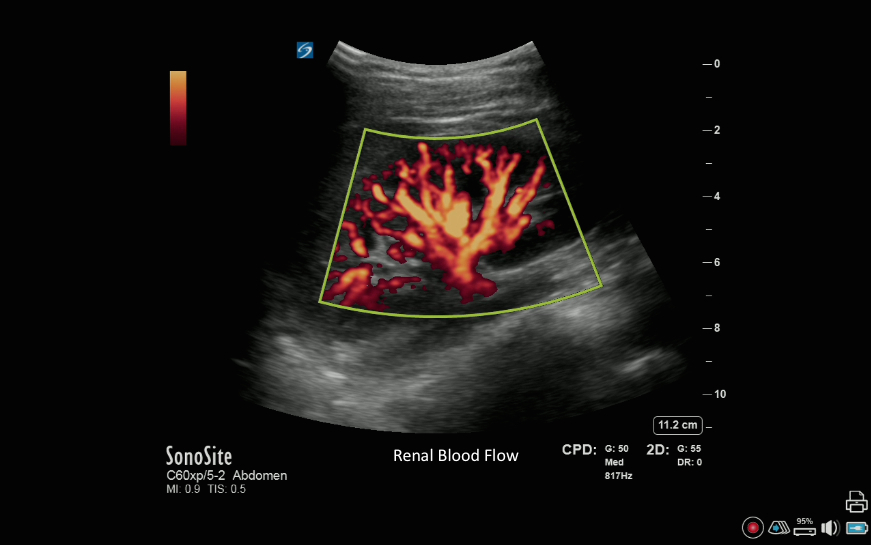
Right Kidney with Color Power Doppler Right Kidney with Color Power Doppler Read more about Right Kidney with Color Power Doppler /sites/default/files/201408_IMAGE_X-PORTE_RIGHT_KIDNEY_COLOR_POWER.jpg Media Library Type Image Media Library Tag Blood Flow Calcification Color Doppler Cortex Hydronephrosis Hypertension Image Kidney Kidney Stone Liver Medium Mild Morrison's Pouch Parachema Power Doppler Renal Pelvis Renals Severe Shadow Spleen Hepatorenal Spleenorenal Ureter Urology X-Porte
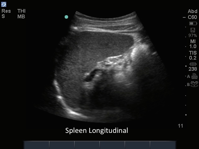
M-Turbo: Abdomen 03 M-Turbo: Abdomen 03 Read more about M-Turbo: Abdomen 03 /sites/default/files/201410_Image_M-Turbo_C60_Spleen_Longitudinal.jpg M-Turbo: Spleen Longitudinal. Media Library Type Image Media Library Tag Abd Abdomen Abdominal Artifacts Ascities Blood Collections Diaphragm Fluid Focused Free Fluid Hypotension Image Kidney Left Upper Quadrant M-Turbo Spleen Spleenorenal Trauma Triage
Liver: Right Kidney Transverse Liver: Right Kidney Transverse Read more about Liver: Right Kidney Transverse /sites/default/files/201408_IMAGE_EDGE_RIGHT_KIDNEY_LIVER_TRANSVERSE.jpg Media Library Type Image Media Library Tag Calcification Cortex Edge Hydronephrosis Hypertension Image Kidney Kidney Stone Medium Parachema Renal Pelvis Renals Servere Shadow Spleen Mild Spleenorenal Ureter Urology