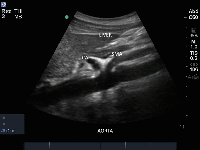
Edge: Aorta Sagital Edge: Aorta Sagital Read more about Edge: Aorta Sagital /sites/default/files/201408_IMAGE_EDGE_AORTA_SAGITAL.jpg Media Library Type Image Media Library Tag Abd Abdomen Abdominal Aneurysm Aorta Arterial Blood Disecting Distal Edge False Lumen Focused Free Fluid Fusiform Hemmorrhage Hypotension Iliacs Image Lumen Mid Proximal Rupture Sacular Soundbytes Cases Thrombus Trauma
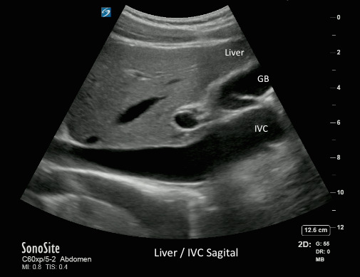
Liver IVC Sagital Liver IVC Sagital Read more about Liver IVC Sagital /sites/default/files/201408_IMAGE_X-PORTE_LIVER_IVC_SAGITAL.jpg Media Library Type Image Media Library Tag Aorta Assessment Blood Pressure Cardiac Cardiac Output Caval Index Central Venous Pressure Collapsibility Index Cvp High Low Dialation Diameter Diaphragm Epigastric Expiration Fluid Heart Failure Inferior Vena Cava Inspiration Jemodynamics Liver M-Mode Preload Responsiveness Respiration Respiratory Variation Right Atrium Size Sniff Spinal Process Spontaneously Breathing Venous Volume Responsiveness Volume Status
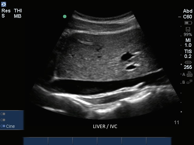
IVC Sagital IVC Sagital Read more about IVC Sagital /sites/default/files/201408_IMAGE_EDGE_IVC_SAGITAL.jpg Media Library Type Image Media Library Tag Aorta Assessment Blood Pressure Cardiac Cardiac Output Caval Index Central Venous Pressure Collapsibility Index Cvp High Low Dialation Diameter Diaphragm Edge Epigastric Expiration Fluid Heart Failure Image Inferior Vena Cava Inspiration Jemodynamics Liver M-Mode Preload Responsiveness Respiration Respiratory Variation Right Atrium Size Sniff Spinal Process Spontaneously Breathing Venous Volume Responsiveness Volume Status
S Series: Proximal Aorta Sagitial View/Diaphraghm S Series: Proximal Aorta Sagitial View/Diaphraghm Read more about S Series: Proximal Aorta Sagitial View/Diaphraghm /sites/default/files/201410_Image_S-System_Prox_Aorta_Sagital_1.jpg S Series: Proximal Aorta Sagitial View/Diaphraghm Clinical Specialties FP/GP Media Library Type Image Media Library Tag S-System Image Aorta Aneurysm Sacular Disecting Fusiform Blood Free Fluid Rupture Lumen Arterial Proximal Mid Distal Iliacs False Lumen Thrombus Focused Trauma Abdominal Abd Abdomen Hypotension Hemmorrhage Body S Series: Proximal Aorta Sagitial View/Diaphraghm