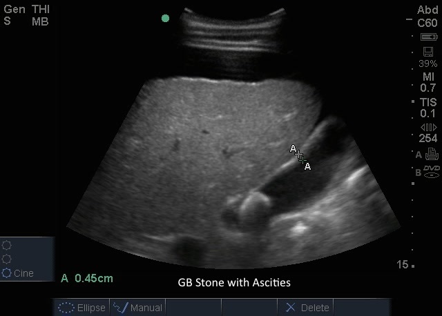
GB Stones GB Stones Read more about GB Stones /sites/default/files/201408_IMAGE_EDGE_GB_STONE_ASCITIES.jpg Media Library Type Image Media Library Tag Abd Abdomen Basic Bile Biliary Cholecystitis Cholelithiasis Disease Edge Gallbladder Gallstones Hepatobillary Image Pain Pathology Right Upper Quadrant Soundbytes Cases Stones
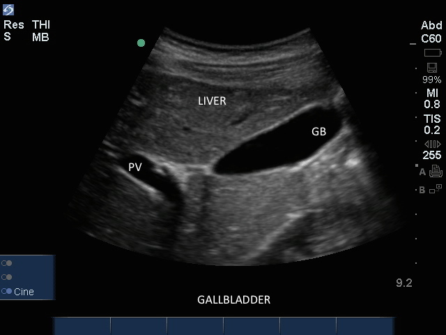
Gallbladder Gallbladder Read more about Gallbladder /sites/default/files/201408_IMAGE_EDGE_GALLBLADDER.jpg Clinical Specialties FP/GP Media Library Type Image Media Library Tag Gallbladder Disease Stones Abdomen Abd Pain Cholecystitis Cholelithiasis Biliary Bile Right Upper Quadrant Basic Pathology Hepatobillary Gallstones Soundbytes Cases Edge Image
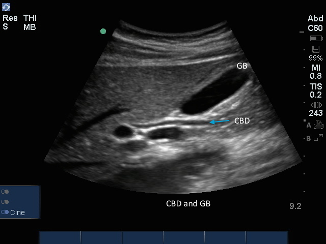
CBD with Gallbladder CBD with Gallbladder Read more about CBD with Gallbladder /sites/default/files/201408_IMAGE_EDGE_CBD_GALLBLADDER.jpg Clinical Specialties FP/GP Media Library Type Image Media Library Tag Gallbladder Disease Stones Abdomen Abd Pain Cholecystitis Cholelithiasis Biliary Bile Right Upper Quadrant Basic Pathology Hepatobillary Gallstones Soundbytes Cases Edge Image Compatible Products Sonosite Edge
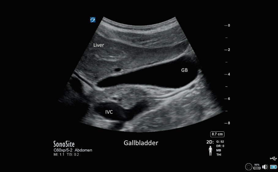
Gallbladder 09 Gallbladder 09 Read more about Gallbladder 09 /sites/default/files/201408_IMAGE_X-PORTE_GALLBLADDER.jpg Clinical Specialties FP/GP Media Library Type Image Media Library Tag Gallbladder Disease Stones Abdomen Abd Pain Cholecystitis Cholelithiasis Biliary Bile Right Upper Quadrant Basic Pathology Hepatobillary Gallstones Image X-Porte Compatible Products Sonosite X-Porte
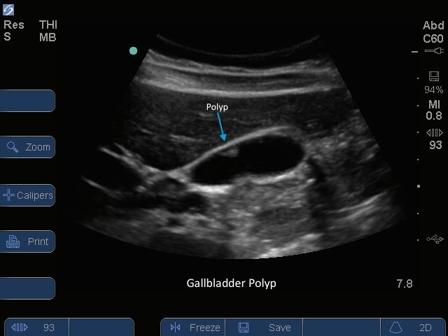
Gallbladder 06 Gallbladder 06 Read more about Gallbladder 06 /sites/default/files/201410_image_S-SysteGallbladder_Polyp_Anterior_Wall.jpg S Series: Gallbladder Polyp Anterior Wall. Media Library Type Image Media Library Tag Abd Abdomen Basic Bile Biliary Cholecystitis Cholelithiasis Disease Gallbladder Gallstones Hepatobillary Image Pain Pathology Right Upper Quadrant S-Series Stones
Gallbladder 08 Gallbladder 08 Read more about Gallbladder 08 /sites/default/files/201410_Image_M-Turbo_Gallbladder_Transverse_View.jpg M-Turbo: Gallbladder Transverse View Clinical Specialties FP/GP Media Library Type Image Media Library Tag Gallbladder Disease Stones Abdomen Abd Pain Cholecystitis Cholelithiasis Biliary Bile Right Upper Quadrant Basic Pathology Hepatobillary Gallstones Image M-Turbo
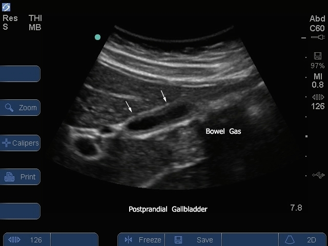
Gallbladder 07 Gallbladder 07 Read more about Gallbladder 07 /sites/default/files/201410_Image_S-System_Gallbladder_Transverse_Post_Prandial.jpg S Series: Gallbladder Transverse Post Prandial. Clinical Specialties FP/GP Media Library Type Image Media Library Tag Gallbladder Disease Stones Abdomen Abd S-System S-Series Pain Cholecystitis Cholelithiasis Biliary Bile Right Upper Quadrant Basic Pathology Hepatobillary Gallstones Image
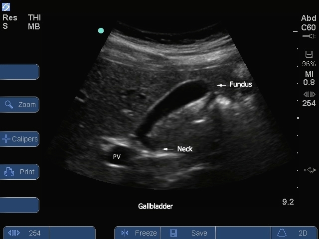
Gallbladder 05 Gallbladder 05 Read more about Gallbladder 05 /sites/default/files/201410_Image_S-System_Gallbladder_Neck_Logitudinal_View.jpg S Series: Gallbladder Neck Longitudinal View. Clinical Specialties FP/GP Media Library Type Image Media Library Tag Gallbladder Disease Stones Abdomen Abd Pain Cholecystitis Cholelithiasis Biliary Bile Right Upper Quadrant Basic Pathology Hepatobillary Gallstones Image S-Series
Gallbladder 04 Gallbladder 04 Read more about Gallbladder 04 /sites/default/files/201410_Image_S_System_Gallbladder_Longitudinal_View_Post_Prandial2.jpg S Series: Gallbladder Longitudinal View Post Prandial. Media Library Type Image Media Library Tag Abd Abdomen Basic Bile Biliary Cholecystitis Cholelithiasis Disease Gallbladder Gallstones Hepatobillary Image Pain Pathology Right Upper Quadrant S-Series Stones