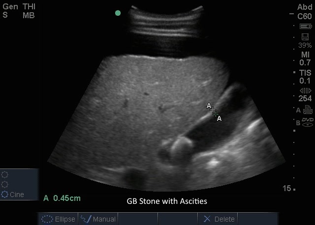
GB Stones GB Stones Read more about GB Stones /sites/default/files/201408_IMAGE_EDGE_GB_STONE_ASCITIES.jpg Media Library Type Image Media Library Tag Abd Abdomen Basic Bile Biliary Cholecystitis Cholelithiasis Disease Edge Gallbladder Gallstones Hepatobillary Image Pain Pathology Right Upper Quadrant Soundbytes Cases Stones
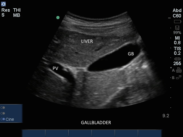
Gallbladder Gallbladder Read more about Gallbladder /sites/default/files/201408_IMAGE_EDGE_GALLBLADDER.jpg Clinical Specialties FP/GP Media Library Type Image Media Library Tag Gallbladder Disease Stones Abdomen Abd Pain Cholecystitis Cholelithiasis Biliary Bile Right Upper Quadrant Basic Pathology Hepatobillary Gallstones Soundbytes Cases Edge Image
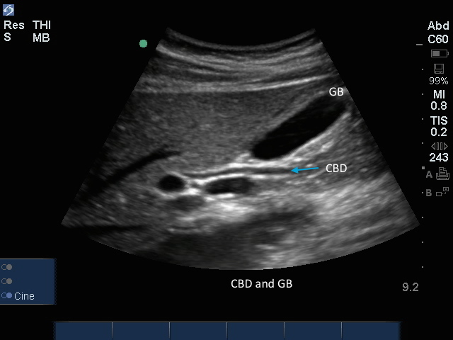
CBD with Gallbladder CBD with Gallbladder Read more about CBD with Gallbladder /sites/default/files/201408_IMAGE_EDGE_CBD_GALLBLADDER.jpg Clinical Specialties FP/GP Media Library Type Image Media Library Tag Gallbladder Disease Stones Abdomen Abd Pain Cholecystitis Cholelithiasis Biliary Bile Right Upper Quadrant Basic Pathology Hepatobillary Gallstones Soundbytes Cases Edge Image Compatible Products Sonosite Edge
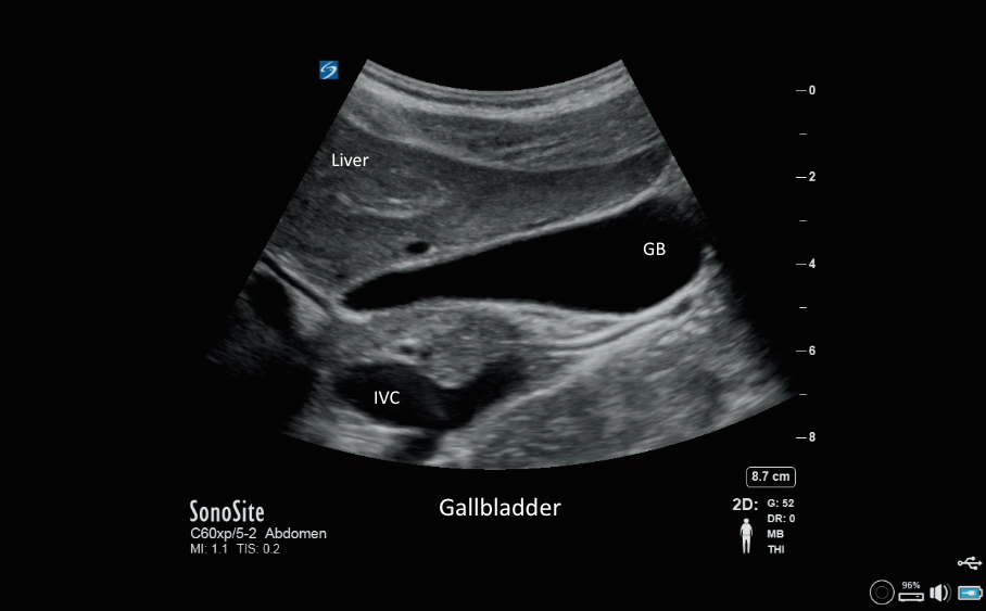
Gallbladder 09 Gallbladder 09 Read more about Gallbladder 09 /sites/default/files/201408_IMAGE_X-PORTE_GALLBLADDER.jpg Clinical Specialties FP/GP Media Library Type Image Media Library Tag Gallbladder Disease Stones Abdomen Abd Pain Cholecystitis Cholelithiasis Biliary Bile Right Upper Quadrant Basic Pathology Hepatobillary Gallstones Image X-Porte Compatible Products Sonosite X-Porte
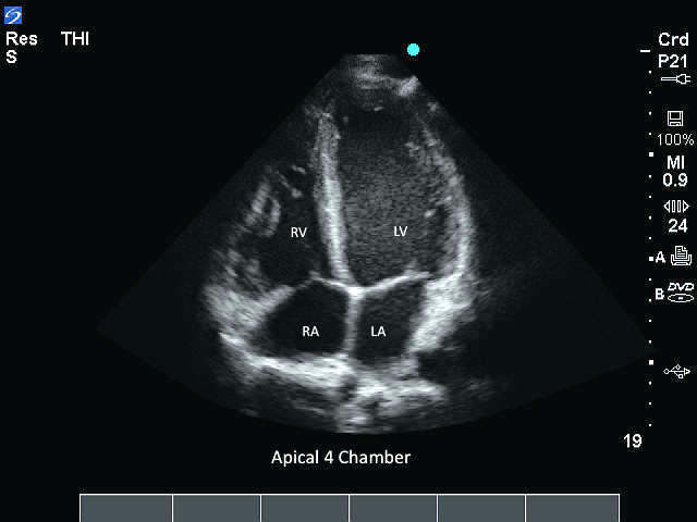
M-Turbo: Apical 4 Chamber M-Turbo: Apical 4 Chamber Read more about M-Turbo: Apical 4 Chamber /sites/default/files/201410_Image_M-Turbo_P21_Apical_4_Chamber.jpg M-Turbo: Apical 4 Chamber. Media Library Type Image Media Library Tag 4 Chamber Aortic Apical Atrium Cardiac Cardiac Output Cardiomyopathy Chronic Heart Disease Congested Heart Disease Ejection Faction Fluid Overload Free Fluid Heart Heart Failure Hypotension Image Left Atrium Left Ventricular Normal Pathology Pericardial Fluid Pericardium Right Ventrical Septal Septum Size Tamponade Trauma Ventrical Ventrical Size Wall Motion Window
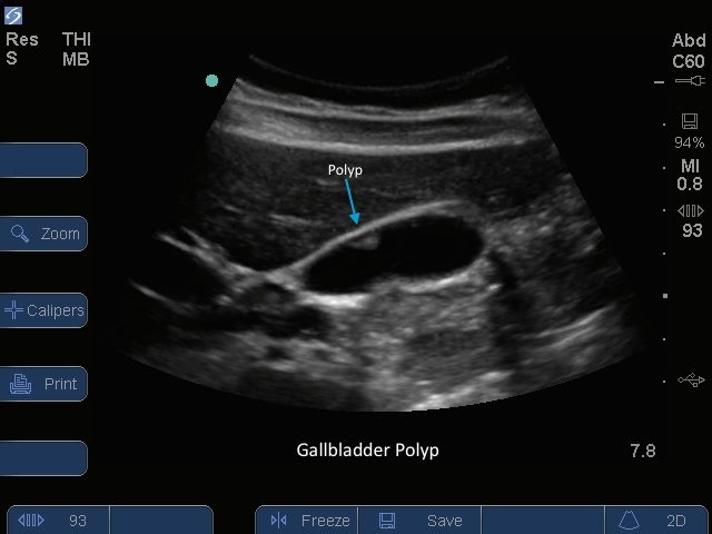
Gallbladder 06 Gallbladder 06 Read more about Gallbladder 06 /sites/default/files/201410_image_S-SysteGallbladder_Polyp_Anterior_Wall.jpg S Series: Gallbladder Polyp Anterior Wall. Media Library Type Image Media Library Tag Abd Abdomen Basic Bile Biliary Cholecystitis Cholelithiasis Disease Gallbladder Gallstones Hepatobillary Image Pain Pathology Right Upper Quadrant S-Series Stones