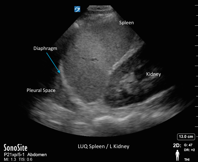
Case: FAST Exam - LUQ Exam

Case: FAST Exam - LUQ Exam

/sites/default/files/Cases_SB_FAST_LUQ_Thumb.jpg
The left upper quadrant (LUQ) ultrasound exam can help clinicians more accurately diagnose intra-abdominal injuries. Topics: infra-diaphragmatic and splenorenal spaces, & how to identify free fluid caused by a trauma.
Media Library Type
Subtitles
<p begin="00:00:17.881" end="00:00:19.619" style="s2">- Hello, my name is Phil Perera,</p>
<p begin="00:00:19.619" end="00:00:21.592" style="s2">and I'm the emergency<br />ultrasound coordinator</p>
<p begin="00:00:21.592" end="00:00:24.440" style="s2">at the New York Presbyterian<br />Hospital in New York City,</p>
<p begin="00:00:24.440" end="00:00:27.107" style="s2">and welcome to SoundBytes Cases.</p>
<p begin="00:00:28.910" end="00:00:30.733" style="s2">In this module, we'll<br />continue our journey,</p>
<p begin="00:00:30.733" end="00:00:33.577" style="s2">looking at the views of<br />the Trauma FAST exam.</p>
<p begin="00:00:33.577" end="00:00:35.597" style="s2">Hopefully you've had a<br />chance to join me prior</p>
<p begin="00:00:35.597" end="00:00:37.538" style="s2">for the views of the right upper quadrant,</p>
<p begin="00:00:37.538" end="00:00:38.558" style="s2">and in this module,</p>
<p begin="00:00:38.558" end="00:00:39.677" style="s2">we're going to look specifically</p>
<p begin="00:00:39.677" end="00:00:42.935" style="s2">at the left upper quadrant<br />views of the Trauma FAST exam,</p>
<p begin="00:00:42.935" end="00:00:44.796" style="s2">known traditionally by two terms,</p>
<p begin="00:00:44.796" end="00:00:48.639" style="s2">the splenorenal, or the perisplenic views.</p>
<p begin="00:00:48.639" end="00:00:49.882" style="s2">In an upcoming module,</p>
<p begin="00:00:49.882" end="00:00:51.864" style="s2">we'll look specifically<br />at the suprapubic view,</p>
<p begin="00:00:51.864" end="00:00:54.831" style="s2">or bladder view, of the Trauma FAST exam.</p>
<p begin="00:00:54.831" end="00:00:56.576" style="s2">There's a lot of information we can gain</p>
<p begin="00:00:56.576" end="00:00:57.948" style="s2">by looking at the left upper quadrant</p>
<p begin="00:00:57.948" end="00:00:59.190" style="s2">in our trauma patients,</p>
<p begin="00:00:59.190" end="00:01:01.294" style="s2">and we'll need to know that<br />it's not a mirror image</p>
<p begin="00:01:01.294" end="00:01:02.845" style="s2">of the right upper quadrant,</p>
<p begin="00:01:02.845" end="00:01:05.245" style="s2">that the spleen offers<br />less of an acoustic window</p>
<p begin="00:01:05.245" end="00:01:07.413" style="s2">onto the left upper<br />quadrant than the liver does</p>
<p begin="00:01:07.413" end="00:01:09.274" style="s2">on the other side.</p>
<p begin="00:01:09.274" end="00:01:10.694" style="s2">Here's a slide reviewing how to perform</p>
<p begin="00:01:10.694" end="00:01:13.546" style="s2">the left upper quadrant view<br />of the Trauma FAST exam.</p>
<p begin="00:01:13.546" end="00:01:15.876" style="s2">As the spleen offers less<br />of an acoustic window</p>
<p begin="00:01:15.876" end="00:01:17.122" style="s2">on the left upper quadrant,</p>
<p begin="00:01:17.122" end="00:01:18.615" style="s2">we need to bring the probe in</p>
<p begin="00:01:18.615" end="00:01:20.409" style="s2">from a more posterior position.</p>
<p begin="00:01:20.409" end="00:01:22.951" style="s2">Thus, the mantra, knuckles to stretcher.</p>
<p begin="00:01:22.951" end="00:01:25.309" style="s2">Optimally, we're using a<br />smaller footprint probe</p>
<p begin="00:01:25.309" end="00:01:26.948" style="s2">that can get in between the ribs</p>
<p begin="00:01:26.948" end="00:01:29.719" style="s2">and get a good view into the<br />left upper quadrant area.</p>
<p begin="00:01:29.719" end="00:01:31.565" style="s2">Position the probe in the long axis view</p>
<p begin="00:01:31.565" end="00:01:34.002" style="s2">with the probe marker<br />towards the patient's head,</p>
<p begin="00:01:34.002" end="00:01:37.718" style="s2">at about the midaxillary line,<br />or posterior axillary line,</p>
<p begin="00:01:37.718" end="00:01:40.798" style="s2">with your knuckles almost<br />touching down to the bedside.</p>
<p begin="00:01:40.798" end="00:01:43.344" style="s2">We'll concentrate on two<br />areas, most importantly,</p>
<p begin="00:01:43.344" end="00:01:46.047" style="s2">the area above the spleen<br />and below the diaphragm,</p>
<p begin="00:01:46.047" end="00:01:48.063" style="s2">where fluid will<br />preferentially accumulate,</p>
<p begin="00:01:48.063" end="00:01:49.232" style="s2">but rounding out our exam,</p>
<p begin="00:01:49.232" end="00:01:52.561" style="s2">we'll look inferior at<br />that spleno-renal space.</p>
<p begin="00:01:52.561" end="00:01:53.524" style="s2">Now that we know how to perform</p>
<p begin="00:01:53.524" end="00:01:56.167" style="s2">the left upper quadrant view<br />of the Trauma FAST exam,</p>
<p begin="00:01:56.167" end="00:01:59.262" style="s2">let's take a look at a<br />normal ultrasound image.</p>
<p begin="00:01:59.262" end="00:02:01.439" style="s2">I have the probe oriented<br />towards the patient's head,</p>
<p begin="00:02:01.439" end="00:02:03.725" style="s2">so superior chest cavity<br />is towards the left,</p>
<p begin="00:02:03.725" end="00:02:06.193" style="s2">inferior abdominal<br />cavity towards the right.</p>
<p begin="00:02:06.193" end="00:02:07.324" style="s2">Notice the spleen,</p>
<p begin="00:02:07.324" end="00:02:09.416" style="s2">the large organ in the<br />middle of the image here,</p>
<p begin="00:02:09.416" end="00:02:11.828" style="s2">and the kidney, the football shaped organ,</p>
<p begin="00:02:11.828" end="00:02:14.536" style="s2">as seen inferior and<br />posterior to the spleen.</p>
<p begin="00:02:14.536" end="00:02:17.212" style="s2">Notice the curving white<br />line just above the spleen,</p>
<p begin="00:02:17.212" end="00:02:19.078" style="s2">which is the diaphragm.</p>
<p begin="00:02:19.078" end="00:02:20.858" style="s2">Recall that in the left upper quadrant,</p>
<p begin="00:02:20.858" end="00:02:23.091" style="s2">that fluid will accumulate preferentially</p>
<p begin="00:02:23.091" end="00:02:25.331" style="s2">in between the spleen and the diaphragm</p>
<p begin="00:02:25.331" end="00:02:30.023" style="s2">and will be a dark or anechoic<br />stripe positioned there.</p>
<p begin="00:02:30.023" end="00:02:31.522" style="s2">Here's another normal video clip</p>
<p begin="00:02:31.522" end="00:02:33.132" style="s2">taken from the left upper quadrant.</p>
<p begin="00:02:33.132" end="00:02:35.566" style="s2">In this case, I'm swinging<br />the probe from inferior,</p>
<p begin="00:02:35.566" end="00:02:37.316" style="s2">looking at the spleno-renal interface,</p>
<p begin="00:02:37.316" end="00:02:41.148" style="s2">to superior, looking at that<br />infra-diaphragmatic space.</p>
<p begin="00:02:41.148" end="00:02:42.576" style="s2">And here, as I freeze the image,</p>
<p begin="00:02:42.576" end="00:02:44.731" style="s2">we see the spleen right in<br />the middle of the image,</p>
<p begin="00:02:44.731" end="00:02:46.894" style="s2">the curving white line<br />making up the diaphragm,</p>
<p begin="00:02:46.894" end="00:02:48.748" style="s2">and notice the thoracic cavity</p>
<p begin="00:02:48.748" end="00:02:51.812" style="s2">as seen just left, or<br />superior, to the diaphragm.</p>
<p begin="00:02:51.812" end="00:02:54.267" style="s2">If the patient had a<br />significant hemothorax,</p>
<p begin="00:02:54.267" end="00:02:56.265" style="s2">or fluid collection in<br />the thoracic cavity,</p>
<p begin="00:02:56.265" end="00:02:57.598" style="s2">that would be represented by</p>
<p begin="00:02:57.598" end="00:02:59.813" style="s2">a dark or anechoic fluid collection</p>
<p begin="00:02:59.813" end="00:03:03.009" style="s2">just above the diaphragm<br />in the thoracic cavity.</p>
<p begin="00:03:03.009" end="00:03:03.934" style="s2">Now that we've had a chance</p>
<p begin="00:03:03.934" end="00:03:05.702" style="s2">to examine several normal video clips</p>
<p begin="00:03:05.702" end="00:03:07.371" style="s2">as taken from the left upper quadrant,</p>
<p begin="00:03:07.371" end="00:03:09.037" style="s2">let's look at a pictorial here,</p>
<p begin="00:03:09.037" end="00:03:12.330" style="s2">showing a positive left<br />upper quadrant FAST exam.</p>
<p begin="00:03:12.330" end="00:03:14.480" style="s2">Here we see superior located to the left,</p>
<p begin="00:03:14.480" end="00:03:15.838" style="s2">inferior to the right.</p>
<p begin="00:03:15.838" end="00:03:17.648" style="s2">We see the spleen in<br />the middle of the image,</p>
<p begin="00:03:17.648" end="00:03:19.370" style="s2">the kidney inferiorly to the right.</p>
<p begin="00:03:19.370" end="00:03:21.579" style="s2">The thoracic cavity with the diaphragm</p>
<p begin="00:03:21.579" end="00:03:24.062" style="s2">to the left of the spleen, or superior.</p>
<p begin="00:03:24.062" end="00:03:25.499" style="s2">We see the area of fresh fluid</p>
<p begin="00:03:25.499" end="00:03:27.517" style="s2">as demarcated by the orange color,</p>
<p begin="00:03:27.517" end="00:03:28.873" style="s2">and notice that it layers out</p>
<p begin="00:03:28.873" end="00:03:30.463" style="s2">predominantly below the diaphragm</p>
<p begin="00:03:30.463" end="00:03:32.675" style="s2">and above the spleen,</p>
<p begin="00:03:32.675" end="00:03:33.928" style="s2">and this is the area where fluid</p>
<p begin="00:03:33.928" end="00:03:36.919" style="s2">will preferentially deposit<br />in the left upper quadrant.</p>
<p begin="00:03:36.919" end="00:03:38.194" style="s2">There are ligaments that sling</p>
<p begin="00:03:38.194" end="00:03:40.207" style="s2">from the diaphragm all<br />the way to the colon</p>
<p begin="00:03:40.207" end="00:03:41.484" style="s2">that prevent the flow of fluid</p>
<p begin="00:03:41.484" end="00:03:43.961" style="s2">into that area between<br />the spleen and the kidney</p>
<p begin="00:03:43.961" end="00:03:45.815" style="s2">until the fluid is relatively large</p>
<p begin="00:03:45.815" end="00:03:48.303" style="s2">within the left upper quadrant.</p>
<p begin="00:03:48.303" end="00:03:50.544" style="s2">So, now let's take a<br />look at a positive exam</p>
<p begin="00:03:50.544" end="00:03:51.885" style="s2">from a trauma patient,</p>
<p begin="00:03:51.885" end="00:03:54.340" style="s2">and we see here the spleen<br />in the middle of the image,</p>
<p begin="00:03:54.340" end="00:03:57.103" style="s2">the kidney inferiorly<br />located to the spleen,</p>
<p begin="00:03:57.103" end="00:03:59.426" style="s2">and notice the large<br />amount of fresh fluid,</p>
<p begin="00:03:59.426" end="00:04:01.376" style="s2">that dark or anechoic fluid collection</p>
<p begin="00:04:01.376" end="00:04:03.160" style="s2">that layers out above the spleen</p>
<p begin="00:04:03.160" end="00:04:05.273" style="s2">in the infra-diaphragmatic location,</p>
<p begin="00:04:05.273" end="00:04:08.044" style="s2">and anterior to the spleen.</p>
<p begin="00:04:08.044" end="00:04:10.206" style="s2">This indicates a large<br />amount of fresh blood</p>
<p begin="00:04:10.206" end="00:04:11.589" style="s2">in the left upper quadrant,</p>
<p begin="00:04:11.589" end="00:04:12.905" style="s2">and we also see a blood clot,</p>
<p begin="00:04:12.905" end="00:04:14.748" style="s2">that echogenic material waving around</p>
<p begin="00:04:14.748" end="00:04:17.152" style="s2">anteriorly to the spleen.</p>
<p begin="00:04:17.152" end="00:04:21.187" style="s2">So a positive exam in a trauma patient.</p>
<p begin="00:04:21.187" end="00:04:23.597" style="s2">Here's another positive<br />left upper quadrant view.</p>
<p begin="00:04:23.597" end="00:04:25.209" style="s2">Notice here, there's a larger amount</p>
<p begin="00:04:25.209" end="00:04:28.026" style="s2">of fresh fluid present<br />on this examination.</p>
<p begin="00:04:28.026" end="00:04:29.952" style="s2">We see the spleen in<br />the middle of the image,</p>
<p begin="00:04:29.952" end="00:04:32.182" style="s2">the kidney inferiorly there to the right,</p>
<p begin="00:04:32.182" end="00:04:33.958" style="s2">and all the dark, fresh fluid,</p>
<p begin="00:04:33.958" end="00:04:37.132" style="s2">as indicated by the dark, or<br />anechoic, fluid collection,</p>
<p begin="00:04:37.132" end="00:04:41.221" style="s2">as seen infra-diaphragmatic<br />and above the spleen.</p>
<p begin="00:04:41.221" end="00:04:43.079" style="s2">Notice again that the fluid is not</p>
<p begin="00:04:43.079" end="00:04:44.817" style="s2">preferentially layering out in between</p>
<p begin="00:04:44.817" end="00:04:47.286" style="s2">the spleen and the kidney,<br />reinforcing the point that</p>
<p begin="00:04:47.286" end="00:04:51.535" style="s2">this is not a mirror image<br />of the right upper quadrant.</p>
<p begin="00:04:51.535" end="00:04:53.377" style="s2">Here's another positive examination</p>
<p begin="00:04:53.377" end="00:04:55.502" style="s2">in a patient who comes in hypotensive</p>
<p begin="00:04:55.502" end="00:04:57.417" style="s2">after being hit by a car.</p>
<p begin="00:04:57.417" end="00:04:58.624" style="s2">Notice I'm swinging the probe</p>
<p begin="00:04:58.624" end="00:05:01.144" style="s2">between the kidney, up superiorly,</p>
<p begin="00:05:01.144" end="00:05:02.417" style="s2">to look at the spleen.</p>
<p begin="00:05:02.417" end="00:05:04.843" style="s2">Notice the absence of<br />fluid in between the spleen</p>
<p begin="00:05:04.843" end="00:05:05.676" style="s2">and the kidney,</p>
<p begin="00:05:05.676" end="00:05:07.081" style="s2">but the presence of free fluid</p>
<p begin="00:05:07.081" end="00:05:09.472" style="s2">right above the spleen<br />and below the diaphragm</p>
<p begin="00:05:09.472" end="00:05:12.270" style="s2">as indicated by that dark stripe.</p>
<p begin="00:05:12.270" end="00:05:14.697" style="s2">Here's an interesting video<br />clip from a trauma patient.</p>
<p begin="00:05:14.697" end="00:05:16.253" style="s2">Again, we're looking at<br />the left upper quadrant,</p>
<p begin="00:05:16.253" end="00:05:18.543" style="s2">and we delineate the spleen and kidney.</p>
<p begin="00:05:18.543" end="00:05:20.645" style="s2">Notice the presence here of fresh fluid,</p>
<p begin="00:05:20.645" end="00:05:22.886" style="s2">the dark or anechoic fluid stripe</p>
<p begin="00:05:22.886" end="00:05:25.168" style="s2">as seen layering out<br />superior, or in anterior,</p>
<p begin="00:05:25.168" end="00:05:26.389" style="s2">to the spleen there,</p>
<p begin="00:05:26.389" end="00:05:28.514" style="s2">but let's look above the diaphragm here,</p>
<p begin="00:05:28.514" end="00:05:30.095" style="s2">which we see as the curving white line</p>
<p begin="00:05:30.095" end="00:05:32.363" style="s2">moving up and down as<br />the patient breathes,</p>
<p begin="00:05:32.363" end="00:05:33.740" style="s2">and what we notice here is the presence</p>
<p begin="00:05:33.740" end="00:05:37.430" style="s2">of a dark fluid collection<br />within the thoracic compartment.</p>
<p begin="00:05:37.430" end="00:05:39.256" style="s2">So we're able to diagnose in this patient</p>
<p begin="00:05:39.256" end="00:05:41.010" style="s2">an associated hemothorax</p>
<p begin="00:05:41.010" end="00:05:44.214" style="s2">in addition to the hemoperitoneum.</p>
<p begin="00:05:44.214" end="00:05:45.906" style="s2">So the left upper quadrant view</p>
<p begin="00:05:45.906" end="00:05:48.838" style="s2">also helpful for looking<br />into the thoracic compartment</p>
<p begin="00:05:48.838" end="00:05:52.481" style="s2">as well as diagnosing<br />interabdominal injury.</p>
<p begin="00:05:52.481" end="00:05:53.598" style="s2">One maneuver that can help you</p>
<p begin="00:05:53.598" end="00:05:56.025" style="s2">uncover fresh fluid within<br />the left upper quadrant</p>
<p begin="00:05:56.025" end="00:05:57.743" style="s2">is to have the patient take a deep breath</p>
<p begin="00:05:57.743" end="00:06:00.124" style="s2">and analyze that infra-diaphragmatic space</p>
<p begin="00:06:00.124" end="00:06:03.200" style="s2">as the diaphragm moves<br />upward, off of the spleen.</p>
<p begin="00:06:03.200" end="00:06:05.286" style="s2">Notice here that we uncovered the amount</p>
<p begin="00:06:05.286" end="00:06:07.906" style="s2">of fresh fluid that's present<br />right above the spleen</p>
<p begin="00:06:07.906" end="00:06:09.708" style="s2">and below the diaphragm as the patient</p>
<p begin="00:06:09.708" end="00:06:10.794" style="s2">takes a deep breath,</p>
<p begin="00:06:10.794" end="00:06:13.494" style="s2">and that diaphragm moves superiorly.</p>
<p begin="00:06:13.494" end="00:06:15.224" style="s2">So, in conclusion, I'm<br />glad I could share with you</p>
<p begin="00:06:15.224" end="00:06:17.841" style="s2">this SoundBytes module, going<br />over the trauma FAST exam,</p>
<p begin="00:06:17.841" end="00:06:21.296" style="s2">specifically the left upper<br />quadrant, or perisplenic view.</p>
<p begin="00:06:21.296" end="00:06:23.071" style="s2">There's a great deal of<br />information that we can gain</p>
<p begin="00:06:23.071" end="00:06:24.748" style="s2">by looking into the left upper quadrant</p>
<p begin="00:06:24.748" end="00:06:25.925" style="s2">in our trauma patients,</p>
<p begin="00:06:25.925" end="00:06:27.744" style="s2">and it's important to realize that</p>
<p begin="00:06:27.744" end="00:06:29.795" style="s2">the left upper quadrant<br />is not a mirror view</p>
<p begin="00:06:29.795" end="00:06:31.145" style="s2">of the right upper quadrant,</p>
<p begin="00:06:31.145" end="00:06:33.070" style="s2">and that fluid will<br />preferentially layer out</p>
<p begin="00:06:33.070" end="00:06:36.608" style="s2">in the area above the spleen<br />and below the diaphragm,</p>
<p begin="00:06:36.608" end="00:06:38.726" style="s2">in contrast to the hepatorenal space</p>
<p begin="00:06:38.726" end="00:06:41.050" style="s2">on the right upper quadrant.</p>
<p begin="00:06:41.050" end="00:06:43.555" style="s2">So, I hope to see you back<br />as SoundBytes continues</p>
<p begin="00:06:43.555" end="00:06:45.551" style="s2">and we move on to look at the suprapubic,</p>
<p begin="00:06:45.551" end="00:06:48.968" style="s2">or bladder view, of the Trauma FAST exam.</p>
Brightcove ID
5726805223001
https://youtube.com/watch?v=VBHCmw8iHCc

Case: FAST RUQ Exam - Hemorrhage

Case: FAST RUQ Exam - Hemorrhage

/sites/default/files/Cases_SB_FAST_RUQ2_Thumb.jpg
This video (part 2 of 2) focuses on positive findings found during right upper quadrant (RUQ) ultrasound examinations; it also includes instructions for performing the trauma FAST RUQ-hepatorenal ultrasound exams
Media Library Type
Subtitles
<p begin="00:00:18.274" end="00:00:19.681" style="s2">- Hello, my name is Phil Perera</p>
<p begin="00:00:19.681" end="00:00:21.698" style="s2">and I'm the Emergency<br />Ultrasound Coordinator</p>
<p begin="00:00:21.698" end="00:00:24.668" style="s2">at the New York Presbyterian<br />Hospital in New York City</p>
<p begin="00:00:24.668" end="00:00:27.335" style="s2">and welcome to SoundBytes Cases.</p>
<p begin="00:00:28.292" end="00:00:29.381" style="s2">In this module entitled,</p>
<p begin="00:00:29.381" end="00:00:31.324" style="s2">Part Two of the Right Upper Quadrant View</p>
<p begin="00:00:31.324" end="00:00:33.018" style="s2">of the Trauma Fast Exam,</p>
<p begin="00:00:33.018" end="00:00:35.829" style="s2">we're going to focus<br />entirely on positive findings</p>
<p begin="00:00:35.829" end="00:00:37.117" style="s2">from this view.</p>
<p begin="00:00:37.117" end="00:00:38.549" style="s2">Hopefully you've had a<br />chance to join me prior</p>
<p begin="00:00:38.549" end="00:00:40.975" style="s2">for Part One of the<br />Right Upper Quadrant View</p>
<p begin="00:00:40.975" end="00:00:43.605" style="s2">of the Trauma Fast Exam<br />where we focused entirely</p>
<p begin="00:00:43.605" end="00:00:46.967" style="s2">on normal findings from<br />the right upper quadrant.</p>
<p begin="00:00:46.967" end="00:00:48.796" style="s2">And hopefully have a<br />chance to join me back</p>
<p begin="00:00:48.796" end="00:00:50.346" style="s2">as we go through the left upper quadrant</p>
<p begin="00:00:50.346" end="00:00:53.836" style="s2">and suprapubic views of<br />the Trauma Fast Exam.</p>
<p begin="00:00:53.836" end="00:00:56.135" style="s2">So let's begin this<br />module by going through a</p>
<p begin="00:00:56.135" end="00:00:57.736" style="s2">review of how to perform</p>
<p begin="00:00:57.736" end="00:01:01.200" style="s2">the right upper quadrant<br />view of the Trauma Fast Exam.</p>
<p begin="00:01:01.200" end="00:01:02.853" style="s2">Remember that the probe<br />is positioned in the</p>
<p begin="00:01:02.853" end="00:01:05.887" style="s2">long axis configuration with<br />the marker dot superiorally</p>
<p begin="00:01:05.887" end="00:01:08.204" style="s2">and that we're going to place<br />the probe in between the ribs</p>
<p begin="00:01:08.204" end="00:01:10.836" style="s2">at about the anterior axillary line.</p>
<p begin="00:01:10.836" end="00:01:12.922" style="s2">Here we'll want to<br />focus on the three areas</p>
<p begin="00:01:12.922" end="00:01:15.338" style="s2">as I emphasized in Part<br />One of this module.</p>
<p begin="00:01:15.338" end="00:01:17.987" style="s2">Number one, the infra-diaphragmatic space</p>
<p begin="00:01:17.987" end="00:01:19.388" style="s2">looking at that region<br />just below the diaphragm</p>
<p begin="00:01:19.388" end="00:01:21.619" style="s2">and above the liver,</p>
<p begin="00:01:21.619" end="00:01:25.422" style="s2">and actually looking into the<br />chest cavity for a hemothorax.</p>
<p begin="00:01:25.422" end="00:01:28.084" style="s2">Position two, which is the<br />traditional Morison's Pouch</p>
<p begin="00:01:28.084" end="00:01:29.756" style="s2">or hepatorenal interface.</p>
<p begin="00:01:29.756" end="00:01:31.793" style="s2">And position three, to round out the exam</p>
<p begin="00:01:31.793" end="00:01:34.033" style="s2">looking at the caudal<br />liver tip representing the</p>
<p begin="00:01:34.033" end="00:01:37.306" style="s2">beginning of the right paracolic gutter.</p>
<p begin="00:01:37.306" end="00:01:39.680" style="s2">Here's a nice illustration<br />showing a positive examination</p>
<p begin="00:01:39.680" end="00:01:43.088" style="s2">from the right upper quadrant<br />view of the Trauma Fast Exam.</p>
<p begin="00:01:43.088" end="00:01:45.365" style="s2">We see the liver smack in<br />the middle of the image</p>
<p begin="00:01:45.365" end="00:01:48.981" style="s2">and the kidney slightly posterior<br />and inferior to the liver.</p>
<p begin="00:01:48.981" end="00:01:51.533" style="s2">Let's look at the preferential<br />deposition of fluid</p>
<p begin="00:01:51.533" end="00:01:53.081" style="s2">as shown here in the orange color,</p>
<p begin="00:01:53.081" end="00:01:55.156" style="s2">and we see that it layers<br />out predominantly around</p>
<p begin="00:01:55.156" end="00:01:57.231" style="s2">the caudal tip of the<br />liver as shown towards the</p>
<p begin="00:01:57.231" end="00:01:59.674" style="s2">inferior edge of the image here.</p>
<p begin="00:01:59.674" end="00:02:02.437" style="s2">Notice that it flows from<br />the caudal tip of the liver</p>
<p begin="00:02:02.437" end="00:02:05.439" style="s2">into that potential space<br />which is Morison's Pouch</p>
<p begin="00:02:05.439" end="00:02:07.817" style="s2">in between the liver and the kidney.</p>
<p begin="00:02:07.817" end="00:02:10.397" style="s2">Now this is very important<br />to emphasize as many times,</p>
<p begin="00:02:10.397" end="00:02:12.487" style="s2">fresh fluid can only be seen around that</p>
<p begin="00:02:12.487" end="00:02:13.823" style="s2">caudal tip of the liver,</p>
<p begin="00:02:13.823" end="00:02:16.881" style="s2">and then it would move into<br />that hepatorenal interface</p>
<p begin="00:02:16.881" end="00:02:18.752" style="s2">or Morison's Pouch.</p>
<p begin="00:02:18.752" end="00:02:21.083" style="s2">Here's a positive ultrasound<br />examination from a patient</p>
<p begin="00:02:21.083" end="00:02:23.553" style="s2">who was hit by a car<br />while crossing the street</p>
<p begin="00:02:23.553" end="00:02:24.947" style="s2">and we see here the liver</p>
<p begin="00:02:24.947" end="00:02:26.823" style="s2">smack in the middle of the image here.</p>
<p begin="00:02:26.823" end="00:02:29.772" style="s2">Notice the kidney located<br />inferior and posterior</p>
<p begin="00:02:29.772" end="00:02:30.794" style="s2">to the liver.</p>
<p begin="00:02:30.794" end="00:02:32.597" style="s2">And in between the liver<br />and the kidney in that</p>
<p begin="00:02:32.597" end="00:02:34.966" style="s2">potential space known as Morison's Pouch,</p>
<p begin="00:02:34.966" end="00:02:37.712" style="s2">we see a dark or anechoic<br />fluid collection.</p>
<p begin="00:02:37.712" end="00:02:38.904" style="s2">Not insignificant,</p>
<p begin="00:02:38.904" end="00:02:41.351" style="s2">actually a pretty large<br />stripe in this patient.</p>
<p begin="00:02:41.351" end="00:02:44.143" style="s2">This represents the positive examination,</p>
<p begin="00:02:44.143" end="00:02:45.618" style="s2">or the presence of fresh fluid,</p>
<p begin="00:02:45.618" end="00:02:48.786" style="s2">in this case blood within Morison's Pouch.</p>
<p begin="00:02:48.786" end="00:02:50.717" style="s2">And notice that it layers out in between</p>
<p begin="00:02:50.717" end="00:02:52.851" style="s2">that liver and kidney.</p>
<p begin="00:02:52.851" end="00:02:55.158" style="s2">To emphasize the point that a<br />complete right upper quadrant</p>
<p begin="00:02:55.158" end="00:02:57.687" style="s2">view includes looking at<br />the caudal tip of the liver,</p>
<p begin="00:02:57.687" end="00:03:00.905" style="s2">let's examine this video clip<br />from another trauma patient.</p>
<p begin="00:03:00.905" end="00:03:02.994" style="s2">If we look at the<br />hepatorenal interface there,</p>
<p begin="00:03:02.994" end="00:03:05.333" style="s2">we fail to see the presence of any fluid,</p>
<p begin="00:03:05.333" end="00:03:07.439" style="s2">but as we move the probe inferiorially,</p>
<p begin="00:03:07.439" end="00:03:10.530" style="s2">we see that the caudal tip of<br />the liver as shown right there</p>
<p begin="00:03:10.530" end="00:03:12.643" style="s2">is well outlined by fluid.</p>
<p begin="00:03:12.643" end="00:03:15.267" style="s2">And here we see the<br />inferior pole of the kidney,</p>
<p begin="00:03:15.267" end="00:03:18.300" style="s2">the caudal tip of the liver<br />extending above the kidney,</p>
<p begin="00:03:18.300" end="00:03:19.688" style="s2">and notice all the fresh fluid</p>
<p begin="00:03:19.688" end="00:03:23.230" style="s2">both anterior and posterior to<br />the caudal tip of the liver.</p>
<p begin="00:03:23.230" end="00:03:25.335" style="s2">Making the point that this<br />is a very important area</p>
<p begin="00:03:25.335" end="00:03:28.583" style="s2">to look for in the right<br />upper quadrant exam.</p>
<p begin="00:03:28.583" end="00:03:30.565" style="s2">Here's a video clip from<br />a patient who was stabbed</p>
<p begin="00:03:30.565" end="00:03:33.680" style="s2">with a really big knife<br />to the epigastric region.</p>
<p begin="00:03:33.680" end="00:03:35.362" style="s2">And as we look at the<br />right upper quadrant,</p>
<p begin="00:03:35.362" end="00:03:38.070" style="s2">we can actually see a<br />linear liver laceration</p>
<p begin="00:03:38.070" end="00:03:41.307" style="s2">extending through the<br />medial aspect of the liver.</p>
<p begin="00:03:41.307" end="00:03:43.222" style="s2">We also appreciate the presence of a large</p>
<p begin="00:03:43.222" end="00:03:44.441" style="s2">amount of fresh fluid.</p>
<p begin="00:03:44.441" end="00:03:46.764" style="s2">That dark or anechoic fluid collection</p>
<p begin="00:03:46.764" end="00:03:48.893" style="s2">layering out posteriorally<br />there to the liver</p>
<p begin="00:03:48.893" end="00:03:51.027" style="s2">and extending into Morison's Pouch,</p>
<p begin="00:03:51.027" end="00:03:53.194" style="s2">the hepatorenal interface.</p>
<p begin="00:03:54.048" end="00:03:56.660" style="s2">So as this patent had<br />unstable hemodynamics,</p>
<p begin="00:03:56.660" end="00:03:59.865" style="s2">the next move was to package<br />for the operating room.</p>
<p begin="00:03:59.865" end="00:04:02.804" style="s2">And here we can actually see<br />the caudal tip of the liver</p>
<p begin="00:04:02.804" end="00:04:04.937" style="s2">waving around in all the fresh fluid,</p>
<p begin="00:04:04.937" end="00:04:06.466" style="s2">in this case blood.</p>
<p begin="00:04:06.466" end="00:04:08.332" style="s2">And I obtained this<br />view by moving the probe</p>
<p begin="00:04:08.332" end="00:04:11.699" style="s2">one intercostal space<br />inferior from the last view</p>
<p begin="00:04:11.699" end="00:04:14.210" style="s2">to well delineate the caudal tip of liver</p>
<p begin="00:04:14.210" end="00:04:17.735" style="s2">which we can see here moving<br />around in all the fresh fluid.</p>
<p begin="00:04:17.735" end="00:04:20.328" style="s2">In the last few video clips<br />the appearance of fresh fluid</p>
<p begin="00:04:20.328" end="00:04:23.161" style="s2">on bedside sonography<br />was darker, anechoic.</p>
<p begin="00:04:23.161" end="00:04:26.028" style="s2">However as blood clots it<br />can take on a more echogenic</p>
<p begin="00:04:26.028" end="00:04:27.586" style="s2">or lighter appearance.</p>
<p begin="00:04:27.586" end="00:04:29.431" style="s2">In this video clip a patient was stabbed</p>
<p begin="00:04:29.431" end="00:04:31.383" style="s2">and had a significant<br />down time before being</p>
<p begin="00:04:31.383" end="00:04:33.156" style="s2">brought to the Emergency Department.</p>
<p begin="00:04:33.156" end="00:04:35.099" style="s2">As we look at the hepatorenal interface,</p>
<p begin="00:04:35.099" end="00:04:38.357" style="s2">we see a large amount<br />of clotted blood there</p>
<p begin="00:04:38.357" end="00:04:39.651" style="s2">made up by that echogenic</p>
<p begin="00:04:39.651" end="00:04:41.668" style="s2">or lighter appearance of the blood there.</p>
<p begin="00:04:41.668" end="00:04:44.129" style="s2">Notice that the large<br />amount of blood extends into</p>
<p begin="00:04:44.129" end="00:04:47.615" style="s2">the right paracolic gutter<br />as we scan inferiorially.</p>
<p begin="00:04:47.615" end="00:04:48.950" style="s2">And here we can see the liver,</p>
<p begin="00:04:48.950" end="00:04:49.946" style="s2">the kidney,</p>
<p begin="00:04:49.946" end="00:04:51.079" style="s2">and the clotted blood,</p>
<p begin="00:04:51.079" end="00:04:54.524" style="s2">that more echogenic or lighter<br />blood in Morison's Pouch.</p>
<p begin="00:04:54.524" end="00:04:55.421" style="s2">So in conclusion,</p>
<p begin="00:04:55.421" end="00:04:57.889" style="s2">I'm glad I could share with<br />you this Part Two Module</p>
<p begin="00:04:57.889" end="00:05:01.202" style="s2">on the Right Upper Quadrant<br />View of the Trauma Fast Exam.</p>
<p begin="00:05:01.202" end="00:05:02.767" style="s2">I hope now you have a better understanding</p>
<p begin="00:05:02.767" end="00:05:04.762" style="s2">on how to perform a complete exam</p>
<p begin="00:05:04.762" end="00:05:07.234" style="s2">looking into the three<br />areas as we discussed.</p>
<p begin="00:05:07.234" end="00:05:08.530" style="s2">Infradiaphragmatic,</p>
<p begin="00:05:08.530" end="00:05:09.651" style="s2">Morison's Pouch,</p>
<p begin="00:05:09.651" end="00:05:12.246" style="s2">and inferior tip of the liver.</p>
<p begin="00:05:12.246" end="00:05:13.986" style="s2">And hopefully now you'll<br />be able to discern</p>
<p begin="00:05:13.986" end="00:05:16.560" style="s2">the presence of fresh<br />blood versus clotted blood</p>
<p begin="00:05:16.560" end="00:05:18.445" style="s2">on bedside sonography.</p>
<p begin="00:05:18.445" end="00:05:21.397" style="s2">So I hope to see you back<br />as Sono access continues</p>
<p begin="00:05:21.397" end="00:05:24.553" style="s2">and as we move further<br />through the other views of the</p>
<p begin="00:05:24.553" end="00:05:25.970" style="s2">Trauma Fast Exam.</p>
Brightcove ID
5508109915001
https://youtube.com/watch?v=Gj5IioG7SyM