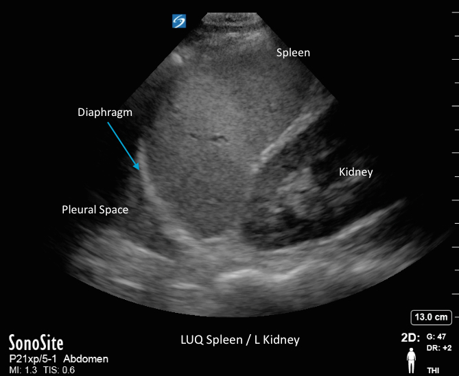
LUQ Spleen/L Kidney LUQ Spleen/L Kidney Read more about LUQ Spleen/L Kidney /sites/default/files/201408_IMAGE_X-PORTE_SPLEEN_LUQ.jpg Media Library Type Image Media Library Tag Abdomen Abdominal Ascities Blood Diaphragm Fluid Fluid Collection Focused Free Fluid Hemorragic Hemorrhage Hypotension Image Kidney Left Upper Quadrant Soundbytes Cases Spleen Trauma Triage X-Porte
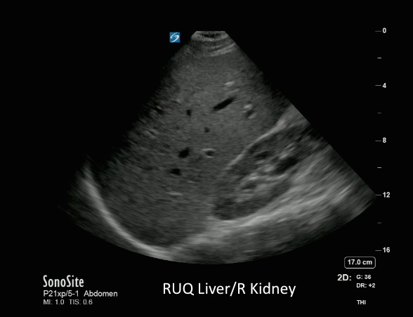
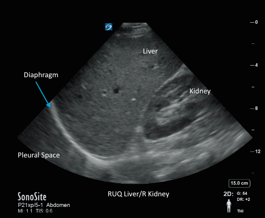
RUQ Liver/R Kidney1 RUQ Liver/R Kidney1 Read more about RUQ Liver/R Kidney1 /sites/default/files/201408_IMAGE_X-PORTE_RUQ_LIVER_R_KIDNEY.jpg Media Library Type Image Media Library Tag Abdomen Abdominal Ascities Blood Collections Diaphragm Fluid Focused Free Fluid Hemorragic Hypotension Image Kidney Liver Morison's Pouch Right Upper Quadrant Trauma Triage X-Porte
RUQ Liver/R Kidney RUQ Liver/R Kidney Read more about RUQ Liver/R Kidney /sites/default/files/201408_IMAGE_X-PORTE_LIVER_RT_KIDNEY_RUQ.jpg Media Library Type Image Media Library Tag Abdomen Abdominal Ascities Blood Collections Diaphragm Fluid Focused Free Fluid Hemorragic Hypotension Image Kidney Liver Morison's Pouch Right Upper Quadrant Trauma Triage X-Porte
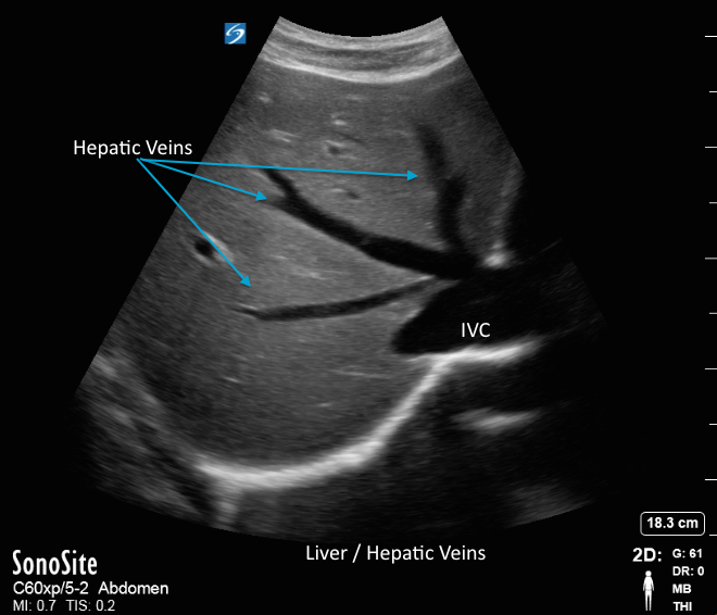
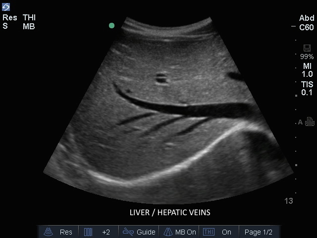
Liver/Hepatic Veins Trv Liver/Hepatic Veins Trv Read more about Liver/Hepatic Veins Trv /sites/default/files/201408_IMAGE_X-PORTE_LIVER_HEPATIC_VEINS.jpg Media Library Type Image Media Library Tag Abdomen Abdominal Ascities Blood Collections Diaphragm Fluid Focused Free Fluid Hemorragic Hypotension Image Kidney Liver Morison's Pouch Right Upper Quadrant Trauma Triage X-Porte Compatible Products Sonosite X-Porte
Liver: Hepatic Veins Liver: Hepatic Veins Read more about Liver: Hepatic Veins /sites/default/files/201408_IMAGE_EDGE_LIVER_HEPATIC_VEINS.jpg Clinical Specialties FP/GP Media Library Type Image Media Library Tag Abdominal Abdomen Trauma Focused Blood Triage Right Upper Quadrant Collections Ascities Liver Diaphragm Hypotension Hemorragic Hepatic Veins Fluid Responsiviness Increased Central Venous Pressure Image Edge Compatible Products Sonosite Edge
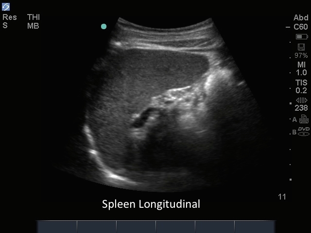
M-Turbo: Abdomen 03 M-Turbo: Abdomen 03 Read more about M-Turbo: Abdomen 03 /sites/default/files/201410_Image_M-Turbo_C60_Spleen_Longitudinal.jpg M-Turbo: Spleen Longitudinal. Media Library Type Image Media Library Tag Abd Abdomen Abdominal Artifacts Ascities Blood Collections Diaphragm Fluid Focused Free Fluid Hypotension Image Kidney Left Upper Quadrant M-Turbo Spleen Spleenorenal Trauma Triage
3D How To: eFAST RUQ 3D animation demonstrating a Right Upper Quadrant (RUQ) view while performing the eFAST ultrasound exam.