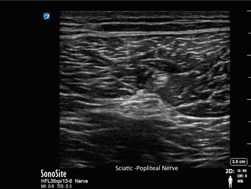
Sciatic-Popliteal Nerve
Sciatic-Popliteal Nerve
/sites/default/files/201408_IMAGE_X-PORTE_SCIATIC_NERVE.jpg
Clinical Specialties
Media Library Type
Media Library Tag


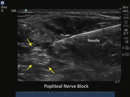
M-Turbo: Popliteal nerve block with Needle.
M-Turbo: Popliteal nerve block with Needle.