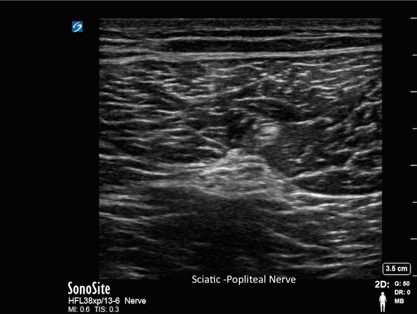
Sciatic-Popliteal Nerve Sciatic-Popliteal Nerve Read more about Sciatic-Popliteal Nerve /sites/default/files/201408_IMAGE_X-PORTE_SCIATIC_NERVE.jpg Clinical Specialties Anesthesiology (Regional) Media Library Type Image Media Library Tag A Anesthesia Anesthesiology Anesthetic Biceps Femoris Block Blockade Common Peroneal Nerve Hyperechoic Image Injection Insertion Knee Lower Extremity Needle Nerve Nerve Bundles Popliteal Popliteal Artery Popliteal Fossa Popliteal Vein Scanning Technique Sciatic Semimembranosus Muscual Semitendinosus Muscle Spread Surgery Tibia Nerve Ultrasound Guidance X-Porte
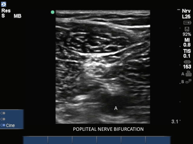
Popliteal Nerve Bifurcation1 Popliteal Nerve Bifurcation1 Read more about Popliteal Nerve Bifurcation1 /sites/default/files/201408_IMAGE_EDGE_POPLITEAL_NERVE_BIFURCATION1.jpg Clinical Specialties Anesthesiology (Regional) Media Library Type Image Media Library Tag A Anesthesia Anesthesiology Anesthetic Biceps Femoris Block Blockade Common Peroneal Nerve Edge Hyperechoic Image Injection Insertion Knee Lower Extremity Needle Nerve Nerve Bundles Popliteal Popliteal Artery Popliteal Fossa Popliteal Vein Scanning Technique Sciatic Sciatic Nerve Semimembranosus Muscual Semitendinosus Muscle Spread Surgery Tibia Nerve Ultrasound Guidance
Popliteal Nerve Bifurcation Popliteal Nerve Bifurcation Read more about Popliteal Nerve Bifurcation /sites/default/files/201408_IMAGE_EDGE_POPLITEAL_NERVE_BIFURCTION_0.jpg Clinical Specialties Anesthesiology (Regional) Media Library Type Image Media Library Tag A Anesthesia Anesthesiology Anesthetic Biceps Femoris Block Blockade Common Peroneal Nerve Edge Hyperechoic Image Injection Insertion Knee Lower Extremity Needle Nerve Nerve Bundles Popliteal Popliteal Artery Popliteal Fossa Popliteal Vein Scanning Technique Sciatic Sciatic Nerve Semimembranosus Muscual Semitendinosus Muscle Spread Surgery Tibia Nerve Ultrasound Guidance
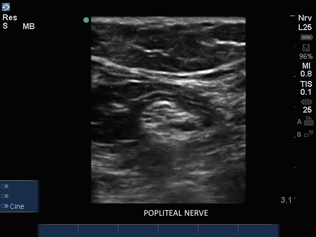
Popliteal Nerve1 Popliteal Nerve1 Read more about Popliteal Nerve1 /sites/default/files/201408_IMAGE_EDGE_SCIATIC_POPLITEAL_NERVE1.jpg Clinical Specialties Anesthesiology (Regional) Media Library Type Image Media Library Tag A Anesthesia Anesthesiology Anesthetic Biceps Femoris Block Blockade Common Peroneal Nerve Edge Hyperechoic Image Injection Insertion Knee Lower Extremity Needle Nerve Nerve Bundles Popliteal Popliteal Artery Popliteal Fossa Popliteal Vein Scanning Technique Sciatic Sciatic Nerve Semimembranosus Muscual Semitendinosus Muscle Spread Surgery Tibia Nerve Ultrasound Guidance
Popliteal Nerve Popliteal Nerve Read more about Popliteal Nerve /sites/default/files/201408_IMAGE_EDGE_SCIATIC_POPLITEAL_NERVE.jpg Clinical Specialties Anesthesiology (Regional) Media Library Type Image Media Library Tag A Anesthesia Anesthesiology Anesthetic Biceps Femoris Block Blockade Common Peroneal Nerve Edge Hyperechoic Image Injection Insertion Knee Lower Extremity Needle Nerve Nerve Bundles Popliteal Popliteal Artery Popliteal Fossa Popliteal Vein Scanning Technique Sciatic Sciatic Nerve Semimembranosus Muscual Semitendinosus Muscle Spread Surgery Tibia Nerve Ultrasound Guidance
3D How To: Popliteal Sciatic Nerve Block 3D animation demonstrating an ultrasound guided Popliteal nerve block.
3D How To: Saphenous Nerve Block 3D animation demonstrating an ultrasound guided saphenous nerve block.