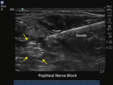
M-Turbo: Screen Layout

M-Turbo: Screen Layout

/sites/default/files/Sonosite_M-Turbo_Screen_Layout_Thumb.jpg
Learn to navigate the screen layout in your Sonosite M-Turbo® Ultrasound System.
Media Library Type
Compatible Products
Subtitles
<p begin="00:00:24.565" end="00:00:27.308" style="s2">- [Voiceover] The screen layout<br />includes the patient header</p>
<p begin="00:00:27.308" end="00:00:28.881" style="s2">at the top of the screen,</p>
<p begin="00:00:28.881" end="00:00:32.457" style="s2">which displays the current<br />patient name, ID number,</p>
<p begin="00:00:32.457" end="00:00:36.124" style="s2">institution, and date<br />and time of the study.</p>
<p begin="00:00:36.966" end="00:00:39.207" style="s2">The symbols displayed on<br />the right of the screen</p>
<p begin="00:00:39.207" end="00:00:42.230" style="s2">provide information about system status.</p>
<p begin="00:00:42.230" end="00:00:45.237" style="s2">There is information on<br />the exam type, transducer,</p>
<p begin="00:00:45.237" end="00:00:49.222" style="s2">AC connected, battery level,<br />and a number indicating</p>
<p begin="00:00:49.222" end="00:00:53.333" style="s2">the amount of internal storage remaining.</p>
<p begin="00:00:53.333" end="00:00:56.789" style="s2">The system meets the AIUM<br />standard by providing</p>
<p begin="00:00:56.789" end="00:01:00.948" style="s2">a continuous display of<br />the mechanical index.</p>
<p begin="00:01:00.948" end="00:01:03.861" style="s2">When the image is frozen,<br />you can select a frame</p>
<p begin="00:01:03.861" end="00:01:08.126" style="s2">by scrolling with the gain<br />knob, touch pad, or arrow keys.</p>
<p begin="00:01:08.126" end="00:01:11.868" style="s2">This number indicates the frame displayed.</p>
<p begin="00:01:11.868" end="00:01:15.088" style="s2">If the A and B shortcut keys<br />have functions assigned,</p>
<p begin="00:01:15.088" end="00:01:18.993" style="s2">an icon for each key<br />will be displayed here.</p>
<p begin="00:01:18.993" end="00:01:22.685" style="s2">If a USB memory stick<br />is in use, a USB icon</p>
<p begin="00:01:22.685" end="00:01:26.268" style="s2">will be displayed to<br />indicate it is active.</p>
<p begin="00:01:27.131" end="00:01:29.739" style="s2">Just to the left of the<br />symbols, the depth marker</p>
<p begin="00:01:29.739" end="00:01:33.970" style="s2">displays marks in 0.5<br />centimeters, 1 centimeter,</p>
<p begin="00:01:33.970" end="00:01:38.433" style="s2">and 5 centimeter increments,<br />depending on the depth.</p>
<p begin="00:01:38.433" end="00:01:42.797" style="s2">The mode data is displayed at<br />the top left of the screen.</p>
<p begin="00:01:42.797" end="00:01:47.349" style="s2">The current image mode<br />information, such as Gen, Res, Pen,</p>
<p begin="00:01:47.349" end="00:01:51.266" style="s2">THI, SonoHD, or SonoMB,<br />will be displayed here.</p>
<p begin="00:01:53.611" end="00:01:57.297" style="s2">When color Doppler is active,<br />the pulse repetition frequency</p>
<p begin="00:01:57.297" end="00:01:58.797" style="s2">will be displayed.</p>
<p begin="00:02:00.610" end="00:02:04.245" style="s2">When pulse Doppler is active,<br />the pulse repetition frequency</p>
<p begin="00:02:04.245" end="00:02:07.169" style="s2">will be displayed and<br />information regarding the angle</p>
<p begin="00:02:07.169" end="00:02:10.167" style="s2">of the ROI box, the sample line angle,</p>
<p begin="00:02:10.167" end="00:02:13.917" style="s2">and the sample volume<br />size will be displayed.</p>
<p begin="00:02:15.193" end="00:02:18.033" style="s2">If a calculation package<br />has been selected,</p>
<p begin="00:02:18.033" end="00:02:20.735" style="s2">the calculation menu<br />and measurement results</p>
<p begin="00:02:20.735" end="00:02:24.995" style="s2">will be displayed to the<br />bottom left of the screen.</p>
<p begin="00:02:24.995" end="00:02:27.999" style="s2">This completes the overview<br />on the system control panel</p>
<p begin="00:02:27.999" end="00:02:30.099" style="s2">and screen layout.</p>
<p begin="00:02:30.099" end="00:02:33.057" style="s2">Now that you are finished with<br />the overview of the system,</p>
<p begin="00:02:33.057" end="00:02:34.890" style="s2">you are ready to scan.</p>
Brightcove ID
5508121173001
https://youtube.com/watch?v=HNRPRrMlg80

M-Turbo: Performing an Ultrasound Exam

M-Turbo: Performing an Ultrasound Exam

/sites/default/files/Sonosite_M-Turbo_Performing_an_Exam_Thumb.jpg
Learn basic system functions for scanning with your Sonosite M-Turbo® Ultrasound System.
Media Library Type
Compatible Products
Subtitles
<p begin="00:00:24.251" end="00:00:26.530" style="s2">- [Voiceover] This sort<br />video will outline the steps</p>
<p begin="00:00:26.530" end="00:00:29.017" style="s2">to optimize the ultrasound system</p>
<p begin="00:00:29.017" end="00:00:31.925" style="s2">to perform an examination.</p>
<p begin="00:00:31.925" end="00:00:35.598" style="s2">To start this study,<br />press the Patient Key.</p>
<p begin="00:00:35.598" end="00:00:38.781" style="s2">Enter the information into<br />the appropriate fields,</p>
<p begin="00:00:38.781" end="00:00:41.491" style="s2">including patient name, ID number,</p>
<p begin="00:00:41.491" end="00:00:44.574" style="s2">date of birth, gender, and exam type.</p>
<p begin="00:00:45.446" end="00:00:47.907" style="s2">When you have completed<br />patient information entry,</p>
<p begin="00:00:47.907" end="00:00:48.824" style="s2">press Done.</p>
<p begin="00:00:50.751" end="00:00:52.899" style="s2">The Exam Type symbol will now indicate</p>
<p begin="00:00:52.899" end="00:00:56.119" style="s2">the exam type that has been selected.</p>
<p begin="00:00:56.119" end="00:00:58.460" style="s2">Use the coupling gel to remove any air</p>
<p begin="00:00:58.460" end="00:01:02.543" style="s2">between the transducer<br />face and the skin surface.</p>
<p begin="00:01:07.149" end="00:01:10.641" style="s2">The system has advanced<br />image optimization technology</p>
<p begin="00:01:10.641" end="00:01:14.005" style="s2">that greatly simplifies user controls.</p>
<p begin="00:01:14.005" end="00:01:17.201" style="s2">Many of the controls such<br />as Tissue Harmonic Imaging,</p>
<p begin="00:01:17.201" end="00:01:18.509" style="s2">SonoMB,</p>
<p begin="00:01:18.509" end="00:01:20.092" style="s2">and SonoHD</p>
<p begin="00:01:20.092" end="00:01:23.867" style="s2">are automatically<br />activated with exam type.</p>
<p begin="00:01:23.867" end="00:01:25.823" style="s2">Therefore only a few controls</p>
<p begin="00:01:25.823" end="00:01:30.233" style="s2">may need to be adjusted to<br />obtain the best image quality.</p>
<p begin="00:01:30.233" end="00:01:32.805" style="s2">This tutorial will<br />review the key functions</p>
<p begin="00:01:32.805" end="00:01:37.164" style="s2">that are used most frequently<br />when optimizing an image.</p>
<p begin="00:01:37.164" end="00:01:40.523" style="s2">Please refer to the user manual<br />for a complete explanation</p>
<p begin="00:01:40.523" end="00:01:44.385" style="s2">of all controls that may be adjusted.</p>
<p begin="00:01:44.385" end="00:01:47.087" style="s2">Once you have located<br />the area of interest,</p>
<p begin="00:01:47.087" end="00:01:50.478" style="s2">adjust the depth of image to<br />center the area of interest</p>
<p begin="00:01:50.478" end="00:01:53.907" style="s2">in the middle of the image display.</p>
<p begin="00:01:53.907" end="00:01:55.655" style="s2">As you adjust the depth,</p>
<p begin="00:01:55.655" end="00:01:57.812" style="s2">the maximum depth number changes</p>
<p begin="00:01:57.812" end="00:02:00.666" style="s2">in the lower right corner of the screen.</p>
<p begin="00:02:00.666" end="00:02:03.376" style="s2">Select to the desired optimization setting</p>
<p begin="00:02:03.376" end="00:02:05.437" style="s2">from the onscreen menu.</p>
<p begin="00:02:05.437" end="00:02:09.056" style="s2">Res provides the best resolution possible.</p>
<p begin="00:02:09.056" end="00:02:13.754" style="s2">Gen provides a balance between<br />resolution and penetration.</p>
<p begin="00:02:13.754" end="00:02:17.337" style="s2">Pen provides the best<br />possible penetration.</p>
<p begin="00:02:18.534" end="00:02:21.777" style="s2">For quick optimization of the<br />gain settings on the system,</p>
<p begin="00:02:21.777" end="00:02:24.244" style="s2">press the Auto Gain key.</p>
<p begin="00:02:24.244" end="00:02:27.694" style="s2">The system will evaluate the<br />image and apply the appropriate</p>
<p begin="00:02:27.694" end="00:02:30.444" style="s2">gain level settings to the image.</p>
<p begin="00:02:31.381" end="00:02:34.433" style="s2">If you would prefer to change<br />the gain settings manually,</p>
<p begin="00:02:34.433" end="00:02:37.960" style="s2">turn the Near, Far, or Gain<br />knobs on the lower left side</p>
<p begin="00:02:37.960" end="00:02:41.224" style="s2">of the control panel<br />to increase or decrease</p>
<p begin="00:02:41.224" end="00:02:43.882" style="s2">the amount of gain<br />applied to the Near field,</p>
<p begin="00:02:43.882" end="00:02:46.549" style="s2">Far field, or the Overall image.</p>
<p begin="00:02:48.272" end="00:02:50.416" style="s2">You may save the image you have frozen</p>
<p begin="00:02:50.416" end="00:02:54.393" style="s2">or review individual<br />frames in the cine buffer.</p>
<p begin="00:02:54.393" end="00:02:56.320" style="s2">The cine buffer can be accessed</p>
<p begin="00:02:56.320" end="00:02:59.173" style="s2">using the touch pad or Gain knob.</p>
<p begin="00:02:59.173" end="00:03:02.058" style="s2">Moving across the touch pad<br />or turning the Gain knob</p>
<p begin="00:03:02.058" end="00:03:06.039" style="s2">to the left reviews<br />images retrospectively.</p>
<p begin="00:03:06.039" end="00:03:10.373" style="s2">And moving to the right<br />reviews images prospectively.</p>
<p begin="00:03:10.373" end="00:03:13.792" style="s2">The frame number will change<br />dynamically as the touch pad</p>
<p begin="00:03:13.792" end="00:03:16.125" style="s2">or Gain knob is manipulated.</p>
<p begin="00:03:17.453" end="00:03:21.206" style="s2">A measurement may be<br />performed on a frozen image.</p>
<p begin="00:03:21.206" end="00:03:25.055" style="s2">Activate calipers by<br />pressing the Caliper key.</p>
<p begin="00:03:25.055" end="00:03:27.298" style="s2">Move the caliper to the point of interest</p>
<p begin="00:03:27.298" end="00:03:29.215" style="s2">by using the touch pad.</p>
<p begin="00:03:30.059" end="00:03:31.982" style="s2">An active measurement will be displayed</p>
<p begin="00:03:31.982" end="00:03:34.768" style="s2">in the bottom left corner.</p>
<p begin="00:03:34.768" end="00:03:36.962" style="s2">To perform a linear measurement,</p>
<p begin="00:03:36.962" end="00:03:39.697" style="s2">place the caliper at the point of interest</p>
<p begin="00:03:39.697" end="00:03:41.801" style="s2">and press the Set key.</p>
<p begin="00:03:41.801" end="00:03:44.392" style="s2">Another caliper will be activated.</p>
<p begin="00:03:44.392" end="00:03:46.657" style="s2">Move the caliper to the point of interest</p>
<p begin="00:03:46.657" end="00:03:49.052" style="s2">to complete the measurement.</p>
<p begin="00:03:49.052" end="00:03:50.736" style="s2">Area measurements may be obtained</p>
<p begin="00:03:50.736" end="00:03:54.486" style="s2">by using the Ellipse or<br />Manual Trace methods.</p>
<p begin="00:03:56.166" end="00:04:00.916" style="s2">To label the image, select<br />the Text key on the keyboard.</p>
<p begin="00:04:00.916" end="00:04:04.177" style="s2">You may select a text label<br />from the annotation lists</p>
<p begin="00:04:04.177" end="00:04:06.844" style="s2">or type the label on the screen.</p>
<p begin="00:04:09.322" end="00:04:11.199" style="s2">When you are ready to store your image,</p>
<p begin="00:04:11.199" end="00:04:15.218" style="s2">select Save to save the image<br />to the internal storage.</p>
<p begin="00:04:15.218" end="00:04:18.544" style="s2">Approximately 64,000 images may be stored</p>
<p begin="00:04:18.544" end="00:04:20.544" style="s2">on the internal storage.</p>
<p begin="00:04:21.400" end="00:04:24.582" style="s2">Press the Freeze key or 2D Mode key</p>
<p begin="00:04:24.582" end="00:04:26.749" style="s2">to return to live imaging.</p>
<p begin="00:04:27.605" end="00:04:30.332" style="s2">Press the Color key to<br />activate Color Doppler</p>
<p begin="00:04:30.332" end="00:04:32.249" style="s2">or Color Power Doppler.</p>
<p begin="00:04:33.881" end="00:04:36.359" style="s2">A region of interest, ROI box,</p>
<p begin="00:04:36.359" end="00:04:39.117" style="s2">will appear in the center of the screen.</p>
<p begin="00:04:39.117" end="00:04:42.364" style="s2">Use the touch pad or arrow<br />keys on the control panel</p>
<p begin="00:04:42.364" end="00:04:46.197" style="s2">to change the position<br />or size of the ROI box.</p>
<p begin="00:04:47.202" end="00:04:49.086" style="s2">Use the Select key to toggle</p>
<p begin="00:04:49.086" end="00:04:53.169" style="s2">between changing position<br />or size of the ROI box.</p>
<p begin="00:04:54.479" end="00:04:56.221" style="s2">The settings for color will be displayed</p>
<p begin="00:04:56.221" end="00:04:59.304" style="s2">in the top left corner of the screen.</p>
<p begin="00:05:01.952" end="00:05:03.837" style="s2">Select the desired sensitivity</p>
<p begin="00:05:03.837" end="00:05:06.693" style="s2">from the onscreen context menu.</p>
<p begin="00:05:06.693" end="00:05:10.134" style="s2">Low optimizes the system<br />for low flow state.</p>
<p begin="00:05:10.134" end="00:05:14.550" style="s2">Med optimizes the system<br />for medium flow states.</p>
<p begin="00:05:14.550" end="00:05:18.467" style="s2">High optimizes the system<br />for high flow states.</p>
<p begin="00:05:19.749" end="00:05:23.012" style="s2">Use the Gain knob to adjust<br />the overall color gain level</p>
<p begin="00:05:23.012" end="00:05:27.178" style="s2">for Color Power Doppler or Color Doppler.</p>
<p begin="00:05:27.178" end="00:05:29.490" style="s2">You may freeze a color image to save</p>
<p begin="00:05:29.490" end="00:05:33.148" style="s2">or review individual<br />frames in the cine buffer.</p>
<p begin="00:05:33.148" end="00:05:35.289" style="s2">Just as in 2D imaging,</p>
<p begin="00:05:35.289" end="00:05:38.337" style="s2">the cine buffer can be<br />accessed using the touch pad,</p>
<p begin="00:05:38.337" end="00:05:40.420" style="s2">Gain knob, or arrow keys.</p>
<p begin="00:05:41.748" end="00:05:45.915" style="s2">Press the 2D or Color key<br />to return to 2D imaging.</p>
<p begin="00:05:47.511" end="00:05:50.467" style="s2">Color Doppler may be<br />used to quickly identify</p>
<p begin="00:05:50.467" end="00:05:54.486" style="s2">where blood flow is<br />located in a structure.</p>
<p begin="00:05:54.486" end="00:05:56.843" style="s2">Pulsed Wave Doppler may be used to detect</p>
<p begin="00:05:56.843" end="00:06:01.377" style="s2">and measure the velocity of<br />blood flow through a vessel.</p>
<p begin="00:06:01.377" end="00:06:03.614" style="s2">To activate Pulsed Wave Doppler,</p>
<p begin="00:06:03.614" end="00:06:05.727" style="s2">press the Doppler key.</p>
<p begin="00:06:05.727" end="00:06:08.578" style="s2">A Doppler sample line will appear.</p>
<p begin="00:06:08.578" end="00:06:10.317" style="s2">Use the touch pad or arrow keys</p>
<p begin="00:06:10.317" end="00:06:13.487" style="s2">to place the sample line<br />in the blood vessel.</p>
<p begin="00:06:13.487" end="00:06:15.349" style="s2">Note that the color ROI box</p>
<p begin="00:06:15.349" end="00:06:18.895" style="s2">is tied to the Doppler sample line.</p>
<p begin="00:06:18.895" end="00:06:20.404" style="s2">Press the Doppler key again</p>
<p begin="00:06:20.404" end="00:06:23.700" style="s2">to activate the Doppler Spectral Trace.</p>
<p begin="00:06:23.700" end="00:06:25.476" style="s2">Press the Update key to toggle</p>
<p begin="00:06:25.476" end="00:06:30.162" style="s2">between the Doppler Spectral<br />Trace and 2D imaging.</p>
<p begin="00:06:30.162" end="00:06:33.422" style="s2">Select Freeze to stop the<br />Doppler Spectral Trace</p>
<p begin="00:06:33.422" end="00:06:37.006" style="s2">when an adequate sample is obtained.</p>
<p begin="00:06:37.006" end="00:06:39.863" style="s2">To review the trace at<br />different points in time,</p>
<p begin="00:06:39.863" end="00:06:42.881" style="s2">use the touch pad,<br />arrow keys, or Gain knob</p>
<p begin="00:06:42.881" end="00:06:45.048" style="s2">to access the cine buffer.</p>
<p begin="00:06:46.365" end="00:06:49.578" style="s2">Measurements may be performed<br />on the Doppler Spectral Trace</p>
<p begin="00:06:49.578" end="00:06:53.495" style="s2">by selecting the Caliper<br />key or Calcs menu key.</p>
<p begin="00:06:54.883" end="00:06:58.810" style="s2">If desired, press the<br />Save key to save the image</p>
<p begin="00:06:58.810" end="00:07:01.757" style="s2">with the measurements displayed.</p>
<p begin="00:07:01.757" end="00:07:03.550" style="s2">Press the Freeze key to return</p>
<p begin="00:07:03.550" end="00:07:05.791" style="s2">to the live Doppler Spectral Trace</p>
<p begin="00:07:05.791" end="00:07:09.791" style="s2">or press 2D Imaging to<br />exit Pulsed Wave Doppler.</p>
<p begin="00:07:11.055" end="00:07:14.036" style="s2">When you have acquired<br />all images for the study,</p>
<p begin="00:07:14.036" end="00:07:17.538" style="s2">press the Patient key and choose End Exam</p>
<p begin="00:07:17.538" end="00:07:19.103" style="s2">from the onscreen menu</p>
<p begin="00:07:19.103" end="00:07:22.353" style="s2">to finalize the exam and end the study.</p>
<p begin="00:07:23.478" end="00:07:26.273" style="s2">This concludes this brief<br />review of imaging modes</p>
<p begin="00:07:26.273" end="00:07:30.870" style="s2">and system controls to<br />optimize the ultrasound image.</p>
<p begin="00:07:30.870" end="00:07:34.103" style="s2">For an extensive review of<br />system features and functions,</p>
<p begin="00:07:34.103" end="00:07:37.436" style="s2">please refer to the system user manual.</p>
Brightcove ID
5508120226001
https://youtube.com/watch?v=S-wojEALpW8

M-Turbo: Image Review and Export

M-Turbo: Image Review and Export

/sites/default/files/Sonosite_M-Turbo_Image_Review_and_Export_Thumb.jpg
Learn how to review and export images on your Sonosite M-Turbo® Ultrasound System.
Media Library Type
Compatible Products
Subtitles
<p begin="00:00:25.464" end="00:00:27.155" style="s2">- [Voiceover] This video<br />will demonstrate how to</p>
<p begin="00:00:27.155" end="00:00:31.157" style="s2">save images in clips, recall<br />these images for review,</p>
<p begin="00:00:31.157" end="00:00:34.657" style="s2">archive, or export images to a USB device.</p>
<p begin="00:00:36.152" end="00:00:38.313" style="s2">Images or clips of the ultrasound study,</p>
<p begin="00:00:38.313" end="00:00:39.819" style="s2">may be obtained and saved</p>
<p begin="00:00:39.819" end="00:00:42.652" style="s2">to internal storage on the system.</p>
<p begin="00:00:43.515" end="00:00:45.145" style="s2">To save a frozen image,</p>
<p begin="00:00:45.145" end="00:00:47.336" style="s2">select the save button.</p>
<p begin="00:00:47.336" end="00:00:50.352" style="s2">Images of the screen<br />display or patient reports</p>
<p begin="00:00:50.352" end="00:00:53.019" style="s2">can be saved to print or export.</p>
<p begin="00:00:55.031" end="00:00:56.261" style="s2">To save a clip,</p>
<p begin="00:00:56.261" end="00:00:58.418" style="s2">press the save clip key.</p>
<p begin="00:00:58.418" end="00:01:02.501" style="s2">A clip length of 2 to 60<br />seconds can be selected.</p>
<p begin="00:01:06.815" end="00:01:09.673" style="s2">To view a report of all<br />patient data and measurements,</p>
<p begin="00:01:09.673" end="00:01:11.689" style="s2">select the report key.</p>
<p begin="00:01:11.689" end="00:01:13.449" style="s2">This information can be saved</p>
<p begin="00:01:13.449" end="00:01:17.898" style="s2">with a patient exam by<br />pressing the save key.</p>
<p begin="00:01:17.898" end="00:01:19.928" style="s2">Each page of the report can be viewed</p>
<p begin="00:01:19.928" end="00:01:23.918" style="s2">by pressing the next key<br />in the on-screen menu.</p>
<p begin="00:01:23.918" end="00:01:26.709" style="s2">Save each page of the report to include</p>
<p begin="00:01:26.709" end="00:01:29.542" style="s2">with the patient exam information.</p>
<p begin="00:01:35.373" end="00:01:38.500" style="s2">Press the review key<br />to view a patient list.</p>
<p begin="00:01:38.500" end="00:01:41.447" style="s2">The patient list displays all exams stored</p>
<p begin="00:01:41.447" end="00:01:44.987" style="s2">on the internal storage<br />of the ultrasound system.</p>
<p begin="00:01:44.987" end="00:01:47.032" style="s2">The list can be displayed alphabetically</p>
<p begin="00:01:47.032" end="00:01:49.385" style="s2">or by the order of archival.</p>
<p begin="00:01:49.385" end="00:01:53.052" style="s2">A to Z, Z to A, or most<br />recent, or earliest.</p>
<p begin="00:01:55.015" end="00:01:56.698" style="s2">The header at the top of the screen</p>
<p begin="00:01:56.698" end="00:02:00.823" style="s2">identifies patient and image<br />information for the study.</p>
<p begin="00:02:00.823" end="00:02:04.010" style="s2">This includes patient name, ID number,</p>
<p begin="00:02:04.010" end="00:02:06.224" style="s2">date and time of the exam,</p>
<p begin="00:02:06.224" end="00:02:09.815" style="s2">the sonographer's<br />initials, and connectivity.</p>
<p begin="00:02:09.815" end="00:02:12.398" style="s2">Completed, suspended, archived.</p>
<p begin="00:02:13.632" end="00:02:15.287" style="s2">To review a patient exam,</p>
<p begin="00:02:15.287" end="00:02:16.900" style="s2">use the up and down arrows</p>
<p begin="00:02:16.900" end="00:02:19.677" style="s2">to select a patient exam from the list.</p>
<p begin="00:02:19.677" end="00:02:23.010" style="s2">The exam highlighted in white is active.</p>
<p begin="00:02:24.136" end="00:02:26.580" style="s2">To select a patient exam to archive,</p>
<p begin="00:02:26.580" end="00:02:28.747" style="s2">check the box on the left.</p>
<p begin="00:02:29.606" end="00:02:31.586" style="s2">To choose additional patients,</p>
<p begin="00:02:31.586" end="00:02:34.963" style="s2">just scroll down and select each exam.</p>
<p begin="00:02:34.963" end="00:02:37.416" style="s2">Notice each patient exam has a check mark</p>
<p begin="00:02:37.416" end="00:02:39.666" style="s2">to indicate it is included.</p>
<p begin="00:02:41.179" end="00:02:43.345" style="s2">Once you have completed the selection,</p>
<p begin="00:02:43.345" end="00:02:45.909" style="s2">select export to send all images</p>
<p begin="00:02:45.909" end="00:02:48.992" style="s2">to the USB device or archival device.</p>
<p begin="00:02:50.026" end="00:02:52.665" style="s2">Up to three USB devices may be connected</p>
<p begin="00:02:52.665" end="00:02:54.975" style="s2">to the system at one time.</p>
<p begin="00:02:54.975" end="00:02:58.975" style="s2">Select the USB device<br />prior to exporting images.</p>
<p begin="00:03:00.347" end="00:03:04.499" style="s2">This is a brief overview of<br />image review and archival.</p>
<p begin="00:03:04.499" end="00:03:06.194" style="s2">For more complete information,</p>
<p begin="00:03:06.194" end="00:03:08.944" style="s2">please refer to the user manual.</p>
Brightcove ID
5508134318001
https://youtube.com/watch?v=ZWCKKi5sQ3Q

How to: FAST Exam: System Set Up

How to: FAST Exam: System Set Up

/sites/default/files/youtube_klqeADRgvkM.jpg
Learn the M-Turbo system startup sequence for the FAST exam.
Media Library Type
Subtitles
<p begin="00:00:15.317" end="00:00:16.610" style="s2">- The FAST exam.</p>
<p begin="00:00:16.610" end="00:00:17.899" style="s2">The FAST stands for</p>
<p begin="00:00:17.899" end="00:00:20.450" style="s2">focus assessment with<br />sonography for trauma.</p>
<p begin="00:00:20.450" end="00:00:23.747" style="s2">It's a classic fore-exam<br />view of the torso.</p>
<p begin="00:00:23.747" end="00:00:25.608" style="s2">There's also what's also described</p>
<p begin="00:00:25.608" end="00:00:28.096" style="s2">as a extended FAST that includes</p>
<p begin="00:00:28.096" end="00:00:30.072" style="s2">a further examination of the lung,</p>
<p begin="00:00:30.072" end="00:00:31.780" style="s2">which we'll take you<br />to in a later segment,</p>
<p begin="00:00:31.780" end="00:00:32.952" style="s2">as well as long bones,</p>
<p begin="00:00:32.952" end="00:00:35.604" style="s2">as well as expanded views of the heart.</p>
<p begin="00:00:35.604" end="00:00:38.274" style="s2">Let's go ahead and start<br />with the basic FAST exam.</p>
<p begin="00:00:38.274" end="00:00:40.856" style="s2">First thing you're going to<br />do, is choose your transducer.</p>
<p begin="00:00:40.856" end="00:00:42.832" style="s2">For the FAST exam you're gonna want to use</p>
<p begin="00:00:42.832" end="00:00:44.601" style="s2">a phased array transducer.</p>
<p begin="00:00:44.601" end="00:00:48.288" style="s2">We've chosen a phased-array<br />transducer for the FAST exam</p>
<p begin="00:00:48.288" end="00:00:50.016" style="s2">for a particular reason.</p>
<p begin="00:00:50.016" end="00:00:52.489" style="s2">The small, tiny footprint<br />of the transducer</p>
<p begin="00:00:52.489" end="00:00:55.409" style="s2">allows us to do great<br />intercostal scanning,</p>
<p begin="00:00:55.409" end="00:00:59.255" style="s2">it also provides great<br />echocardiac graphic examinations</p>
<p begin="00:00:59.255" end="00:01:02.103" style="s2">allowing us to get good<br />views of the full torso.</p>
<p begin="00:01:02.103" end="00:01:05.502" style="s2">Now we're gonna choose the<br />exam type on the machine.</p>
<p begin="00:01:05.502" end="00:01:07.422" style="s2">We'll hit the exam button,</p>
<p begin="00:01:07.422" end="00:01:10.023" style="s2">we'll choose the type<br />examine that we want,</p>
<p begin="00:01:10.023" end="00:01:12.166" style="s2">we'll hit select and we will be ready</p>
<p begin="00:01:12.166" end="00:01:14.327" style="s2">to start the FAST exam.</p>
<p begin="00:01:14.327" end="00:01:15.622" style="s2">Who do you do a FAST exam on?</p>
<p begin="00:01:15.622" end="00:01:18.150" style="s2">Anyone that you're worried<br />about serious internal injury.</p>
<p begin="00:01:18.150" end="00:01:22.297" style="s2">That would be a blunt trauma<br />or a penetrating trauma.</p>
<p begin="00:01:22.297" end="00:01:24.423" style="s2">Now the areas I'm gonna examine carefully</p>
<p begin="00:01:24.423" end="00:01:26.719" style="s2">would be the peritoneal space,</p>
<p begin="00:01:26.719" end="00:01:29.144" style="s2">the pericardial space,<br />and the pleural space.</p>
<p begin="00:01:29.144" end="00:01:31.775" style="s2">And I'm gonna take you<br />through these one-by-one.</p>
<p begin="00:01:31.775" end="00:01:34.980" style="s2">Often what comes up is which<br />space do you evaluate first?</p>
<p begin="00:01:34.980" end="00:01:36.732" style="s2">Certainly if it's blunt<br />trauma we're gonna look</p>
<p begin="00:01:36.732" end="00:01:38.239" style="s2">at the peritoneal space first,</p>
<p begin="00:01:38.239" end="00:01:40.716" style="s2">so we're gonna start<br />really at Morison's pouch,</p>
<p begin="00:01:40.716" end="00:01:43.037" style="s2">and if it's a penetrating injury</p>
<p begin="00:01:43.037" end="00:01:44.534" style="s2">I like to start at the heart first.</p>
<p begin="00:01:44.534" end="00:01:48.701" style="s2">So we're gonna examine the<br />pericardial space first.</p>
Brightcove ID
5726740432001
https://youtube.com/watch?v=klqeADRgvkM

Advanced Needle Visualization for Primary Care

Advanced Needle Visualization for Primary Care

/sites/default/files/1.4.5_ANV_PC_iphone_thumbnail.jpg
Sonosite's Advanced Needle Visualization brightens the appearance of the needle at the touch of a button. This video discusses the technology from the perspective of a primary care physician.
Media Library Type
Subtitles
<p begin="00:00:02.881" end="00:00:04.710" style="s2">- Soon as we turn on the function of the</p>
<p begin="00:00:04.710" end="00:00:09.042" style="s2">advanced neo visualization, the<br />image quality stays the same</p>
<p begin="00:00:09.042" end="00:00:13.209" style="s2">and we just see greater echos<br />when we insert the needle.</p>
<p begin="00:00:19.386" end="00:00:21.172" style="s2">So I haven't noted any decrement to the</p>
<p begin="00:00:21.172" end="00:00:23.864" style="s2">image resolution and quality.</p>
<p begin="00:00:23.864" end="00:00:26.957" style="s2">In my practice it's really<br />added increased speed.</p>
<p begin="00:00:26.957" end="00:00:30.492" style="s2">I'm getting to the target<br />more directly with less time.</p>
<p begin="00:00:30.492" end="00:00:33.267" style="s2">Getting in and out, which<br />always makes the patient happier</p>
<p begin="00:00:33.267" end="00:00:35.589" style="s2">and certainly it reduces<br />the amount of pain</p>
<p begin="00:00:35.589" end="00:00:38.063" style="s2">that might be involved in a procedure.</p>
<p begin="00:00:38.063" end="00:00:40.614" style="s2">- So the ability to see the<br />needle going through tissue</p>
<p begin="00:00:40.614" end="00:00:43.197" style="s2">makes their clinical procedures safer,</p>
<p begin="00:00:43.197" end="00:00:46.169" style="s2">makes it faster, obviously<br />better for the patient.</p>
<p begin="00:00:46.169" end="00:00:49.649" style="s2">- It appears maybe two,<br />three, four times brighter.</p>
<p begin="00:00:49.649" end="00:00:52.315" style="s2">Seems to have a better echo<br />during the entire procedure.</p>
<p begin="00:00:52.315" end="00:00:55.471" style="s2">- Basically, the enhanced<br />visualization technology</p>
<p begin="00:00:55.471" end="00:00:58.939" style="s2">is a softer grade, and<br />it's very easy to use.</p>
<p begin="00:00:58.939" end="00:01:01.561" style="s2">There's no extra equipment<br />or specialty needles</p>
<p begin="00:01:01.561" end="00:01:04.168" style="s2">that have to be part of the procedure.</p>
<p begin="00:01:04.168" end="00:01:06.587" style="s2">Hard to close, it works<br />with any needle out there,</p>
<p begin="00:01:06.587" end="00:01:09.099" style="s2">and it's basically available<br />at the push of a button.</p>
<p begin="00:01:09.099" end="00:01:11.309" style="s2">- The beauty of it is that<br />there really is no setup time.</p>
<p begin="00:01:11.309" end="00:01:12.997" style="s2">It actually speeds up the procedure</p>
<p begin="00:01:12.997" end="00:01:14.621" style="s2">'cause it's easier to see the needle.</p>
<p begin="00:01:14.621" end="00:01:17.660" style="s2">So if anything it's a<br />time saving technique.</p>
<p begin="00:01:17.660" end="00:01:20.502" style="s2">I think it really can enhance learning</p>
<p begin="00:01:20.502" end="00:01:23.453" style="s2">and becoming competent<br />in performing aspirations</p>
<p begin="00:01:23.453" end="00:01:24.979" style="s2">and biopsies and a variety of different</p>
<p begin="00:01:24.979" end="00:01:26.977" style="s2">needle localization techniques</p>
<p begin="00:01:26.977" end="00:01:29.690" style="s2">in a much quicker, safer fashion.</p>
<p begin="00:01:29.690" end="00:01:31.370" style="s2">- There's really no<br />other competitor that has</p>
<p begin="00:01:31.370" end="00:01:33.840" style="s2">anything quite like what we're doing.</p>
<p begin="00:01:33.840" end="00:01:35.199" style="s2">There's a few technologies out there</p>
<p begin="00:01:35.199" end="00:01:37.569" style="s2">that try to help see the needle tip.</p>
<p begin="00:01:37.569" end="00:01:39.070" style="s2">The bigger issue I see with those</p>
<p begin="00:01:39.070" end="00:01:41.149" style="s2">is that they take a long time to set up,</p>
<p begin="00:01:41.149" end="00:01:42.569" style="s2">and they're also expensive.</p>
<p begin="00:01:42.569" end="00:01:44.094" style="s2">There's hardware involved.</p>
<p begin="00:01:44.094" end="00:01:46.232" style="s2">This was a proprietary software algorithm</p>
<p begin="00:01:46.232" end="00:01:49.735" style="s2">that we can upgrade any turbo<br />or S system on the market.</p>
<p begin="00:01:49.735" end="00:01:51.977" style="s2">- You get all the visualization<br />that you really need</p>
<p begin="00:01:51.977" end="00:01:53.971" style="s2">and you don't need to<br />use any special needles,</p>
<p begin="00:01:53.971" end="00:01:56.041" style="s2">you don't need to use a larger needle.</p>
<p begin="00:01:56.041" end="00:01:57.848" style="s2">The larger the caliber of the needle,</p>
<p begin="00:01:57.848" end="00:02:00.593" style="s2">the more painful and the more<br />potential for complications</p>
<p begin="00:02:00.593" end="00:02:04.488" style="s2">so I get great visualization<br />with a 25 gauge needle.</p>
<p begin="00:02:04.488" end="00:02:07.485" style="s2">- The reason we're excited<br />is because now we can</p>
<p begin="00:02:07.485" end="00:02:10.264" style="s2">for the first time say<br />that stick needle angles</p>
<p begin="00:02:10.264" end="00:02:13.201" style="s2">are no longer a problem<br />for ultrasound imaging.</p>
<p begin="00:02:13.201" end="00:02:14.587" style="s2">- I think it's one of our biggest</p>
<p begin="00:02:14.587" end="00:02:16.362" style="s2">breakthroughs in a long time,</p>
<p begin="00:02:16.362" end="00:02:17.843" style="s2">and again it meets the criteria.</p>
<p begin="00:02:17.843" end="00:02:19.925" style="s2">It addresses a real<br />problem that our customers</p>
<p begin="00:02:19.925" end="00:02:22.706" style="s2">and our clinicians are<br />dealing with every day.</p>
<p begin="00:02:22.706" end="00:02:26.026" style="s2">It's inexpensive, it's<br />simple, and it works.</p>
<p begin="00:02:26.026" end="00:02:28.350" style="s2">- It is a game changer in the market</p>
<p begin="00:02:28.350" end="00:02:31.017" style="s2">of ultrasound guided procedures.</p>
<p begin="00:02:31.886" end="00:02:34.553" style="s2">It's the speed, the ease of use,</p>
<p begin="00:02:35.700" end="00:02:39.602" style="s2">the reduction of time to<br />completion of the procedure,</p>
<p begin="00:02:39.602" end="00:02:42.084" style="s2">the safety features, all of those are</p>
<p begin="00:02:42.084" end="00:02:46.167" style="s2">a win-win situation for<br />doctor and patient alike.</p>
Brightcove ID
5508117994001
https://www.youtube.com/watch?v=uScA7ilzdvM

S-System: Prox Aorta Sagital 2

S-System: Prox Aorta Sagital 2

/sites/default/files/201410_Image_S-System_Prox_Aorta_Sagital_2_0.jpg
S-System: Proximal Aorta Sagital View 2.
Clinical Specialties
Media Library Type
Compatible Products

M-Turbo: Distal Aorta Sagital

M-Turbo: Distal Aorta Sagital

/sites/default/files/201410_Image_M-Turbo_Distal_Aorta_Sagital.jpg
M-Turbo: Distal Aorta Longitudinal View.
Clinical Specialties
Media Library Type
Compatible Products