Gallbladder 08 Gallbladder 08 Read more about Gallbladder 08 /sites/default/files/201410_Image_M-Turbo_Gallbladder_Transverse_View.jpg M-Turbo: Gallbladder Transverse View Clinical Specialties FP/GP Media Library Type Image Media Library Tag Gallbladder Disease Stones Abdomen Abd Pain Cholecystitis Cholelithiasis Biliary Bile Right Upper Quadrant Basic Pathology Hepatobillary Gallstones Image M-Turbo
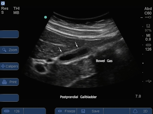
Gallbladder 07 Gallbladder 07 Read more about Gallbladder 07 /sites/default/files/201410_Image_S-System_Gallbladder_Transverse_Post_Prandial.jpg S Series: Gallbladder Transverse Post Prandial. Clinical Specialties FP/GP Media Library Type Image Media Library Tag Gallbladder Disease Stones Abdomen Abd S-System S-Series Pain Cholecystitis Cholelithiasis Biliary Bile Right Upper Quadrant Basic Pathology Hepatobillary Gallstones Image
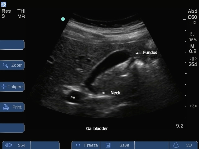
Gallbladder 05 Gallbladder 05 Read more about Gallbladder 05 /sites/default/files/201410_Image_S-System_Gallbladder_Neck_Logitudinal_View.jpg S Series: Gallbladder Neck Longitudinal View. Clinical Specialties FP/GP Media Library Type Image Media Library Tag Gallbladder Disease Stones Abdomen Abd Pain Cholecystitis Cholelithiasis Biliary Bile Right Upper Quadrant Basic Pathology Hepatobillary Gallstones Image S-Series
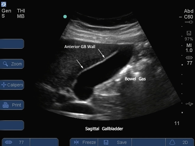
Gallbladder 04 Gallbladder 04 Read more about Gallbladder 04 /sites/default/files/201410_Image_S_System_Gallbladder_Longitudinal_View_Post_Prandial2.jpg S Series: Gallbladder Longitudinal View Post Prandial. Media Library Type Image Media Library Tag Abd Abdomen Basic Bile Biliary Cholecystitis Cholelithiasis Disease Gallbladder Gallstones Hepatobillary Image Pain Pathology Right Upper Quadrant S-Series Stones
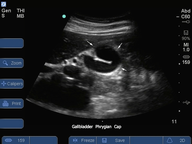
Gallbladder 03 Gallbladder 03 Read more about Gallbladder 03 /sites/default/files/201410_Image_S-System_Gallbladder_Longitudinal_View_Pharyngeal_Cap.jpg S Series: Gallbladder Longitudinal View Pharyngeal Cap. Media Library Type Image Media Library Tag Abd Abdomen Basic Bile Biliary Cholecystitis Cholelithiasis Disease Gallbladder Gallstones Hepatobillary Image Pain Pathology Right Upper Quadrant S-Series Stones
Gallbladder 02 Gallbladder 02 Read more about Gallbladder 02 /sites/default/files/201410_Image_S_System_Gallbladder_Longitudinal_View_3.jpg S Series: Gallbladder Longitudinal View 2. Media Library Type Image Media Library Tag Abd Abdomen Basic Bile Biliary Cholecystitis Cholelithiasis Disease Gallbladder Gallstones Hepatobillary Image Pain Pathology Right Upper Quadrant S-Series Stones
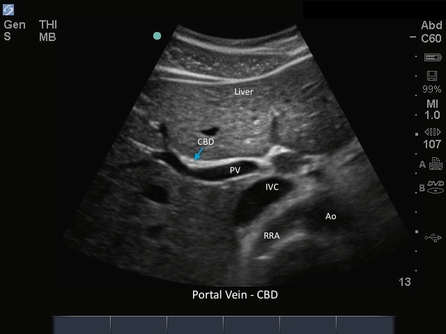
Gallbladder CBD Gallbladder CBD Read more about Gallbladder CBD /sites/default/files/201410_Image_M-Turbo_CBD_Portal_Vein.jpg M-Turbo: CBD Portal Vein. Clinical Specialties FP/GP Media Library Type Image Media Library Tag M-Turbo Comon Bile Duct Biliary Portal Triad Gallbladder Disease Stones Abdomen Abd Pain Cholecystitis Cholelithiasis Bile Right Upper Quadrant Basic Pathology Hepatobillary Gallstones Image Compatible Products Sonosite M-Turbo