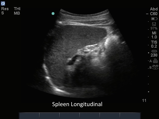
How to: FAST Exam: LUQ

How to: FAST Exam: LUQ

/sites/default/files/06_FAST_Exam_LUQ_View_Scanning_Technique.jpg
Learn to scan the splenorenal recess for fluid collections.
Media Library Type
Subtitles
<p begin="00:00:15.546" end="00:00:16.890" style="s2">- The next view I'm<br />going to take you through</p>
<p begin="00:00:16.890" end="00:00:19.221" style="s2">is a perisplenic or left<br />upper quadrant view.</p>
<p begin="00:00:19.221" end="00:00:21.219" style="s2">This view tends to be a<br />little bit more difficult</p>
<p begin="00:00:21.219" end="00:00:23.222" style="s2">because the spleen is<br />smaller and doesn't provide</p>
<p begin="00:00:23.222" end="00:00:25.156" style="s2">as large an acoustic window.</p>
<p begin="00:00:25.156" end="00:00:25.989" style="s2">We're going to start.</p>
<p begin="00:00:25.989" end="00:00:27.762" style="s2">Again, the patient's going to be supine.</p>
<p begin="00:00:27.762" end="00:00:29.700" style="s2">This is a probe marker right here.</p>
<p begin="00:00:29.700" end="00:00:31.184" style="s2">We're going to go really posterior.</p>
<p begin="00:00:31.184" end="00:00:32.939" style="s2">My hand is going to touch the gurney.</p>
<p begin="00:00:32.939" end="00:00:34.428" style="s2">You can go about four finger breadths</p>
<p begin="00:00:34.428" end="00:00:37.158" style="s2">just above the costal margin.</p>
<p begin="00:00:37.158" end="00:00:38.895" style="s2">You can identify the kidney.</p>
<p begin="00:00:38.895" end="00:00:41.275" style="s2">That's going to be a<br />pretty identifiable organ,</p>
<p begin="00:00:41.275" end="00:00:42.830" style="s2">characteristic bean shape.</p>
<p begin="00:00:42.830" end="00:00:44.445" style="s2">Then we can see it here on the screen.</p>
<p begin="00:00:44.445" end="00:00:47.184" style="s2">Just above it, we're<br />going to see the spleen.</p>
<p begin="00:00:47.184" end="00:00:50.328" style="s2">To the far left of the screen as well</p>
<p begin="00:00:50.328" end="00:00:51.609" style="s2">we see the diaphragm.</p>
<p begin="00:00:51.609" end="00:00:53.113" style="s2">Once you're in this location,</p>
<p begin="00:00:53.113" end="00:00:55.301" style="s2">you want to carefully pan through</p>
<p begin="00:00:55.301" end="00:00:56.973" style="s2">the inferior part of the spleen,</p>
<p begin="00:00:56.973" end="00:00:58.803" style="s2">and then jump over that rib,</p>
<p begin="00:00:58.803" end="00:01:01.066" style="s2">and see the superior part of the spleen,</p>
<p begin="00:01:01.066" end="00:01:04.617" style="s2">because fluid often collects<br />just above the spleen</p>
<p begin="00:01:04.617" end="00:01:06.199" style="s2">and below the diaphragm.</p>
<p begin="00:01:06.199" end="00:01:09.079" style="s2">Once you identify a good view<br />in the left upper quadrant,</p>
<p begin="00:01:09.079" end="00:01:10.808" style="s2">you'll want to do a<br />sweep through that area</p>
<p begin="00:01:10.808" end="00:01:12.416" style="s2">looking for free fluid.</p>
<p begin="00:01:12.416" end="00:01:14.762" style="s2">Again, you see the kidney,<br />you see the spleen.</p>
<p begin="00:01:14.762" end="00:01:16.427" style="s2">Find the interface between.</p>
<p begin="00:01:16.427" end="00:01:18.594" style="s2">Look for fluid between those two organs.</p>
<p begin="00:01:18.594" end="00:01:21.864" style="s2">Then particularly scan above that spleen,</p>
<p begin="00:01:21.864" end="00:01:24.144" style="s2">scanning all the way through the organ,</p>
<p begin="00:01:24.144" end="00:01:26.408" style="s2">looking for free fluid above the spleen</p>
<p begin="00:01:26.408" end="00:01:27.960" style="s2">and below the diaphragm.</p>
<p begin="00:01:27.960" end="00:01:30.249" style="s2">Again, when you're<br />scanning through this area,</p>
<p begin="00:01:30.249" end="00:01:32.431" style="s2">you want to carefully<br />look above the diaphragm</p>
<p begin="00:01:32.431" end="00:01:35.170" style="s2">for evidence of hemothorax.</p>
<p begin="00:01:35.170" end="00:01:36.260" style="s2">When you're scanning in this view,</p>
<p begin="00:01:36.260" end="00:01:38.846" style="s2">also make sure you scan the full kidney.</p>
<p begin="00:01:38.846" end="00:01:43.581" style="s2">Here we see the superior pole<br />to the left of the screen,</p>
<p begin="00:01:43.581" end="00:01:45.337" style="s2">and the inferior pole.</p>
<p begin="00:01:45.337" end="00:01:47.178" style="s2">You want to make sure<br />you get that full kidney</p>
<p begin="00:01:47.178" end="00:01:51.261" style="s2">in view when you examine<br />the left upper quadrant.</p>
Brightcove ID
5508114757001
https://youtube.com/watch?v=IuRklL3cWJU

3D How To: eFAST LUQ

3D How To: eFAST LUQ

/sites/default/files/EFast_Luq_EDU00453_Thumnail.jpg
3D animation demonstrating a Left Upper Quadrant (LUQ) view while performing the eFAST exam.
Media Library Type
Subtitles
<p begin="00:00:07.200" end="00:00:09.566" style="s2">- [Voiceover] A phased array transducer</p>
<p begin="00:00:09.566" end="00:00:11.913" style="s2">with an abdomen exam<br />type is used to perform</p>
<p begin="00:00:11.913" end="00:00:14.758" style="s2">the left upper quadrant<br />view of the FAST Exam.</p>
<p begin="00:00:14.758" end="00:00:16.868" style="s2">The orientation marker is directed</p>
<p begin="00:00:16.868" end="00:00:18.771" style="s2">toward the patient's head.</p>
<p begin="00:00:18.771" end="00:00:22.071" style="s2">the transducer is placed in<br />a long-access orientation</p>
<p begin="00:00:22.071" end="00:00:24.725" style="s2">along the left-posterior axillary line</p>
<p begin="00:00:24.725" end="00:00:28.372" style="s2">between the fifth to<br />seventh intercostal spaces.</p>
<p begin="00:00:28.372" end="00:00:30.923" style="s2">The sonographer's hand<br />will touch the gurney</p>
<p begin="00:00:30.923" end="00:00:33.344" style="s2">with the proper transducer position.</p>
<p begin="00:00:33.344" end="00:00:36.425" style="s2">Rotation and oblique<br />positioning of the transducer</p>
<p begin="00:00:36.425" end="00:00:39.092" style="s2">will help eliminate rib shadows.</p>
<p begin="00:00:43.087" end="00:00:46.518" style="s2">To evaluate the entire area<br />of the spleenorenal recess</p>
<p begin="00:00:46.518" end="00:00:49.173" style="s2">for free fluid, sweep the transducer</p>
<p begin="00:00:49.173" end="00:00:52.423" style="s2">from an anterior to posterior position.</p>
<p begin="00:00:53.723" end="00:00:56.699" style="s2">Carefully examine the spleenorenal recess</p>
<p begin="00:00:56.699" end="00:00:58.567" style="s2">and subphrenic space.</p>
<p begin="00:00:58.567" end="00:01:01.479" style="s2">Fluid will appear dark or anechoic.</p>
<p begin="00:01:01.479" end="00:01:04.925" style="s2">If it is difficult to visualize<br />the spreenorenal recess,</p>
<p begin="00:01:04.925" end="00:01:07.562" style="s2">a deep inspiration will move the diaphragm</p>
<p begin="00:01:07.562" end="00:01:09.580" style="s2">and other structures in this area</p>
<p begin="00:01:09.580" end="00:01:12.581" style="s2">down and below the ribs for easier access.</p>
<p begin="00:01:12.581" end="00:01:15.311" style="s2">Sliding the transducer<br />in an upward direction</p>
<p begin="00:01:15.311" end="00:01:19.478" style="s2">will visualize the diaphragm<br />and plural interface.</p>
Brightcove ID
5508120178001
https://youtube.com/watch?v=foyIEuUlgD0