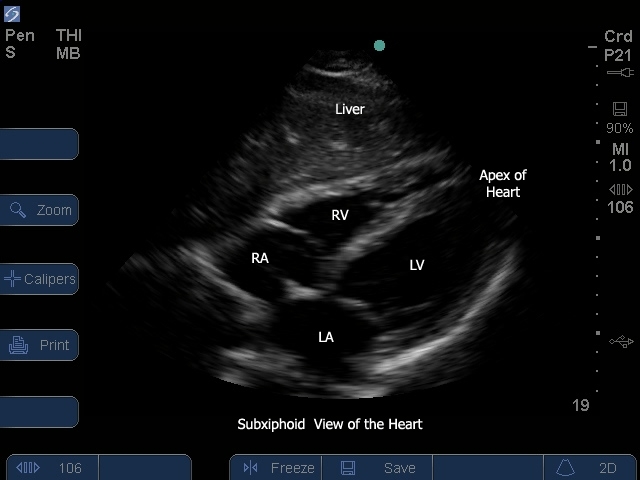
Subtitles
<p begin="00:00:10.942" end="00:00:12.419" style="s2">- Hello, my name is Phil Perera,</p>
<p begin="00:00:12.419" end="00:00:14.395" style="s2">and I'm the Emergency<br />Ultrasound Coordinator</p>
<p begin="00:00:14.395" end="00:00:17.231" style="s2">at the New York Presbyterian<br />Hospital in New York City,</p>
<p begin="00:00:17.231" end="00:00:19.898" style="s2">and welcome to SoundBytes Cases.</p>
<p begin="00:00:20.899" end="00:00:22.392" style="s2">In this module, we'll continue our journey</p>
<p begin="00:00:22.392" end="00:00:24.929" style="s2">through the cardiac<br />echocardiography examinations,</p>
<p begin="00:00:24.929" end="00:00:27.067" style="s2">looking at the four standard views.</p>
<p begin="00:00:27.067" end="00:00:29.289" style="s2">In this module, we're<br />specifically going to focus on</p>
<p begin="00:00:29.289" end="00:00:32.134" style="s2">probe position B, as shown<br />in the pictorial here,</p>
<p begin="00:00:32.134" end="00:00:35.565" style="s2">the subxiphoid view of the heart.</p>
<p begin="00:00:35.565" end="00:00:36.914" style="s2">Hopefully you've joined me prior</p>
<p begin="00:00:36.914" end="00:00:40.170" style="s2">for the parasternal views,<br />as shown in probe position A,</p>
<p begin="00:00:40.170" end="00:00:42.603" style="s2">and in an upcoming module<br />on the apical view,</p>
<p begin="00:00:42.603" end="00:00:45.243" style="s2">as shown in probe position C.</p>
<p begin="00:00:45.243" end="00:00:46.703" style="s2">The subxiphoid view of the heart</p>
<p begin="00:00:46.703" end="00:00:49.287" style="s2">is an excellent way of<br />imaging the patient's heart,</p>
<p begin="00:00:49.287" end="00:00:52.725" style="s2">and getting a lot of information<br />directly at the bedside.</p>
<p begin="00:00:52.725" end="00:00:53.843" style="s2">Now let's learn how to perform</p>
<p begin="00:00:53.843" end="00:00:55.698" style="s2">the subxiphoid view of the heart.</p>
<p begin="00:00:55.698" end="00:00:57.399" style="s2">As shown in the pictorial to the right,</p>
<p begin="00:00:57.399" end="00:00:59.388" style="s2">the probe is coming from<br />an abdominal position,</p>
<p begin="00:00:59.388" end="00:01:02.989" style="s2">placed just inferior to the<br />xiphoid tip of the sternum.</p>
<p begin="00:01:02.989" end="00:01:04.921" style="s2">It's important to lay the probe flat</p>
<p begin="00:01:04.921" end="00:01:06.908" style="s2">and push down and under the sternum,</p>
<p begin="00:01:06.908" end="00:01:09.312" style="s2">aiming towards the<br />patient's left shoulder.</p>
<p begin="00:01:09.312" end="00:01:11.008" style="s2">Now the marker dot on the probe</p>
<p begin="00:01:11.008" end="00:01:13.350" style="s2">should be over towards<br />the patient's right side,</p>
<p begin="00:01:13.350" end="00:01:15.537" style="s2">with a caveat that the ultrasound's screen</p>
<p begin="00:01:15.537" end="00:01:19.162" style="s2">indicator dot is over towards<br />the left of the screen.</p>
<p begin="00:01:19.162" end="00:01:20.511" style="s2">Now it's very important to put your hands</p>
<p begin="00:01:20.511" end="00:01:23.783" style="s2">on top of the probe, and<br />really push down and up</p>
<p begin="00:01:23.783" end="00:01:25.682" style="s2">to get the good imaging plane underneath</p>
<p begin="00:01:25.682" end="00:01:27.937" style="s2">the sternum, to make the angle to get</p>
<p begin="00:01:27.937" end="00:01:31.354" style="s2">a good view of the heart from this plane.</p>
<p begin="00:01:32.452" end="00:01:33.787" style="s2">Let's now take a look at the image</p>
<p begin="00:01:33.787" end="00:01:36.911" style="s2">that you'll obtain from the<br />subxiphoid view of the heart.</p>
<p begin="00:01:36.911" end="00:01:38.459" style="s2">Here's a pictorial to the left, and an</p>
<p begin="00:01:38.459" end="00:01:40.922" style="s2">ultrasound image to the right.</p>
<p begin="00:01:40.922" end="00:01:42.583" style="s2">The first chamber that we'll encounter</p>
<p begin="00:01:42.583" end="00:01:44.374" style="s2">directly below the liver, which is our</p>
<p begin="00:01:44.374" end="00:01:46.324" style="s2">acoustic window in this case, on to</p>
<p begin="00:01:46.324" end="00:01:48.603" style="s2">the heart will be the right ventricle.</p>
<p begin="00:01:48.603" end="00:01:51.019" style="s2">Immediately posterior<br />to the right ventricle</p>
<p begin="00:01:51.019" end="00:01:52.700" style="s2">we'll be seeing the left ventricle,</p>
<p begin="00:01:52.700" end="00:01:54.433" style="s2">and as shown in this pictorial,</p>
<p begin="00:01:54.433" end="00:01:58.653" style="s2">notice that it has more<br />muscular and hypertrophic walls.</p>
<p begin="00:01:58.653" end="00:02:00.267" style="s2">From the subxiphoid plane, we'll also</p>
<p begin="00:02:00.267" end="00:02:02.050" style="s2">be able to image the right atrium</p>
<p begin="00:02:02.050" end="00:02:04.023" style="s2">to the left of the right ventricle,</p>
<p begin="00:02:04.023" end="00:02:06.011" style="s2">and the left atrium, just to the left</p>
<p begin="00:02:06.011" end="00:02:07.673" style="s2">of the left ventricle.</p>
<p begin="00:02:07.673" end="00:02:09.401" style="s2">We can also appreciate the white line</p>
<p begin="00:02:09.401" end="00:02:11.432" style="s2">that is the pericardium circumferentially</p>
<p begin="00:02:11.432" end="00:02:13.265" style="s2">surrounding the heart.</p>
<p begin="00:02:15.126" end="00:02:16.452" style="s2">Now that we know where the chambers are,</p>
<p begin="00:02:16.452" end="00:02:17.853" style="s2">let's take a look at a video clip</p>
<p begin="00:02:17.853" end="00:02:21.393" style="s2">of a normal heart from<br />the subxiphoid plane.</p>
<p begin="00:02:21.393" end="00:02:23.160" style="s2">As we remember, the liver is our</p>
<p begin="00:02:23.160" end="00:02:25.579" style="s2">acoustic window onto the<br />heart from this plane,</p>
<p begin="00:02:25.579" end="00:02:27.720" style="s2">and so the liver will be seen anteriorly,</p>
<p begin="00:02:27.720" end="00:02:29.758" style="s2">just to the top of the screen.</p>
<p begin="00:02:29.758" end="00:02:31.454" style="s2">Just below the liver, we appreciate here</p>
<p begin="00:02:31.454" end="00:02:33.783" style="s2">the right ventricle, and notice here,</p>
<p begin="00:02:33.783" end="00:02:35.552" style="s2">just to the left of the right ventricle,</p>
<p begin="00:02:35.552" end="00:02:38.623" style="s2">we can appreciate the right atrium.</p>
<p begin="00:02:38.623" end="00:02:41.075" style="s2">Notice the tricuspid<br />valve flipping up and down</p>
<p begin="00:02:41.075" end="00:02:44.932" style="s2">in between the right atrium<br />and the right ventricle.</p>
<p begin="00:02:44.932" end="00:02:47.172" style="s2">Now let's look posterior<br />to the right ventricle,</p>
<p begin="00:02:47.172" end="00:02:49.136" style="s2">and we appreciate the left ventricle.</p>
<p begin="00:02:49.136" end="00:02:53.968" style="s2">Notice again, its more muscular<br />and hypertrophic walls.</p>
<p begin="00:02:53.968" end="00:02:55.740" style="s2">Just to the left of the left ventricle</p>
<p begin="00:02:55.740" end="00:02:58.326" style="s2">we appreciate, in this<br />case, the left atrium,</p>
<p begin="00:02:58.326" end="00:03:01.084" style="s2">and we also get a glimpse<br />here of the mitral valve</p>
<p begin="00:03:01.084" end="00:03:02.602" style="s2">flipping up and down in between</p>
<p begin="00:03:02.602" end="00:03:05.382" style="s2">the left atrium and the left ventricle.</p>
<p begin="00:03:05.382" end="00:03:06.794" style="s2">Now let's look at that white line,</p>
<p begin="00:03:06.794" end="00:03:09.159" style="s2">both anteriorly above the right ventricle,</p>
<p begin="00:03:09.159" end="00:03:11.112" style="s2">and posterior, below the left ventricle,</p>
<p begin="00:03:11.112" end="00:03:12.710" style="s2">that is the pericardium.</p>
<p begin="00:03:12.710" end="00:03:14.504" style="s2">Note here the absence of any significant</p>
<p begin="00:03:14.504" end="00:03:16.337" style="s2">pericardial effusions.</p>
<p begin="00:03:17.266" end="00:03:18.904" style="s2">In that last video clip, we noted good</p>
<p begin="00:03:18.904" end="00:03:20.604" style="s2">contractility of the left ventricle</p>
<p begin="00:03:20.604" end="00:03:22.739" style="s2">from diastole to systole.</p>
<p begin="00:03:22.739" end="00:03:24.752" style="s2">Let's contrast that clip to this one</p>
<p begin="00:03:24.752" end="00:03:26.269" style="s2">from a patient who<br />presented with shortness</p>
<p begin="00:03:26.269" end="00:03:29.047" style="s2">of breath, and advanced cardiomyopathy.</p>
<p begin="00:03:29.047" end="00:03:31.272" style="s2">We see the right ventricle<br />just below the liver,</p>
<p begin="00:03:31.272" end="00:03:33.392" style="s2">anterior to the left ventricle.</p>
<p begin="00:03:33.392" end="00:03:35.418" style="s2">And what we see here<br />is a poorly contracting</p>
<p begin="00:03:35.418" end="00:03:37.683" style="s2">and dilated left ventricle, consistent</p>
<p begin="00:03:37.683" end="00:03:40.236" style="s2">with a cardiomyopathy heart.</p>
<p begin="00:03:40.236" end="00:03:42.525" style="s2">However, note the absence<br />of any significant</p>
<p begin="00:03:42.525" end="00:03:44.725" style="s2">dark or anechoic fluid collections</p>
<p begin="00:03:44.725" end="00:03:48.229" style="s2">consistent with a pericardial effusion.</p>
<p begin="00:03:48.229" end="00:03:50.208" style="s2">Here's a patient who<br />presented with renal failure</p>
<p begin="00:03:50.208" end="00:03:51.934" style="s2">and acute shortness of breath.</p>
<p begin="00:03:51.934" end="00:03:54.021" style="s2">We're again looking from<br />the subxiphoid plane,</p>
<p begin="00:03:54.021" end="00:03:56.260" style="s2">so we see a little strip<br />of the liver anteriorly.</p>
<p begin="00:03:56.260" end="00:03:58.413" style="s2">The right ventricle just below the liver,</p>
<p begin="00:03:58.413" end="00:04:00.518" style="s2">and the left ventricle seen posteriorly</p>
<p begin="00:04:00.518" end="00:04:01.885" style="s2">to the right ventricle.</p>
<p begin="00:04:01.885" end="00:04:03.889" style="s2">Notice how hypertrophic the walls of</p>
<p begin="00:04:03.889" end="00:04:06.431" style="s2">the left ventricle are in this patient.</p>
<p begin="00:04:06.431" end="00:04:08.556" style="s2">We also appreciate a dark fluid collection</p>
<p begin="00:04:08.556" end="00:04:10.554" style="s2">both anteriorly, just below the liver</p>
<p begin="00:04:10.554" end="00:04:11.840" style="s2">and above the right ventricle,</p>
<p begin="00:04:11.840" end="00:04:14.000" style="s2">and posterior below the left ventricle,</p>
<p begin="00:04:14.000" end="00:04:15.941" style="s2">consistent with a circumferential,</p>
<p begin="00:04:15.941" end="00:04:19.345" style="s2">or large, pericardial effusion.</p>
<p begin="00:04:19.345" end="00:04:21.142" style="s2">If we see a large pericardial effusion</p>
<p begin="00:04:21.142" end="00:04:22.863" style="s2">on bedside echo, our next move is to</p>
<p begin="00:04:22.863" end="00:04:25.413" style="s2">look for signs of cardiac tamponade.</p>
<p begin="00:04:25.413" end="00:04:26.803" style="s2">Here's a patient who manifests</p>
<p begin="00:04:26.803" end="00:04:30.184" style="s2">all the signs of cardiac<br />tamponade on bedside echo.</p>
<p begin="00:04:30.184" end="00:04:33.384" style="s2">Let's look specifically at<br />the right side of the heart.</p>
<p begin="00:04:33.384" end="00:04:35.779" style="s2">Notice the very large<br />pericardial effusion,</p>
<p begin="00:04:35.779" end="00:04:37.883" style="s2">and note the chaotic movement<br />of the right ventricle</p>
<p begin="00:04:37.883" end="00:04:41.069" style="s2">as it struggles to open during diastole.</p>
<p begin="00:04:41.069" end="00:04:42.618" style="s2">The compression of the right ventricle</p>
<p begin="00:04:42.618" end="00:04:44.288" style="s2">in this patient is consistent with</p>
<p begin="00:04:44.288" end="00:04:46.792" style="s2">advanced cardiac tamponade.</p>
<p begin="00:04:46.792" end="00:04:47.811" style="s2">The right side of the heart is</p>
<p begin="00:04:47.811" end="00:04:49.128" style="s2">preferentially compressed before</p>
<p begin="00:04:49.128" end="00:04:52.766" style="s2">the left ventricle, due to<br />its lower pressure circuit.</p>
<p begin="00:04:52.766" end="00:04:54.311" style="s2">In conclusion, I'm glad<br />I could share with you</p>
<p begin="00:04:54.311" end="00:04:55.677" style="s2">this SoundBytes module going over</p>
<p begin="00:04:55.677" end="00:04:58.896" style="s2">the subxiphoid view of the<br />cardiac echo examination.</p>
<p begin="00:04:58.896" end="00:05:00.207" style="s2">This is a very important exam to</p>
<p begin="00:05:00.207" end="00:05:02.008" style="s2">put into your routine practice</p>
<p begin="00:05:02.008" end="00:05:04.673" style="s2">in looking at your patient's<br />heart at the bedside,</p>
<p begin="00:05:04.673" end="00:05:05.741" style="s2">and will tell you if the patient</p>
<p begin="00:05:05.741" end="00:05:07.575" style="s2">has a pericardial effusion, as well as</p>
<p begin="00:05:07.575" end="00:05:10.793" style="s2">giving a sense of left<br />ventricular contractility.</p>
<p begin="00:05:10.793" end="00:05:12.554" style="s2">Also, the subxiphoid view of the heart</p>
<p begin="00:05:12.554" end="00:05:14.802" style="s2">gives better views of the<br />right side of the heart</p>
<p begin="00:05:14.802" end="00:05:18.837" style="s2">than the more superior<br />parasternal views of the heart.</p>
<p begin="00:05:18.837" end="00:05:21.459" style="s2">So I hope to see you back<br />as SoundBytes continues,</p>
<p begin="00:05:21.459" end="00:05:23.018" style="s2">and as we move on to discuss the other</p>
<p begin="00:05:23.018" end="00:05:25.601" style="s2">echo exam planes of the heart.</p>