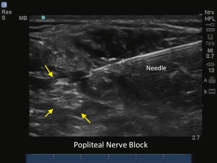
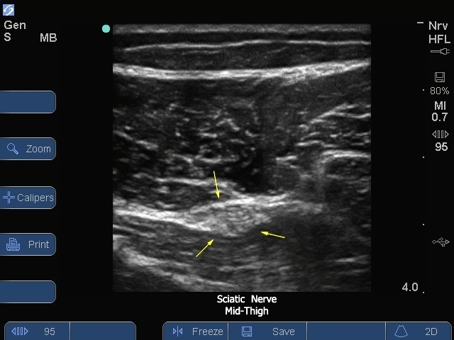
3D How To: Subclavian Vein Catheter Insertion

3D How To: Subclavian Vein Catheter Insertion

/sites/default/files/CentralVenous_SubclavianVein_edu00473_thumbnail.jpg
3D animation demonstrating an ultrasound guided insertion of a Subclavian Vein Catheter.
Applications
Clinical Specialties
Media Library Type
Subtitles
<p begin="00:00:07.010" end="00:00:09.026" style="s2">- [Voiceover] A linear array transducer</p>
<p begin="00:00:09.026" end="00:00:11.485" style="s2">with a venous exam<br />type, is used to perform</p>
<p begin="00:00:11.485" end="00:00:14.424" style="s2">an ultrasound guided<br />insertion of a subclavian</p>
<p begin="00:00:14.424" end="00:00:17.250" style="s2">vein catheter via an transverse approach.</p>
<p begin="00:00:17.250" end="00:00:19.487" style="s2">The patient is in a supine position</p>
<p begin="00:00:19.487" end="00:00:21.144" style="s2">with the head neutral.</p>
<p begin="00:00:21.144" end="00:00:24.198" style="s2">The operator should stand<br />to the patient's right side.</p>
<p begin="00:00:24.198" end="00:00:26.531" style="s2">The transducer is placed transversely</p>
<p begin="00:00:26.531" end="00:00:29.380" style="s2">just inferior to the mid<br />portion of the clavicle,</p>
<p begin="00:00:29.380" end="00:00:31.338" style="s2">with the orientation marker directed</p>
<p begin="00:00:31.338" end="00:00:34.428" style="s2">to the patient's head at<br />a 12 o'clock position.</p>
<p begin="00:00:34.428" end="00:00:36.407" style="s2">The hyperechoic clavicle can be seen</p>
<p begin="00:00:36.407" end="00:00:39.304" style="s2">in the superior portion<br />of the ultrasound image.</p>
<p begin="00:00:39.304" end="00:00:41.387" style="s2">The vein is dark, and anechoic,</p>
<p begin="00:00:41.387" end="00:00:43.977" style="s2">just inferior and deep to the clavicle.</p>
<p begin="00:00:43.977" end="00:00:47.219" style="s2">The transducer should be<br />slowly moved one to two inches</p>
<p begin="00:00:47.219" end="00:00:49.877" style="s2">toward the shoulder, with<br />the face of the transducer</p>
<p begin="00:00:49.877" end="00:00:52.685" style="s2">staying below the clavicle<br />to obtain the best view</p>
<p begin="00:00:52.685" end="00:00:55.397" style="s2">of the subclavian or axillary vein.</p>
<p begin="00:00:55.397" end="00:00:58.063" style="s2">It is important to note<br />that the lung lies directly</p>
<p begin="00:00:58.063" end="00:00:59.861" style="s2">posterior to the vessel.</p>
<p begin="00:00:59.861" end="00:01:02.691" style="s2">So, posterior wall puncture<br />of the axillary vein</p>
<p begin="00:01:02.691" end="00:01:04.214" style="s2">should be avoided.</p>
<p begin="00:01:04.214" end="00:01:07.472" style="s2">Adjust the transducer so it<br />is centered over the vein.</p>
<p begin="00:01:07.472" end="00:01:10.469" style="s2">Follow the needle entry by<br />slowly sliding the transducer</p>
<p begin="00:01:10.469" end="00:01:12.943" style="s2">in the direction of needle advancement.</p>
<p begin="00:01:12.943" end="00:01:16.832" style="s2">The needle will appear as a<br />small, bright, hyperechoic dot.</p>
<p begin="00:01:16.832" end="00:01:19.415" style="s2">When the needle tip<br />appears, the transducer</p>
<p begin="00:01:19.415" end="00:01:21.964" style="s2">should be advanced a<br />short distance distally</p>
<p begin="00:01:21.964" end="00:01:24.265" style="s2">to follow the tip of<br />the needle trajectory,</p>
<p begin="00:01:24.265" end="00:01:26.819" style="s2">and stay in advance of the needle entry.</p>
<p begin="00:01:26.819" end="00:01:28.455" style="s2">The needle is slowly advanced</p>
<p begin="00:01:28.455" end="00:01:30.590" style="s2">under direct ultrasound visualization</p>
<p begin="00:01:30.590" end="00:01:34.266" style="s2">until the tip is seen to<br />puncture the subclavian vein.</p>
<p begin="00:01:34.266" end="00:01:37.399" style="s2">The probe should be moved<br />slightly proximally and distally</p>
<p begin="00:01:37.399" end="00:01:39.357" style="s2">to confirm that the needle tip lies</p>
<p begin="00:01:39.357" end="00:01:41.940" style="s2">in the mid portion of the vein.</p>
Brightcove ID
5508120188001
https://youtube.com/watch?v=IBmbc1ak5fY

3D How To: IJV Catheter Insertion (Transverse)

3D How To: IJV Catheter Insertion (Transverse)

/sites/default/files/CentralVenous_IJ_Transverse_edu00471_thumbnail.jpg
3D animation demonstrating an ultrasound guided insertion of an internal jugular catheter (transverse).
Applications
Clinical Specialties
Media Library Type
Subtitles
<p begin="00:00:07.397" end="00:00:09.087" style="s2">- [Voiceover] A linear array transducer</p>
<p begin="00:00:09.087" end="00:00:10.710" style="s2">with a venous exam type</p>
<p begin="00:00:10.710" end="00:00:13.822" style="s2">is used to perform an<br />ultrasound-guided insertion</p>
<p begin="00:00:13.822" end="00:00:17.827" style="s2">of an internal jugular catheter<br />via a transverse approach.</p>
<p begin="00:00:17.827" end="00:00:19.996" style="s2">The patient is in a supine position</p>
<p begin="00:00:19.996" end="00:00:21.420" style="s2">with the head slightly turned</p>
<p begin="00:00:21.420" end="00:00:23.431" style="s2">toward the contralateral side.</p>
<p begin="00:00:23.431" end="00:00:26.331" style="s2">The operator is positioned<br />at the head of the bed.</p>
<p begin="00:00:26.331" end="00:00:28.712" style="s2">The transducer is placed transversely</p>
<p begin="00:00:28.712" end="00:00:29.970" style="s2">just below the apex</p>
<p begin="00:00:29.970" end="00:00:32.663" style="s2">of the sternocleidomastoid<br />muscle triangle,</p>
<p begin="00:00:32.663" end="00:00:35.671" style="s2">with the orientation marker<br />directed to the patient's left</p>
<p begin="00:00:35.671" end="00:00:37.655" style="s2">at a 9:00 position.</p>
<p begin="00:00:37.655" end="00:00:39.098" style="s2">The most superficial structure</p>
<p begin="00:00:39.098" end="00:00:41.086" style="s2">identified in the ultrasound image</p>
<p begin="00:00:41.086" end="00:00:43.548" style="s2">is the sternocleidomastoid muscle.</p>
<p begin="00:00:43.548" end="00:00:46.077" style="s2">Deeper to this, the internal jugular vein</p>
<p begin="00:00:46.077" end="00:00:49.080" style="s2">will appear as a dark<br />anechoic elliptical shape,</p>
<p begin="00:00:49.080" end="00:00:51.990" style="s2">and is compressible with<br />transducer pressure.</p>
<p begin="00:00:51.990" end="00:00:55.354" style="s2">The artery will be circular and pulsatile.</p>
<p begin="00:00:55.354" end="00:00:58.421" style="s2">The thyroid gland lies<br />medial to these structures,</p>
<p begin="00:00:58.421" end="00:01:01.367" style="s2">and has a light gray echo signature.</p>
<p begin="00:01:01.367" end="00:01:03.420" style="s2">Adjust the transducer so it is centered</p>
<p begin="00:01:03.420" end="00:01:05.804" style="s2">over the internal jugular vein.</p>
<p begin="00:01:05.804" end="00:01:08.867" style="s2">Follow the needle entry by<br />slowly sliding the transducer</p>
<p begin="00:01:08.867" end="00:01:11.272" style="s2">in the direction of needle advancement.</p>
<p begin="00:01:11.272" end="00:01:15.230" style="s2">The needle will appear as a<br />small bright hyperechoic dot.</p>
<p begin="00:01:15.230" end="00:01:16.923" style="s2">When the needle tip appears,</p>
<p begin="00:01:16.923" end="00:01:20.067" style="s2">the transducer should be advanced<br />a short distance distally</p>
<p begin="00:01:20.067" end="00:01:22.537" style="s2">to follow the tip of<br />the needle trajectory,</p>
<p begin="00:01:22.537" end="00:01:25.144" style="s2">and stay in advance of the needle entry.</p>
<p begin="00:01:25.144" end="00:01:26.523" style="s2">The needle is slowly advanced</p>
<p begin="00:01:26.523" end="00:01:28.724" style="s2">under direct ultrasound visualization</p>
<p begin="00:01:28.724" end="00:01:30.616" style="s2">until the tip is seen to indent,</p>
<p begin="00:01:30.616" end="00:01:33.391" style="s2">and then puncture the<br />internal jugular vein.</p>
<p begin="00:01:33.391" end="00:01:34.837" style="s2">The transducer should be moved</p>
<p begin="00:01:34.837" end="00:01:36.859" style="s2">slightly proximally and distally</p>
<p begin="00:01:36.859" end="00:01:38.740" style="s2">to confirm that the needle tip lies</p>
<p begin="00:01:38.740" end="00:01:41.990" style="s2">in the mid-portion of the jugular vein.</p>
Brightcove ID
5742626540001
https://youtube.com/watch?v=eesN9rGoXFM

3D How To: IJV Catheter Insertion (Long)

3D How To: IJV Catheter Insertion (Long)

/sites/default/files/CentralVenous_IJ_Long_edu00472_thumbnail.jpg
3D animation demonstrating an ultrasound guided insertion of an Internal Jugular Catheter (Longitudinal).
Applications
Clinical Specialties
Media Library Type
Subtitles
<p begin="00:00:07.010" end="00:00:09.026" style="s2">- [Voiceover] A linear array transducer</p>
<p begin="00:00:09.026" end="00:00:11.485" style="s2">with a venous exam<br />type, is used to perform</p>
<p begin="00:00:11.485" end="00:00:14.424" style="s2">an ultrasound guided<br />insertion of a subclavian</p>
<p begin="00:00:14.424" end="00:00:17.250" style="s2">vein catheter via an transverse approach.</p>
<p begin="00:00:17.250" end="00:00:19.487" style="s2">The patient is in a supine position</p>
<p begin="00:00:19.487" end="00:00:21.144" style="s2">with the head neutral.</p>
<p begin="00:00:21.144" end="00:00:24.198" style="s2">The operator should stand<br />to the patient's right side.</p>
<p begin="00:00:24.198" end="00:00:26.531" style="s2">The transducer is placed transversely</p>
<p begin="00:00:26.531" end="00:00:29.380" style="s2">just inferior to the mid<br />portion of the clavicle,</p>
<p begin="00:00:29.380" end="00:00:31.338" style="s2">with the orientation marker directed</p>
<p begin="00:00:31.338" end="00:00:34.428" style="s2">to the patient's head at<br />a 12 o'clock position.</p>
<p begin="00:00:34.428" end="00:00:36.407" style="s2">The hyperechoic clavicle can be seen</p>
<p begin="00:00:36.407" end="00:00:39.304" style="s2">in the superior portion<br />of the ultrasound image.</p>
<p begin="00:00:39.304" end="00:00:41.387" style="s2">The vein is dark, and anechoic,</p>
<p begin="00:00:41.387" end="00:00:43.977" style="s2">just inferior and deep to the clavicle.</p>
<p begin="00:00:43.977" end="00:00:47.219" style="s2">The transducer should be<br />slowly moved one to two inches</p>
<p begin="00:00:47.219" end="00:00:49.877" style="s2">toward the shoulder, with<br />the face of the transducer</p>
<p begin="00:00:49.877" end="00:00:52.685" style="s2">staying below the clavicle<br />to obtain the best view</p>
<p begin="00:00:52.685" end="00:00:55.397" style="s2">of the subclavian or axillary vein.</p>
<p begin="00:00:55.397" end="00:00:58.063" style="s2">It is important to note<br />that the lung lies directly</p>
<p begin="00:00:58.063" end="00:00:59.861" style="s2">posterior to the vessel.</p>
<p begin="00:00:59.861" end="00:01:02.691" style="s2">So, posterior wall puncture<br />of the axillary vein</p>
<p begin="00:01:02.691" end="00:01:04.214" style="s2">should be avoided.</p>
<p begin="00:01:04.214" end="00:01:07.472" style="s2">Adjust the transducer so it<br />is centered over the vein.</p>
<p begin="00:01:07.472" end="00:01:10.469" style="s2">Follow the needle entry by<br />slowly sliding the transducer</p>
<p begin="00:01:10.469" end="00:01:12.943" style="s2">in the direction of needle advancement.</p>
<p begin="00:01:12.943" end="00:01:16.832" style="s2">The needle will appear as a<br />small, bright, hyperechoic dot.</p>
<p begin="00:01:16.832" end="00:01:19.415" style="s2">When the needle tip<br />appears, the transducer</p>
<p begin="00:01:19.415" end="00:01:21.964" style="s2">should be advanced a<br />short distance distally</p>
<p begin="00:01:21.964" end="00:01:24.265" style="s2">to follow the tip of<br />the needle trajectory,</p>
<p begin="00:01:24.265" end="00:01:26.819" style="s2">and stay in advance of the needle entry.</p>
<p begin="00:01:26.819" end="00:01:28.455" style="s2">The needle is slowly advanced</p>
<p begin="00:01:28.455" end="00:01:30.590" style="s2">under direct ultrasound visualization</p>
<p begin="00:01:30.590" end="00:01:34.266" style="s2">until the tip is seen to<br />puncture the subclavian vein.</p>
<p begin="00:01:34.266" end="00:01:37.399" style="s2">The probe should be moved<br />slightly proximally and distally</p>
<p begin="00:01:37.399" end="00:01:39.357" style="s2">to confirm that the needle tip lies</p>
<p begin="00:01:39.357" end="00:01:41.940" style="s2">in the mid portion of the vein.</p>
Brightcove ID
5508120188001
https://youtube.com/watch?v=IBmbc1ak5fY

3D How To: Femoral Vein Catheter Insertion

3D How To: Femoral Vein Catheter Insertion

/sites/default/files/CentralVenous_FemoralVein_edu00503_thumbnail.jpg
3D animation demonstrating an ultrasound guided insertion of a Femoral Vein Catheter.
Applications
Clinical Specialties
Media Library Type
Subtitles
<p begin="00:00:07.545" end="00:00:09.217" style="s2">- [Voiceover] A linear array transducer</p>
<p begin="00:00:09.217" end="00:00:11.689" style="s2">with a venous exam type is used to perform</p>
<p begin="00:00:11.689" end="00:00:15.584" style="s2">an ultrasound guided insertion<br />of a femoral vein catheter</p>
<p begin="00:00:15.584" end="00:00:17.953" style="s2">via a transverse approach.</p>
<p begin="00:00:17.953" end="00:00:20.153" style="s2">The patient is in a supine position</p>
<p begin="00:00:20.153" end="00:00:22.616" style="s2">with the operator at the right side.</p>
<p begin="00:00:22.616" end="00:00:24.913" style="s2">The transducer is placed transversely,</p>
<p begin="00:00:24.913" end="00:00:27.362" style="s2">just inferior to the inguinal crease</p>
<p begin="00:00:27.362" end="00:00:30.502" style="s2">with the orientation marker<br />directed to the patient's right</p>
<p begin="00:00:30.502" end="00:00:32.815" style="s2">at a 9 o'clock position.</p>
<p begin="00:00:32.815" end="00:00:35.934" style="s2">The superficial femoral<br />artery is seen as a dark</p>
<p begin="00:00:35.934" end="00:00:38.806" style="s2">anechoic pulsatile circular structure</p>
<p begin="00:00:38.806" end="00:00:41.503" style="s2">in the mid portion of<br />the ultrasound image.</p>
<p begin="00:00:41.503" end="00:00:44.022" style="s2">Deeper and slightly to<br />the right of the image,</p>
<p begin="00:00:44.022" end="00:00:45.838" style="s2">the femoral vein will appear as a</p>
<p begin="00:00:45.838" end="00:00:48.119" style="s2">dark anechoic elliptical shape</p>
<p begin="00:00:48.119" end="00:00:51.470" style="s2">that is compressible<br />with transducer pressure.</p>
<p begin="00:00:51.470" end="00:00:53.632" style="s2">Adjust the transducer so it is centered</p>
<p begin="00:00:53.632" end="00:00:57.199" style="s2">over the femoral vein<br />medial to the artery.</p>
<p begin="00:00:57.199" end="00:01:00.510" style="s2">Follow the needle entry by<br />slowly sliding the transducer</p>
<p begin="00:01:00.510" end="00:01:02.902" style="s2">in the direction of needle advancement.</p>
<p begin="00:01:02.902" end="00:01:06.317" style="s2">The needle will appear<br />as a small bright dot.</p>
<p begin="00:01:06.317" end="00:01:07.849" style="s2">When the needle tip appears,</p>
<p begin="00:01:07.849" end="00:01:11.318" style="s2">the transducer should be advanced<br />a short distance distally</p>
<p begin="00:01:11.318" end="00:01:14.038" style="s2">to follow the tip of<br />the needle trajectory.</p>
<p begin="00:01:14.038" end="00:01:15.838" style="s2">The needle is slowly advanced under</p>
<p begin="00:01:15.838" end="00:01:17.990" style="s2">direct ultrasound visualization</p>
<p begin="00:01:17.990" end="00:01:19.837" style="s2">until the tip is seen to indent</p>
<p begin="00:01:19.837" end="00:01:22.070" style="s2">and then puncture the femoral vein.</p>
<p begin="00:01:22.070" end="00:01:24.942" style="s2">The transducer should be<br />moved slightly proximally</p>
<p begin="00:01:24.942" end="00:01:27.510" style="s2">and distally to confirm<br />that the needle tip</p>
<p begin="00:01:27.510" end="00:01:31.177" style="s2">lies in the mid portion<br />of the femoral vein.</p>
Brightcove ID
5508120181001
https://youtube.com/watch?v=gNvbxeoUMlc

3D How To: Axillary Nerve Block

3D How To: Axillary Nerve Block

/sites/default/files/Axillary_edu00492_thumbnail.jpg
3D animation demonstrating an ultrasound guided axillary nerve block.
Clinical Specialties
Media Library Type
Subtitles
<p begin="00:00:07.376" end="00:00:09.578" style="s2">- [Voiceover] A linear array<br />transducer with a nerve</p>
<p begin="00:00:09.578" end="00:00:12.337" style="s2">exam type, is used to<br />perform an ultra sound</p>
<p begin="00:00:12.337" end="00:00:15.428" style="s2">guided axillary regional nerve block.</p>
<p begin="00:00:15.428" end="00:00:17.955" style="s2">The target depth is<br />approximately one to two</p>
<p begin="00:00:17.955" end="00:00:21.374" style="s2">centimeters in an 80 kilogram adult.</p>
<p begin="00:00:21.374" end="00:00:23.840" style="s2">The patient is positioned<br />supine with the arm</p>
<p begin="00:00:23.840" end="00:00:28.657" style="s2">abducted 90 degrees, and<br />the elbow bent 90 degrees.</p>
<p begin="00:00:28.657" end="00:00:32.334" style="s2">The transducer is placed high<br />as possible in the axilla,</p>
<p begin="00:00:32.334" end="00:00:35.847" style="s2">with the orientation marker<br />directed to the patient's head.</p>
<p begin="00:00:35.847" end="00:00:38.894" style="s2">Slowly slide the transducer<br />in a lateral to medial</p>
<p begin="00:00:38.894" end="00:00:42.731" style="s2">direction to identify the axillary artery.</p>
<p begin="00:00:42.731" end="00:00:46.632" style="s2">The axillary artery is a dark,<br />round, pulsitile structure</p>
<p begin="00:00:46.632" end="00:00:48.637" style="s2">with a hyperechoic wall.</p>
<p begin="00:00:48.637" end="00:00:52.303" style="s2">The axilalry veins lie around<br />the periphery of the artery</p>
<p begin="00:00:52.303" end="00:00:54.250" style="s2">and are easily compressed.</p>
<p begin="00:00:54.250" end="00:00:57.051" style="s2">Slide the transducer up<br />the arm to a proximal</p>
<p begin="00:00:57.051" end="00:00:59.521" style="s2">position, so the terrace major muscle,</p>
<p begin="00:00:59.521" end="00:01:01.625" style="s2">which helps control the<br />distribution of local</p>
<p begin="00:01:01.625" end="00:01:04.638" style="s2">anesthetic during<br />injection, is postero-medial</p>
<p begin="00:01:04.638" end="00:01:06.707" style="s2">to the artery and nerves.</p>
<p begin="00:01:06.707" end="00:01:10.199" style="s2">The biceps and coracobrachialis<br />muscles, will appear</p>
<p begin="00:01:10.199" end="00:01:12.011" style="s2">lateral to the artery.</p>
<p begin="00:01:12.011" end="00:01:15.411" style="s2">The median, ulner, and<br />radial nerves appear as</p>
<p begin="00:01:15.411" end="00:01:18.289" style="s2">hyperechoic circles, with<br />a honey comb appearance</p>
<p begin="00:01:18.289" end="00:01:20.013" style="s2">surrounding the artery.</p>
<p begin="00:01:20.013" end="00:01:23.539" style="s2">The nerve positions will<br />vary around the artery.</p>
<p begin="00:01:23.539" end="00:01:26.582" style="s2">The transducer is slowly<br />moved laterally over</p>
<p begin="00:01:26.582" end="00:01:29.902" style="s2">the biceps and corocobrachealis muscles.</p>
<p begin="00:01:29.902" end="00:01:32.847" style="s2">The musculocutaneous<br />nerve can be seen between</p>
<p begin="00:01:32.847" end="00:01:36.500" style="s2">these muscles as a small,<br />bright, hyperechoic circle</p>
<p begin="00:01:36.500" end="00:01:38.241" style="s2">or triangle.</p>
<p begin="00:01:38.241" end="00:01:40.923" style="s2">The needle is positioned<br />one to two centimeters</p>
<p begin="00:01:40.923" end="00:01:43.840" style="s2">lateral to the transducer,<br />and advanced using</p>
<p begin="00:01:43.840" end="00:01:45.654" style="s2">an in plane technique.</p>
<p begin="00:01:45.654" end="00:01:48.947" style="s2">The needle path is directed<br />through the biceps muscle,</p>
<p begin="00:01:48.947" end="00:01:51.381" style="s2">toward the musculocutaneous nerve.</p>
<p begin="00:01:51.381" end="00:01:53.924" style="s2">The initial end point for<br />the needle is immediately</p>
<p begin="00:01:53.924" end="00:01:56.682" style="s2">beside the musculocutaneous<br />nerve, where three</p>
<p begin="00:01:56.682" end="00:02:00.299" style="s2">to five CCs of anesthetic<br />should be injected.</p>
<p begin="00:02:00.299" end="00:02:03.252" style="s2">The needle is then advanced<br />to a position immediately</p>
<p begin="00:02:03.252" end="00:02:05.392" style="s2">post-terior to the artery.</p>
<p begin="00:02:05.392" end="00:02:08.968" style="s2">As local anesthetic is<br />injected, the terrace muscle</p>
<p begin="00:02:08.968" end="00:02:11.648" style="s2">is pushed down, and the<br />local anesthetic should</p>
<p begin="00:02:11.648" end="00:02:15.567" style="s2">spread medial and lateral<br />underneath the artery.</p>
<p begin="00:02:15.567" end="00:02:18.450" style="s2">The needle can be advanced<br />through hydrodisection</p>
<p begin="00:02:18.450" end="00:02:21.830" style="s2">to facilitate appropriate<br />spread of local anesthetic.</p>
<p begin="00:02:21.830" end="00:02:25.513" style="s2">Finally, the needle is withdrawn<br />and redirected anterior</p>
<p begin="00:02:25.513" end="00:02:27.297" style="s2">to the artery.</p>
<p begin="00:02:27.297" end="00:02:29.918" style="s2">Local anesthetic should be<br />injected over the artery</p>
<p begin="00:02:29.918" end="00:02:33.249" style="s2">to finish with a circumferential<br />spread of local anesthetic</p>
<p begin="00:02:33.249" end="00:02:34.749" style="s2">around the artery.</p>
Brightcove ID
5765653161001
https://youtube.com/watch?v=3MBmUFMoH7w

3D How To: Ultrasound Guided Abscess Drainage

3D How To: Ultrasound Guided Abscess Drainage

/sites/default/files/AbscessDrainage_edu00483_thumbnail.jpg
3D animation demonstrating an ultrasound guided abscess drainage procedure.
Media Library Type
Subtitles
<p begin="00:00:07.204" end="00:00:08.813" style="s2">- [Voiceover] A linear array transducer</p>
<p begin="00:00:08.813" end="00:00:11.101" style="s2">with a superficial venous exam type</p>
<p begin="00:00:11.101" end="00:00:13.364" style="s2">is used to perform an ultrasound guided</p>
<p begin="00:00:13.364" end="00:00:15.289" style="s2">drainage of an abscess.</p>
<p begin="00:00:15.289" end="00:00:18.603" style="s2">The transducer is placed<br />in a longitudinal direction</p>
<p begin="00:00:18.603" end="00:00:21.380" style="s2">over the area of tenderness or fluctuance,</p>
<p begin="00:00:21.380" end="00:00:23.098" style="s2">with the orientation marker directed</p>
<p begin="00:00:23.098" end="00:00:24.780" style="s2">toward the patient's head.</p>
<p begin="00:00:24.780" end="00:00:27.090" style="s2">It is helpful to have a<br />generous amount of gel</p>
<p begin="00:00:27.090" end="00:00:30.651" style="s2">on the transducer face, to<br />decrease transducer pressure</p>
<p begin="00:00:30.651" end="00:00:33.171" style="s2">and pain with the examination.</p>
<p begin="00:00:33.171" end="00:00:35.625" style="s2">The depth and extent of the abscess cavity</p>
<p begin="00:00:35.625" end="00:00:37.445" style="s2">is determined by scanning the cavity</p>
<p begin="00:00:37.445" end="00:00:39.281" style="s2">in a left-to-right direction.</p>
<p begin="00:00:39.281" end="00:00:42.033" style="s2">Septation of the abscess<br />cavity should be determined</p>
<p begin="00:00:42.033" end="00:00:43.879" style="s2">as it is important to free these areas</p>
<p begin="00:00:43.879" end="00:00:46.948" style="s2">during the drainage procedure<br />for optimal results.</p>
<p begin="00:00:46.948" end="00:00:50.132" style="s2">The superior and inferior<br />extent of the abscess cavity</p>
<p begin="00:00:50.132" end="00:00:52.593" style="s2">can be determined by moving the transducer</p>
<p begin="00:00:52.593" end="00:00:55.357" style="s2">in a superior to inferior direction.</p>
<p begin="00:00:55.357" end="00:00:57.394" style="s2">The drainage incision should be planned</p>
<p begin="00:00:57.394" end="00:01:00.730" style="s2">at an area where the abscess<br />cavity is most superficial</p>
<p begin="00:01:00.730" end="00:01:04.147" style="s2">and likely to promote dependent drainage.</p>
Brightcove ID
5508104678001
https://youtube.com/watch?v=7M26wTrphmA

3D How To: Saphenous Nerve Block

3D How To: Saphenous Nerve Block

/sites/default/files/Saphenous_edu00504_thumbnail.jpg

3D animation demonstrating an ultrasound guided saphenous nerve block.

Clinical Specialties
Media Library Type
Subtitles
<p begin="00:00:07.382" end="00:00:09.372" style="s2">- [Voiceover] A linear array transducer</p>
<p begin="00:00:09.372" end="00:00:11.583" style="s2">with a nerve exam type is used to perform</p>
<p begin="00:00:11.583" end="00:00:14.256" style="s2">an ultrasound-guided<br />saphenous nerve block.</p>
<p begin="00:00:14.256" end="00:00:16.488" style="s2">The target depth is approximately</p>
<p begin="00:00:16.488" end="00:00:19.895" style="s2">one to three centimeters<br />in an 80 kilogram adult.</p>
<p begin="00:00:19.895" end="00:00:22.767" style="s2">The patient is positioned<br />in a supine position</p>
<p begin="00:00:22.767" end="00:00:26.181" style="s2">with the leg slightly abducted<br />and externally rotated.</p>
<p begin="00:00:26.181" end="00:00:29.113" style="s2">The transducer is placed<br />in the middle of the thigh</p>
<p begin="00:00:29.113" end="00:00:32.156" style="s2">in a transverse plane,<br />with the orientation marker</p>
<p begin="00:00:32.156" end="00:00:34.315" style="s2">directed to the patient's right.</p>
<p begin="00:00:34.315" end="00:00:36.942" style="s2">The leg is scanned medially to laterally</p>
<p begin="00:00:36.942" end="00:00:39.876" style="s2">to identify the<br />superficial femoral artery,</p>
<p begin="00:00:39.876" end="00:00:42.604" style="s2">which lies underneath<br />the sartorius muscle.</p>
<p begin="00:00:42.604" end="00:00:44.704" style="s2">The saphenous nerve can lie either</p>
<p begin="00:00:44.704" end="00:00:47.489" style="s2">anterior or posterior to the artery.</p>
<p begin="00:00:47.489" end="00:00:50.222" style="s2">The saphenous nerve may not be visible.</p>
<p begin="00:00:50.222" end="00:00:52.648" style="s2">If it is, it will appear as a bright,</p>
<p begin="00:00:52.648" end="00:00:55.461" style="s2">hyperechoic oval or triangular structure.</p>
<p begin="00:00:55.461" end="00:00:58.688" style="s2">The needle is positioned one<br />to two centimeters lateral</p>
<p begin="00:00:58.688" end="00:01:02.121" style="s2">to the transducer, and<br />advanced under the transducer.</p>
<p begin="00:01:02.121" end="00:01:04.989" style="s2">Local anesthetic is<br />injected incrementally,</p>
<p begin="00:01:04.989" end="00:01:07.315" style="s2">superficial and deep to the artery</p>
<p begin="00:01:07.315" end="00:01:10.482" style="s2">to complete the saphenous nerve block.</p>
Brightcove ID
5508114714001
https://youtube.com/watch?v=54VG2GhJ3w4
Body

3D animation demonstrating an ultrasound guided saphenous nerve block.

3D How To: Supraclavicular Nerve Block

3D How To: Supraclavicular Nerve Block

/sites/default/files/Supraclavicular_edu00495_thumbnail_.jpg

3D animation demonstrating an ultrasound guided Supraclavicular nerve block.

Clinical Specialties
Media Library Type
Subtitles
<p begin="00:00:07.580" end="00:00:09.407" style="s2">- [Voiceover] A linear<br />array transducer with</p>
<p begin="00:00:09.407" end="00:00:13.087" style="s2">a nerve exam type is used to<br />perform an ultrasound guided</p>
<p begin="00:00:13.087" end="00:00:15.690" style="s2">supraclavicular regional nerve block.</p>
<p begin="00:00:15.690" end="00:00:18.986" style="s2">The target depth is approximately<br />one to three centimeters</p>
<p begin="00:00:18.986" end="00:00:21.352" style="s2">in an 80 kilogram adult.</p>
<p begin="00:00:21.352" end="00:00:24.233" style="s2">The patient is positioned in a 45 degree</p>
<p begin="00:00:24.233" end="00:00:27.042" style="s2">reclining position with<br />a pillow under their head</p>
<p begin="00:00:27.042" end="00:00:29.627" style="s2">and the neck exposed<br />on the operative side.</p>
<p begin="00:00:29.627" end="00:00:33.564" style="s2">The patient's head is rotated<br />toward the contralateral side.</p>
<p begin="00:00:33.564" end="00:00:37.068" style="s2">The examination begins by finding<br />the supraclavicular region</p>
<p begin="00:00:37.068" end="00:00:40.404" style="s2">of the brachial plexus<br />as a landmark technique.</p>
<p begin="00:00:40.404" end="00:00:42.977" style="s2">The transducer is placed posterior</p>
<p begin="00:00:42.977" end="00:00:46.300" style="s2">to the midpoint of the<br />clavicle at an acute angle</p>
<p begin="00:00:46.300" end="00:00:47.878" style="s2">with the orientation marker directed</p>
<p begin="00:00:47.878" end="00:00:50.750" style="s2">to the patient's right<br />at a ten o'clock position</p>
<p begin="00:00:50.750" end="00:00:54.250" style="s2">with the transducer aimed into the thorax.</p>
<p begin="00:00:56.045" end="00:00:59.684" style="s2">The subclavian artery is<br />seen as a round pulsal tile</p>
<p begin="00:00:59.684" end="00:01:01.824" style="s2">structure superior to<br />the bright reflection</p>
<p begin="00:01:01.824" end="00:01:03.662" style="s2">of the first rib.</p>
<p begin="00:01:03.662" end="00:01:06.728" style="s2">The plura is seen as a<br />bright hyperechoic reflection</p>
<p begin="00:01:06.728" end="00:01:10.679" style="s2">deep two, or at the same<br />depth as the first rib.</p>
<p begin="00:01:10.679" end="00:01:13.352" style="s2">The nerves of the brachial<br />plexus are posterior</p>
<p begin="00:01:13.352" end="00:01:16.608" style="s2">or superior to the subclavian artery.</p>
<p begin="00:01:16.608" end="00:01:19.723" style="s2">The nerve trunks appear as<br />hypoechoic dark circles</p>
<p begin="00:01:19.723" end="00:01:24.288" style="s2">within the bright hyperechoic<br />fascia of the brachial plexus.</p>
<p begin="00:01:24.288" end="00:01:25.915" style="s2">Colored doppler imaging may be used</p>
<p begin="00:01:25.915" end="00:01:28.343" style="s2">to identify smaller arterial branches</p>
<p begin="00:01:28.343" end="00:01:30.056" style="s2">running through the brachial plexus</p>
<p begin="00:01:30.056" end="00:01:33.394" style="s2">or lying in the path<br />of needle advancement.</p>
<p begin="00:01:33.394" end="00:01:35.861" style="s2">The needle is positioned<br />one to two centimeters</p>
<p begin="00:01:35.861" end="00:01:38.295" style="s2">lateral to the transducer, and advanced</p>
<p begin="00:01:38.295" end="00:01:40.770" style="s2">using an in plane technique.</p>
<p begin="00:01:40.770" end="00:01:42.600" style="s2">The initial end point of the needle</p>
<p begin="00:01:42.600" end="00:01:44.320" style="s2">is just posterior to the artery</p>
<p begin="00:01:44.320" end="00:01:46.747" style="s2">immediately above the first rib.</p>
<p begin="00:01:46.747" end="00:01:48.937" style="s2">It is important to distinguish the plura</p>
<p begin="00:01:48.937" end="00:01:52.676" style="s2">from the first rib to<br />avoid a pneumothorax.</p>
<p begin="00:01:52.676" end="00:01:54.378" style="s2">The local anesthetic is injected</p>
<p begin="00:01:54.378" end="00:01:56.994" style="s2">incrementally close to the nerves.</p>
<p begin="00:01:56.994" end="00:01:58.640" style="s2">The needle can be redirected towards</p>
<p begin="00:01:58.640" end="00:02:00.757" style="s2">the upper trunks of the brachial plexus</p>
<p begin="00:02:00.757" end="00:02:04.924" style="s2">if the spread of local anesthetic<br />is not deemed adequate.</p>
Brightcove ID
5750031878001
https://youtube.com/watch?v=9vW1uo7mKDc
Body

3D animation demonstrating an ultrasound guided Supraclavicular nerve block.

Advanced Needle Visualization for Primary Care

Advanced Needle Visualization for Primary Care

/sites/default/files/1.4.5_ANV_PC_iphone_thumbnail.jpg
Sonosite's Advanced Needle Visualization brightens the appearance of the needle at the touch of a button. This video discusses the technology from the perspective of a primary care physician.
Media Library Type
Subtitles
<p begin="00:00:02.881" end="00:00:04.710" style="s2">- Soon as we turn on the function of the</p>
<p begin="00:00:04.710" end="00:00:09.042" style="s2">advanced neo visualization, the<br />image quality stays the same</p>
<p begin="00:00:09.042" end="00:00:13.209" style="s2">and we just see greater echos<br />when we insert the needle.</p>
<p begin="00:00:19.386" end="00:00:21.172" style="s2">So I haven't noted any decrement to the</p>
<p begin="00:00:21.172" end="00:00:23.864" style="s2">image resolution and quality.</p>
<p begin="00:00:23.864" end="00:00:26.957" style="s2">In my practice it's really<br />added increased speed.</p>
<p begin="00:00:26.957" end="00:00:30.492" style="s2">I'm getting to the target<br />more directly with less time.</p>
<p begin="00:00:30.492" end="00:00:33.267" style="s2">Getting in and out, which<br />always makes the patient happier</p>
<p begin="00:00:33.267" end="00:00:35.589" style="s2">and certainly it reduces<br />the amount of pain</p>
<p begin="00:00:35.589" end="00:00:38.063" style="s2">that might be involved in a procedure.</p>
<p begin="00:00:38.063" end="00:00:40.614" style="s2">- So the ability to see the<br />needle going through tissue</p>
<p begin="00:00:40.614" end="00:00:43.197" style="s2">makes their clinical procedures safer,</p>
<p begin="00:00:43.197" end="00:00:46.169" style="s2">makes it faster, obviously<br />better for the patient.</p>
<p begin="00:00:46.169" end="00:00:49.649" style="s2">- It appears maybe two,<br />three, four times brighter.</p>
<p begin="00:00:49.649" end="00:00:52.315" style="s2">Seems to have a better echo<br />during the entire procedure.</p>
<p begin="00:00:52.315" end="00:00:55.471" style="s2">- Basically, the enhanced<br />visualization technology</p>
<p begin="00:00:55.471" end="00:00:58.939" style="s2">is a softer grade, and<br />it's very easy to use.</p>
<p begin="00:00:58.939" end="00:01:01.561" style="s2">There's no extra equipment<br />or specialty needles</p>
<p begin="00:01:01.561" end="00:01:04.168" style="s2">that have to be part of the procedure.</p>
<p begin="00:01:04.168" end="00:01:06.587" style="s2">Hard to close, it works<br />with any needle out there,</p>
<p begin="00:01:06.587" end="00:01:09.099" style="s2">and it's basically available<br />at the push of a button.</p>
<p begin="00:01:09.099" end="00:01:11.309" style="s2">- The beauty of it is that<br />there really is no setup time.</p>
<p begin="00:01:11.309" end="00:01:12.997" style="s2">It actually speeds up the procedure</p>
<p begin="00:01:12.997" end="00:01:14.621" style="s2">'cause it's easier to see the needle.</p>
<p begin="00:01:14.621" end="00:01:17.660" style="s2">So if anything it's a<br />time saving technique.</p>
<p begin="00:01:17.660" end="00:01:20.502" style="s2">I think it really can enhance learning</p>
<p begin="00:01:20.502" end="00:01:23.453" style="s2">and becoming competent<br />in performing aspirations</p>
<p begin="00:01:23.453" end="00:01:24.979" style="s2">and biopsies and a variety of different</p>
<p begin="00:01:24.979" end="00:01:26.977" style="s2">needle localization techniques</p>
<p begin="00:01:26.977" end="00:01:29.690" style="s2">in a much quicker, safer fashion.</p>
<p begin="00:01:29.690" end="00:01:31.370" style="s2">- There's really no<br />other competitor that has</p>
<p begin="00:01:31.370" end="00:01:33.840" style="s2">anything quite like what we're doing.</p>
<p begin="00:01:33.840" end="00:01:35.199" style="s2">There's a few technologies out there</p>
<p begin="00:01:35.199" end="00:01:37.569" style="s2">that try to help see the needle tip.</p>
<p begin="00:01:37.569" end="00:01:39.070" style="s2">The bigger issue I see with those</p>
<p begin="00:01:39.070" end="00:01:41.149" style="s2">is that they take a long time to set up,</p>
<p begin="00:01:41.149" end="00:01:42.569" style="s2">and they're also expensive.</p>
<p begin="00:01:42.569" end="00:01:44.094" style="s2">There's hardware involved.</p>
<p begin="00:01:44.094" end="00:01:46.232" style="s2">This was a proprietary software algorithm</p>
<p begin="00:01:46.232" end="00:01:49.735" style="s2">that we can upgrade any turbo<br />or S system on the market.</p>
<p begin="00:01:49.735" end="00:01:51.977" style="s2">- You get all the visualization<br />that you really need</p>
<p begin="00:01:51.977" end="00:01:53.971" style="s2">and you don't need to<br />use any special needles,</p>
<p begin="00:01:53.971" end="00:01:56.041" style="s2">you don't need to use a larger needle.</p>
<p begin="00:01:56.041" end="00:01:57.848" style="s2">The larger the caliber of the needle,</p>
<p begin="00:01:57.848" end="00:02:00.593" style="s2">the more painful and the more<br />potential for complications</p>
<p begin="00:02:00.593" end="00:02:04.488" style="s2">so I get great visualization<br />with a 25 gauge needle.</p>
<p begin="00:02:04.488" end="00:02:07.485" style="s2">- The reason we're excited<br />is because now we can</p>
<p begin="00:02:07.485" end="00:02:10.264" style="s2">for the first time say<br />that stick needle angles</p>
<p begin="00:02:10.264" end="00:02:13.201" style="s2">are no longer a problem<br />for ultrasound imaging.</p>
<p begin="00:02:13.201" end="00:02:14.587" style="s2">- I think it's one of our biggest</p>
<p begin="00:02:14.587" end="00:02:16.362" style="s2">breakthroughs in a long time,</p>
<p begin="00:02:16.362" end="00:02:17.843" style="s2">and again it meets the criteria.</p>
<p begin="00:02:17.843" end="00:02:19.925" style="s2">It addresses a real<br />problem that our customers</p>
<p begin="00:02:19.925" end="00:02:22.706" style="s2">and our clinicians are<br />dealing with every day.</p>
<p begin="00:02:22.706" end="00:02:26.026" style="s2">It's inexpensive, it's<br />simple, and it works.</p>
<p begin="00:02:26.026" end="00:02:28.350" style="s2">- It is a game changer in the market</p>
<p begin="00:02:28.350" end="00:02:31.017" style="s2">of ultrasound guided procedures.</p>
<p begin="00:02:31.886" end="00:02:34.553" style="s2">It's the speed, the ease of use,</p>
<p begin="00:02:35.700" end="00:02:39.602" style="s2">the reduction of time to<br />completion of the procedure,</p>
<p begin="00:02:39.602" end="00:02:42.084" style="s2">the safety features, all of those are</p>
<p begin="00:02:42.084" end="00:02:46.167" style="s2">a win-win situation for<br />doctor and patient alike.</p>
Brightcove ID
5508117994001
https://www.youtube.com/watch?v=uScA7ilzdvM

Case: Central Line Bundle: Improving Patient Safety

Case: Central Line Bundle: Improving Patient Safety

/sites/default/files/Cases_Central_Line_Bundle_edu00449.jpg
Video case study covering the 6-point central line bundle.
Applications
Clinical Specialties
Media Library Type
Subtitles
<p begin="00:00:15.573" end="00:00:17.038" style="s2">- [Voiceover] In this<br />Soundbytes module, we'll discuss</p>
<p begin="00:00:17.038" end="00:00:19.563" style="s2">how we can improve patient<br />safety through a concept</p>
<p begin="00:00:19.563" end="00:00:22.099" style="s2">known as a central line bundle.</p>
<p begin="00:00:22.099" end="00:00:24.924" style="s2">Now the central line bundle<br />is a six step checklist</p>
<p begin="00:00:24.924" end="00:00:28.016" style="s2">of initiatives that can<br />decrease both the infectious,</p>
<p begin="00:00:28.016" end="00:00:32.183" style="s2">and mechanical complications<br />of central line placement.</p>
<p begin="00:00:33.620" end="00:00:35.340" style="s2">Let's begin this module<br />by going over some of the</p>
<p begin="00:00:35.340" end="00:00:39.054" style="s2">potential patient benefits<br />of central venous access.</p>
<p begin="00:00:39.054" end="00:00:41.940" style="s2">Central venous access allows more secure</p>
<p begin="00:00:41.940" end="00:00:44.462" style="s2">vascular access in our sickest patients,</p>
<p begin="00:00:44.462" end="00:00:46.324" style="s2">and gives us the ability to deliver</p>
<p begin="00:00:46.324" end="00:00:49.407" style="s2">high flow infusions in these patients.</p>
<p begin="00:00:49.407" end="00:00:52.787" style="s2">Central venous access is also<br />a safer administration route</p>
<p begin="00:00:52.787" end="00:00:56.248" style="s2">of vasopressors as opposed<br />to the peripheral route.</p>
<p begin="00:00:56.248" end="00:00:59.137" style="s2">A central line allows for<br />better hemodynamic monitoring</p>
<p begin="00:00:59.137" end="00:01:01.537" style="s2">of our patients, allowing you to monitor</p>
<p begin="00:01:01.537" end="00:01:04.305" style="s2">central venous pressure, or CVP, and also</p>
<p begin="00:01:04.305" end="00:01:06.888" style="s2">mixed venous oxygen saturation.</p>
<p begin="00:01:07.895" end="00:01:10.166" style="s2">However there are some<br />serious patient risks involved</p>
<p begin="00:01:10.166" end="00:01:12.559" style="s2">with placement of a<br />central venous catheter.</p>
<p begin="00:01:12.559" end="00:01:15.366" style="s2">The two main groups of<br />complications are the mechanical,</p>
<p begin="00:01:15.366" end="00:01:17.102" style="s2">and the infectious.</p>
<p begin="00:01:17.102" end="00:01:19.751" style="s2">Those included under<br />mechanical complications are</p>
<p begin="00:01:19.751" end="00:01:23.518" style="s2">pneumothorax formation,<br />hemothorax formation,</p>
<p begin="00:01:23.518" end="00:01:28.481" style="s2">and inadvertent arterial<br />puncture with hematoma formation.</p>
<p begin="00:01:28.481" end="00:01:31.639" style="s2">The second main category are<br />the infectious complications</p>
<p begin="00:01:31.639" end="00:01:33.854" style="s2">and central associated<br />bloodstream infections</p>
<p begin="00:01:33.854" end="00:01:36.144" style="s2">are increasingly recognized<br />cause of increased</p>
<p begin="00:01:36.144" end="00:01:39.972" style="s2">morbidity and mortality<br />in our sickest patients.</p>
<p begin="00:01:39.972" end="00:01:41.812" style="s2">Because of these<br />recognized complications of</p>
<p begin="00:01:41.812" end="00:01:45.197" style="s2">central line placement, bedside<br />ultrasound has stepped up</p>
<p begin="00:01:45.197" end="00:01:47.980" style="s2">to help us lower the complication rate.</p>
<p begin="00:01:47.980" end="00:01:50.084" style="s2">Bedside ultrasound dramatically decreases</p>
<p begin="00:01:50.084" end="00:01:53.124" style="s2">the mechanical complications<br />of central line placement,</p>
<p begin="00:01:53.124" end="00:01:55.508" style="s2">allowing real time guidance<br />of the cannulating needle</p>
<p begin="00:01:55.508" end="00:01:57.484" style="s2">into the central vein.</p>
<p begin="00:01:57.484" end="00:02:00.440" style="s2">Bedside ultrasound is now recommended by</p>
<p begin="00:02:00.440" end="00:02:03.420" style="s2">governmental agencies and<br />multiple medical societies</p>
<p begin="00:02:03.420" end="00:02:06.994" style="s2">as an aid in placement of central lines.</p>
<p begin="00:02:06.994" end="00:02:09.219" style="s2">And over recent years there's<br />been increasing momentum</p>
<p begin="00:02:09.219" end="00:02:10.597" style="s2">in initiatives to decrease</p>
<p begin="00:02:10.597" end="00:02:13.373" style="s2">central line associated infections.</p>
<p begin="00:02:13.373" end="00:02:17.753" style="s2">Two major initiatives were<br />the IHI 100,000 Lives Campaign</p>
<p begin="00:02:17.753" end="00:02:20.514" style="s2">which came out in 2005,<br />with the aim to improve</p>
<p begin="00:02:20.514" end="00:02:23.584" style="s2">patient safety in all USA hospitals.</p>
<p begin="00:02:23.584" end="00:02:26.969" style="s2">Also in 2006, the Joint Commissions, JCAHO</p>
<p begin="00:02:26.969" end="00:02:29.536" style="s2">came out with the Six<br />National Safety Goals,</p>
<p begin="00:02:29.536" end="00:02:31.529" style="s2">also with the aim of reducing risk of</p>
<p begin="00:02:31.529" end="00:02:34.265" style="s2">health care associated infections.</p>
<p begin="00:02:34.265" end="00:02:36.849" style="s2">The Institute for Health<br />Care Improvement, or IHI</p>
<p begin="00:02:36.849" end="00:02:39.639" style="s2">recommendations for central<br />venous access include</p>
<p begin="00:02:39.639" end="00:02:41.590" style="s2">five major initiatives.</p>
<p begin="00:02:41.590" end="00:02:44.797" style="s2">The first is increasing<br />attention to hand hygiene.</p>
<p begin="00:02:44.797" end="00:02:48.086" style="s2">Number two, adequate skin antisepsis,</p>
<p begin="00:02:48.086" end="00:02:51.190" style="s2">number three, maximal barrier precautions,</p>
<p begin="00:02:51.190" end="00:02:54.272" style="s2">number four, catheter site selection,</p>
<p begin="00:02:54.272" end="00:02:56.694" style="s2">and number five, daily review of the need</p>
<p begin="00:02:56.694" end="00:02:58.774" style="s2">for a central line.</p>
<p begin="00:02:58.774" end="00:03:01.838" style="s2">If one adds ultrasound<br />guidance of line placement</p>
<p begin="00:03:01.838" end="00:03:05.605" style="s2">to the five point IHI<br />recommendations of hand hygiene,</p>
<p begin="00:03:05.605" end="00:03:08.722" style="s2">skin antisepsis, maximal<br />barrier precautions,</p>
<p begin="00:03:08.722" end="00:03:11.463" style="s2">catheter site selection,<br />and daily review of the need</p>
<p begin="00:03:11.463" end="00:03:14.062" style="s2">for central line, one<br />gets to the central line</p>
<p begin="00:03:14.062" end="00:03:16.622" style="s2">six point bundle, the current standard</p>
<p begin="00:03:16.622" end="00:03:20.865" style="s2">for decreasing complications<br />of central line placement.</p>
<p begin="00:03:20.865" end="00:03:22.600" style="s2">Before performing central venous access,</p>
<p begin="00:03:22.600" end="00:03:25.587" style="s2">it's mandatory to perform a<br />checklist prior to the procedure</p>
<p begin="00:03:25.587" end="00:03:27.865" style="s2">to decrease the complication rate.</p>
<p begin="00:03:27.865" end="00:03:29.664" style="s2">The first thing one should do is to review</p>
<p begin="00:03:29.664" end="00:03:32.730" style="s2">the patient charts for those<br />increased procedural risks</p>
<p begin="00:03:32.730" end="00:03:35.553" style="s2">to our patients, such as coagulopathy,</p>
<p begin="00:03:35.553" end="00:03:38.520" style="s2">thrombocytopenia, the presence of a DVT</p>
<p begin="00:03:38.520" end="00:03:41.409" style="s2">within the upper extremity<br />or lower extremity veins,</p>
<p begin="00:03:41.409" end="00:03:43.649" style="s2">or a known latex allergy.</p>
<p begin="00:03:43.649" end="00:03:46.368" style="s2">One should obtain informed<br />consent from our patients,</p>
<p begin="00:03:46.368" end="00:03:49.416" style="s2">also performing a prescan<br />ultrasound to look for a clot</p>
<p begin="00:03:49.416" end="00:03:51.404" style="s2">in the targeted veins.</p>
<p begin="00:03:51.404" end="00:03:54.008" style="s2">Last but not least, it's<br />optimal and mandatory</p>
<p begin="00:03:54.008" end="00:03:56.136" style="s2">to perform a time out procedure together</p>
<p begin="00:03:56.136" end="00:03:57.988" style="s2">with the nursing staff.</p>
<p begin="00:03:57.988" end="00:04:00.432" style="s2">Going through the IHI<br />guidelines for decreasing</p>
<p begin="00:04:00.432" end="00:04:02.904" style="s2">the complication rate for<br />central venous access,</p>
<p begin="00:04:02.904" end="00:04:05.129" style="s2">the first step is to wash<br />your hands thoroughly</p>
<p begin="00:04:05.129" end="00:04:06.640" style="s2">prior to the procedure.</p>
<p begin="00:04:06.640" end="00:04:09.120" style="s2">As an alternative, one can<br />consider application of</p>
<p begin="00:04:09.120" end="00:04:12.273" style="s2">alcohol based, waterless<br />hand cleansers which offer</p>
<p begin="00:04:12.273" end="00:04:16.715" style="s2">additional disinfection benefit<br />over conventional washing.</p>
<p begin="00:04:16.715" end="00:04:19.092" style="s2">The second step for decreasing<br />the complication rate</p>
<p begin="00:04:19.092" end="00:04:21.403" style="s2">of central venous access,<br />is adequate attention</p>
<p begin="00:04:21.403" end="00:04:23.170" style="s2">to skin antisepsis.</p>
<p begin="00:04:23.170" end="00:04:26.474" style="s2">For this initiative, Chlorhexidine<br />is going to be optimal.</p>
<p begin="00:04:26.474" end="00:04:28.693" style="s2">Chlorhexidine offers<br />benefits over traditional</p>
<p begin="00:04:28.693" end="00:04:31.989" style="s2">Povidine-iodine with<br />regard to skin antisepsis,</p>
<p begin="00:04:31.989" end="00:04:34.132" style="s2">and it's best to scrub<br />the Chlorhexidine sponge</p>
<p begin="00:04:34.132" end="00:04:37.246" style="s2">vigorously across your<br />patient's skin for 20 seconds,</p>
<p begin="00:04:37.246" end="00:04:40.067" style="s2">applying three Chlorhexidine<br />scrubs sequentially</p>
<p begin="00:04:40.067" end="00:04:43.564" style="s2">to a wide field area<br />over the patient's skin.</p>
<p begin="00:04:43.564" end="00:04:45.361" style="s2">The third step is adequate attention to</p>
<p begin="00:04:45.361" end="00:04:47.329" style="s2">maximal barrier precautions during the</p>
<p begin="00:04:47.329" end="00:04:49.625" style="s2">central venous placement procedure.</p>
<p begin="00:04:49.625" end="00:04:52.681" style="s2">The operator and all<br />assistants should wear a cap,</p>
<p begin="00:04:52.681" end="00:04:54.939" style="s2">mask, sterile gown and sterile gloves</p>
<p begin="00:04:54.939" end="00:04:56.593" style="s2">throughout the procedure.</p>
<p begin="00:04:56.593" end="00:04:58.617" style="s2">It's important to place<br />a wide field barrier</p>
<p begin="00:04:58.617" end="00:05:00.321" style="s2">over the patient during the procedure</p>
<p begin="00:05:00.321" end="00:05:03.129" style="s2">to decrease the infectious<br />risk to our patient.</p>
<p begin="00:05:03.129" end="00:05:05.298" style="s2">The patient should be<br />covered from head to toe</p>
<p begin="00:05:05.298" end="00:05:08.113" style="s2">with this wide field barrier,<br />with only a small opening</p>
<p begin="00:05:08.113" end="00:05:11.075" style="s2">for the insertion site<br />of the central line.</p>
<p begin="00:05:11.075" end="00:05:13.361" style="s2">The fourth main step<br />within the IHI guidelines,</p>
<p begin="00:05:13.361" end="00:05:15.346" style="s2">is adequate attention to site selection</p>
<p begin="00:05:15.346" end="00:05:17.738" style="s2">for placement of a<br />central venous catheter.</p>
<p begin="00:05:17.738" end="00:05:20.146" style="s2">In general, high lines are preferred.</p>
<p begin="00:05:20.146" end="00:05:22.591" style="s2">The internal jugular<br />vein and subclavian vein</p>
<p begin="00:05:22.591" end="00:05:24.443" style="s2">are associated with a decreased risk of</p>
<p begin="00:05:24.443" end="00:05:27.477" style="s2">infectious complications to our patients.</p>
<p begin="00:05:27.477" end="00:05:29.867" style="s2">In general, low lines are less preferred,</p>
<p begin="00:05:29.867" end="00:05:32.292" style="s2">as placement of a catheter<br />into the femoral vein</p>
<p begin="00:05:32.292" end="00:05:34.555" style="s2">is associated with<br />higher risk of infection,</p>
<p begin="00:05:34.555" end="00:05:38.603" style="s2">and also a higher risk<br />of DVT in our patients.</p>
<p begin="00:05:38.603" end="00:05:40.371" style="s2">Critical actions following placement of a</p>
<p begin="00:05:40.371" end="00:05:43.611" style="s2">central venous catheter<br />include using sterile technique</p>
<p begin="00:05:43.611" end="00:05:46.253" style="s2">to flush all lines of the<br />catheter, and then putting</p>
<p begin="00:05:46.253" end="00:05:49.035" style="s2">sterile catheter caps on all lumens.</p>
<p begin="00:05:49.035" end="00:05:50.714" style="s2">We'll then place a sterile dressing,</p>
<p begin="00:05:50.714" end="00:05:53.268" style="s2">like the Tegaderm shown in<br />the picture to the upper right</p>
<p begin="00:05:53.268" end="00:05:56.699" style="s2">over the access site, and<br />obtain a chest radiograph</p>
<p begin="00:05:56.699" end="00:05:58.906" style="s2">after all high lines,<br />to look for placement</p>
<p begin="00:05:58.906" end="00:06:00.532" style="s2">of the tip of the catheter,</p>
<p begin="00:06:00.532" end="00:06:03.306" style="s2">and also to rule out a pneumothorax.</p>
<p begin="00:06:03.306" end="00:06:05.376" style="s2">An optimal approach to<br />facilitate compliance</p>
<p begin="00:06:05.376" end="00:06:07.665" style="s2">with the central line<br />bundle, is to create a</p>
<p begin="00:06:07.665" end="00:06:09.978" style="s2">dedicated central line<br />bundle cart that moves</p>
<p begin="00:06:09.978" end="00:06:13.256" style="s2">to the patient during<br />the actual procedure.</p>
<p begin="00:06:13.256" end="00:06:15.577" style="s2">On this dedicated<br />central line bundle cart,</p>
<p begin="00:06:15.577" end="00:06:17.600" style="s2">can be included all the supplies essential</p>
<p begin="00:06:17.600" end="00:06:21.053" style="s2">to central venous access, to<br />facilitate easy compliance</p>
<p begin="00:06:21.053" end="00:06:22.843" style="s2">with the steps.</p>
<p begin="00:06:22.843" end="00:06:26.065" style="s2">In the cart can be included<br />the chlorhexidine swabs,</p>
<p begin="00:06:26.065" end="00:06:28.905" style="s2">all the sterile barrier<br />supplies for the operator,</p>
<p begin="00:06:28.905" end="00:06:31.584" style="s2">such as the cap, gown and sterile gloves,</p>
<p begin="00:06:31.584" end="00:06:34.121" style="s2">the wide field barrier for our patient,</p>
<p begin="00:06:34.121" end="00:06:37.556" style="s2">sterile caps to go onto the<br />central venous catheter,</p>
<p begin="00:06:37.556" end="00:06:40.305" style="s2">and the dressing cover, the<br />Tegaderm to cover the site</p>
<p begin="00:06:40.305" end="00:06:42.731" style="s2">after the procedure is completed.</p>
<p begin="00:06:42.731" end="00:06:44.681" style="s2">One should also have the ultrasound probe</p>
<p begin="00:06:44.681" end="00:06:47.194" style="s2">sterile sheath cover,<br />to facilitate the use of</p>
<p begin="00:06:47.194" end="00:06:50.779" style="s2">ultrasound in a sterile<br />manner during the procedure.</p>
<p begin="00:06:50.779" end="00:06:52.428" style="s2">A crucial step that's<br />more relevant for the</p>
<p begin="00:06:52.428" end="00:06:55.004" style="s2">critical care units, is<br />a daily review of all</p>
<p begin="00:06:55.004" end="00:06:58.843" style="s2">central venous lines to see<br />if the line is truly needed.</p>
<p begin="00:06:58.843" end="00:07:01.489" style="s2">All unessential lines should<br />be immediately removed</p>
<p begin="00:07:01.489" end="00:07:04.456" style="s2">from the patient, if not essential<br />for optimal patient care,</p>
<p begin="00:07:04.456" end="00:07:07.729" style="s2">to decrease the risk of<br />infections to our patients.</p>
<p begin="00:07:07.729" end="00:07:10.761" style="s2">So in conclusion, the central<br />venous access six point bundle</p>
<p begin="00:07:10.761" end="00:07:13.329" style="s2">can potentially decrease<br />the complication rate</p>
<p begin="00:07:13.329" end="00:07:16.139" style="s2">for our patients<br />undergoing this procedure.</p>
<p begin="00:07:16.139" end="00:07:18.348" style="s2">Remember that we get<br />to the six point bundle</p>
<p begin="00:07:18.348" end="00:07:21.032" style="s2">by adding ultrasound<br />guidance of line placement</p>
<p begin="00:07:21.032" end="00:07:24.753" style="s2">to the IHI five point<br />recommendations as shown below.</p>
<p begin="00:07:24.753" end="00:07:28.760" style="s2">Hand hygiene, skin antisepsis,<br />maximal barrier precautions,</p>
<p begin="00:07:28.760" end="00:07:31.471" style="s2">catheter site selection,<br />going for those high lines</p>
<p begin="00:07:31.471" end="00:07:33.809" style="s2">over the low lines, and<br />a daily review of the</p>
<p begin="00:07:33.809" end="00:07:36.040" style="s2">need for a central line.</p>
<p begin="00:07:36.040" end="00:07:37.015" style="s2">Through adherence to the</p>
<p begin="00:07:37.015" end="00:07:39.224" style="s2">central venous access six point bundle,</p>
<p begin="00:07:39.224" end="00:07:42.167" style="s2">we can potentially make the<br />central venous access procedure</p>
<p begin="00:07:42.167" end="00:07:44.425" style="s2">a safer one for our patients.</p>
<p begin="00:07:44.425" end="00:07:46.447" style="s2">Remember that, number<br />one, we can potentially</p>
<p begin="00:07:46.447" end="00:07:48.713" style="s2">lower the rate of mechanical complications</p>
<p begin="00:07:48.713" end="00:07:51.688" style="s2">by using ultrasound guidance<br />throughout the procedure.</p>
<p begin="00:07:51.688" end="00:07:53.855" style="s2">And number two, we can<br />potentially lower the rate of</p>
<p begin="00:07:53.855" end="00:07:56.129" style="s2">infectious complications of the procedure,</p>
<p begin="00:07:56.129" end="00:08:00.110" style="s2">by close adherence to the IHI guidelines.</p>
<p begin="00:08:00.110" end="00:08:02.815" style="s2">In conclusion, hopefully<br />we can make hospitalization</p>
<p begin="00:08:02.815" end="00:08:05.143" style="s2">a potentially safer<br />experience for the most ill</p>
<p begin="00:08:05.143" end="00:08:07.897" style="s2">of our patients who are<br />receiving central venous access,</p>
<p begin="00:08:07.897" end="00:08:10.230" style="s2">for their treatments.</p>
<p begin="00:08:10.230" end="00:08:12.137" style="s2">So I hope to see you back in the future,</p>
<p begin="00:08:12.137" end="00:08:14.220" style="s2">as Soundbytes continues.</p>
Brightcove ID
5508123477001
https://youtube.com/watch?v=hUH-B7qy-fc