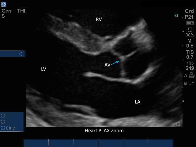
Heart PLAX Zoomed Heart PLAX Zoomed Read more about Heart PLAX Zoomed /sites/default/files/201408_IMAGE_EDGE_HEART_PLAX_ZOOMED.jpg Media Library Type Image Media Library Tag 4 Chamber Aortic Valve Cardiac Cardiac Arrest Cardiac Output Cardiomyopathy Chest Pain Contractility Echo Ejection Fraction Fluid Focused Hypotension Image Left Atrium Left Ventrical Left Ventrical Outflow Track Liver Mitral Valves Parasternal Long Axis Pericardial Pericardial Effusion Pulmonary Emboli Right Ventrical Rt Atrium Septal Soundbyte Cases Tamponde Trauma Volume Window X-Porte
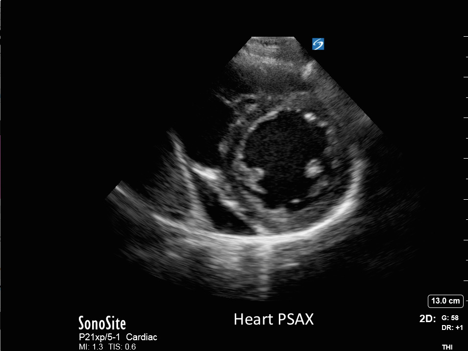
Heart - PSAX View Heart - PSAX View Read more about Heart - PSAX View /sites/default/files/201408_IMAGE_X-PORTE_HEART_PSAX.jpg Left ventricular short-axis image of the left sternum Clinical Specialties Cardiology Media Library Type Image Media Library Tag 4 Chamber Aortic Valve Apex Cardiac Cardiac Arrest Cardiac Output Cardiomyopathy Chest Pain Contractility Echo Ejection Fraction Fluid Focused Hypotension Left Atrium Left Ventrical Mitral Valves Papillary Muscles Parasternal Short Axis Pericardial Pericardial Effusion Pulmonary Emboli Right Ventrical Rt Atrium Septal Tamponde Trauma Volume Wall Motion Window
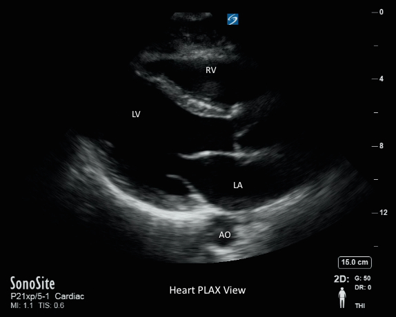
Heart - PLAX View Heart - PLAX View Read more about Heart - PLAX View /sites/default/files/201408_IMAGE_X-PORTE_HEART_PLAX.jpg Media Library Type Image Media Library Tag 4 Chamber Aortic Valve Cardiac Cardiac Arrest Cardiac Output Cardiomyopathy Chest Pain Contractility Echo Ejection Fraction Fluid Focused Hypotension Image Left Atrium Left Ventrical Left Ventrical Outflow Track Liver Mitral Valves Parasternal Long Axis Pericardial Pericardial Effusion Pulmonary Emboli Right Ventrical Rt Atrium Septal Soundbyte Cases Tamponde Trauma Volume Window X-Porte
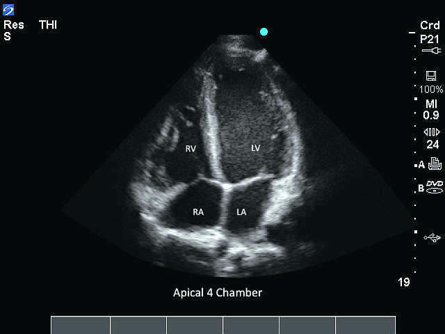
M-Turbo: Apical 4 Chamber M-Turbo: Apical 4 Chamber Read more about M-Turbo: Apical 4 Chamber /sites/default/files/201410_Image_M-Turbo_P21_Apical_4_Chamber.jpg M-Turbo: Apical 4 Chamber. Media Library Type Image Media Library Tag 4 Chamber Aortic Apical Atrium Cardiac Cardiac Output Cardiomyopathy Chronic Heart Disease Congested Heart Disease Ejection Faction Fluid Overload Free Fluid Heart Heart Failure Hypotension Image Left Atrium Left Ventricular Normal Pathology Pericardial Fluid Pericardium Right Ventrical Septal Septum Size Tamponade Trauma Ventrical Ventrical Size Wall Motion Window
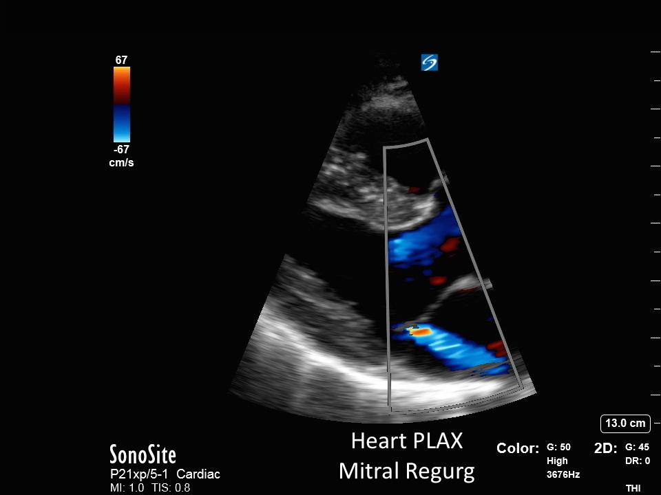
Heart - PLAX Mitral Regurg Heart - PLAX Mitral Regurg Read more about Heart - PLAX Mitral Regurg /sites/default/files/201408_IMAGE_X-PORTE_HEART_PLAX_MITRAL_REGURG.jpg Media Library Type Image Media Library Tag 4 Chamber Aortic Valve Cardiac Cardiac Arrest Cardiac Output Cardiomyopathy Chest Pain Contractility Echo Ejection Fraction Fluid Focused Hypotension Image Left Atrium Left Ventrical Left Ventrical Outflow Track Liver Mitral Valves Parasternal Long Axis Pericardial Pericardial Effusion Pulmonary Emboli Right Ventrical Rt Atrium Septal Soundbyte Cases Tamponde Trauma Volume Window X-Porte
Case: Cardiac Ultrasound - Parasternal Short Axis This video details the use of bedside cardiac ultrasound imaging, specifically the parasternal short-axis view, with a phased array probe to evaluate cardiac health and anatomy, especially when looking at a patient's left ventricular contractility.